Keywords
Potassium; Phosphorous; White blood cells; Red blood cells; Haemoglobin; Packed cell volume
Introduction
Generally, plant parts (seeds, leaves, bark, fruits, stems) contain bioactive agents with medicinal properties but require proper assessment, including toxicity studies, before possible use in animals, including human, as conventional drugs. Avocado plant (Persea americana), a plant belonging to the family of Lauraceae and genus, Persea bears fruit known as avocado pear or alligator pear that contains the avocado pear seed. Reported uses of avocado pear seed include use in the management of hypertension, diabetes, cancer and inflammation [1-4]. Avocado pear seeds, usually discarded as food wastes, could in particular affect monosodium glutamate-induced effects in animals. Monosodium glutamate (MSG) is a sodium salt of L-glutamic acid frequently used as a food flavour enhancer [5]. It is common in packaged foods without appearing on the label [6]. The inadvertent use and possible abuse of MSG could elicit some adverse effects on especially MSG-sensitive individuals, and studies have either established or suggested toxic effects of MSG on some vital organs in experimental animals [7-9]. These reports not withstanding, there was no regulation against the continued use of monosodium glutamate as flavour enhancer. These warranted continued search for natural products that could mitigate the adverse effects of MSG and necessitated this study aimed at determining some mineral contents in avocado pear seed flour and the effect of the ethanolic extract on some haematological parameters in normal and monosodium glutamate-intoxicated rats. The objectives to achieving this aim was by determining in the sample flour some minerals content including sodium, potassium, calcium, magnesium, phosphorus, zinc and iron as well as by determining in the whole blood of exposed rats some hematological parameters, including total leucocyte/white blood cell count (WBC), erythrocyte/red blood cell count (RBC), packed cell volume (PCV) and haemoglobin concentration (HB). In particular, Atasie and Egbuonu [10] recognized hematological parameters as useful bioindicator of health and physiological functions.
Materials and Methods
Collection, identification, preparation and extraction of plant materials
A commercially available brand of MSG (99% purity) used in this study was procured from Ubani market, a daily food condiments market in Umuahia, south east Nigeria. Chemicals and solvents used in this study were products of reputable companies procured from reputable chemical dealers and were used without further purification. Matured avocado pear fruits were bought in a local market in Umuahia, a town close to Michael Okpara University of Agriculture Umudike, during the fruiting season of June, 2015. The fruit was identified as Persea americana mill (Lauraceae) in the Plant Science Department of Michael Okpara University of Agriculture Umudike, Abia State, Nigeria. The fruits were deseeded, the seeds washed with clean tap water, crushed into smaller pieces with the help of manual grater and sun-dried for three days. The dried seeds were milled into powder using a laboratory miller (ED-5, USA) and stored in an air tight container until used for the determination of sodium, potassium, calcium, magnesium, phosphorus, zinc and iron content. The avocado seed flour was extracted by cold maceration method using ethanol as the extracting solvent. The extraction involved weighing 700 g of the avocado pear seed flour into a volumetric flask, soaking in 1400 mL of 90% ethanol with intermittent shaking and stirring for three days and thereafter filtering with No 1 Whatmann filter paper. The filtrate was concentrated using water bath at 60oC and was further dried in an oven at 50oC. The extract was packed into a sample bottle and stored in a refrigerator until used as in the animal study design to assess the effect on normal and monosodium glutamate-intoxicated rats’ hematology.
Animal experimentation
The MSG-intoxicating dose for the rats was 8000 mg/kg body weight for 14 days according to Mariyamma et al. [7] as supported by other studies [11-14]. The ethanolic extract of avocado pear seed (1 g) was dissolved in 10 mL of distilled water as the stock solution and three graded doses were selected as follows: low, medium and high doses (100 mg/kg body weight, 300 mg/kg body weight and 500 mg/kg body weight), respectively. Twenty-four albino rats (Rattus norvegicus) of either sex (mean body weight, 96.00 ± 10.00 g) used in this study were obtained from the animal breeding unit of the College of Veterinary Medicine, University of Nigeria, Nsukka. The animals were acclimatized for 1 week and then randomized (based on weight) to six experimentation groups with sample size of four rats as described below. Rats in the normal control group were sham-dosed with distilled water (without either the extract or MSG) while rats in the MSG group (negative control) were fed intoxicating dose (8000 mg/kg body weight) of MSG according to Mariyamma et al. [7]. Rats in the extract group (Extract control group) were fed ethanolic extract of avocado pear seed flour at 300 mg/kg body weight while rats in the MSG + low extract group were concomitantly exposed to ethanolic extract of avocado pear seed flour (100 mg/kg body weight) and intoxicating dose of MSG (8000 mg/kg body weight) whereas rats in the MSG + medium extract group were co-administered 300 mg/kg body weight of ethanolic extract of avocado pear seed flour and intoxicating dose of MSG (8000 mg/kg body weight). Rats in the MSG+high extract group were concomitantly exposed to ethanolic extract of avocado pear seed flour (500 mg/kg bw) and intoxicating dose of MSG (8000 mg/kg body weight). The exposure was per oral using orogastric tube and daily for 2 weeks (14 days).
Ethical consideration
The animals were placed in rat cages kept in a well-ventilated room and allowed free access to standard feed and clean tap water throughout the experimentation period. Animals were exposed to natural room temperature with a 12 hour day/night cycle. This study considered and adhered to the standard ethical use of experimental animals. Throughout out the experimentation (acclimatization and exposure periods), all rats were housed at 25oC in stainless steel cages under normal daylight/dark cycle and humid tropical conditions. The rats were allowed free access to rat feed (Vital feed, Jos Nigeria) and tap water, and generally received humane care in accordance with the guidelines of the National institute of Health, USA for ethical treatment of laboratory animals as approved by the various (departmental and college) ethical committees of Michael Okpara University of Agriculture Umudike, Nigeria.
Sacrifice and blood sample collection
After 2 weeks (14 days) exposure, the rats were sacrificed the next day after overnight fast by cervical dislocation and the blood sample of the respective rats was collected individually from the heart using a syringe into a clean anti-coagulated polystyrene tube. The whole blood samples thus collected were respectively stored in deep freezer for the determination of hematological parameters, including total leucocyte/white blood cell count (WBC), erythrocyte/red blood cell count (RBC), packed cell volume (PCV) and haemoglobin concentration (HB).
Determination of Studied Parameters
Determination mineral contents in the sample flour
The sodium and potassium contents of the sample flour were determined by flame photometric method, using Jaway Digital Flame Photometer. Briefly, serial dilution of standard sodium and potassium were separately prepared and the flame photometer equilibrated. Then, 1 mL each of the standards was aspirated into the flame photometer and sprayed over the non-luminous flame. The optical density of the resulting emission from each standard solution was recorded against time and corresponding graphs plotted to obtain the standard curve used in extrapolating the content of each test element in the sample. The calcium and magnesium contents of the sample flour were determined by Vasernate Ethylenediaminetetraaceticacid (EDTA) titrimetric method. A solution of the sample flour (20 mL) was placed into a conical flask and treated with pinches of hydroxylamine, hydrochloride, sodium cyanide and sodium potassium perocyanide. The flask was shaken and the mixture dissolved. The, 20 mL of ammonia buffer was added to raise the pH to 10.00 (appoint at which calcium and magnesium form complexes with EDTA). The mixture was titrated against 0.02 N EDTA solution using Eriochrome black-T as indicator. A reagent blank was also titrated, and titration in each case was done from deep red to a permanent blue end point. The titration value represented both Ca2+ and Mg2+ in the test sample. A repeat titration was done to determine Ca2+ alone in the test sample. This was done in similarity with the above titration. However, 10% NaOH was used in place of ammonia buffer and Solechrome dark blue indicator in place of Eriochrome black-T. From the values obtained, the Ca2+ and Mg2+ content were calculated. Phosphorus content in the sample flour was determined by molybdovanadate method involving thus of ammonium molybdenum and based on the inorganic phosphorus-ammonium molybdovanadate colored complex formation [15]. In brief, a measured volume of dry ash digest (2 mg) of sample was dispersed into a 50 mL volumetric flask. The same volume of distilled water and standard P solution were measured into different flask to serve as a regent blank and standard respectively. Then, 2 mL of phosphorus colour reagent was added to each of the flasks and allowed to stand at room temperature for 15 minutes. The content of the flask was diluted to the 50 mL mark with distilled water and its absorbance was measured using a spectrophotometer at a wavelength of 540 nm with the reagent blank set at zero. Zinc and iron contents in the sample flour were determined by spectrophotometric method. Pre-weighed sample flour (0.5 g) was placed into a 100 mL pyrex conical flask and 5 mL of the wet acid digestion reagent (H2SO4- selenium-salicyclic acid) was added and allowed to stand at ambient temperature for about16 hours. The sample was placed on a digestion block and heated at 20°C for 2 hours. The sample was removed from the block and 5 mL of concentrated perchloric acid added and then placed back on the digestion stand with the temperature raised to range of 80°C-150°C. The digestion continued until a white fumes emerged showing a clear digest to indicate the completion of digestion. The sample was removed from the digestion stand, allowed to cool and made up in a 100 mL volumetric flask with distilled water and the concentration of zinc and iron determined with a spectrophometer (Jenway 6305, Jenway Equipment Company, England).
Determination of the hematological parameters of rats’ whole blood
Erythrocyte/red blood cell count (RBC) of the rats exposed as in the study design was determined by haemocytometric method as described by Ochei and Kolhatkar [16]. The method involved diluting the blood specimen to 1:200 with RBC diluting fluid and the RBC cells counted under high power (40X) objective, using a counting chamber and then calculated and reported as concentration of RBC per whole blood.
Total leucocyte/white blood cell count (WBC) was determined by haemocytometric method as described by Ochei and Kolhatkar [16]. The procedure involved diluting the blood specimen to 1:20 in a WBC pipette with the diluting fluid and the counting the cells under low power microscope using a counting chamber and calculated and reported as total WBC per whole blood. The packed cell volume (PCV) was estimated as described by Ochei and Kolhatkar [16]. Blood sample was taken with a heparinised capillary tube, cleaned and sealed with plasticine. The filled tubes were placed in the microhaematocrit centrifuge and spun at 12,000 g for 5 minutes. The tubes were then placed into a specially designed scale and the PCV was read as percentage content in a whole blood, using a hematocrit reader (Hawskey, England). The haemoglobin concentration (HB) was determined using cyanomethaglobin method as described by Ochei and Kolhatkar [16]. This was based on the principle that hemoglobin when mixed with Drabkin’s solution (that contains potassium ferricyanide, potassium cyanide and potassium dihydrogen phosphate) the ferricyanide form methaemoglobin is converted to cyanmethaemoglobin by the cyanide and the cyanmethaemoglobin produces a colour which is measured colorimetrically.
Statistical Analysis
Descriptive statistics and test for significance difference in mean were carried out on the data generated by analysis of variance (ANOVA) with the statistical package for social sciences (SPSS) for Windows version 16. The turkey HSD post hoc test was used to identify the means that differ significantly at p ≤ 0.05. Results were expressed as mean ± standard error of mean, SEM (Table 1). As shown on Table 1, the value of the determined minerals content in the sample flour was potassium (43.73%)>phosphorus (28.06%)>sodium (22.11%)>calcium (21.12%)>magnesium (10.23%)>zinc (2.04%)>iron (1.04%).
Table 1 Some minerals composition of avocado pear (Persea americana) seed flour.
| Minerals |
Concentration (mg/100 g) |
| Sodium |
22.11 ± 0.01 |
| Calcium |
21. 12 ± 0.02 |
| Magnesium |
10.23 ± 0.01 |
| Potassium |
43.73 ± 0.01 |
| Phosphorus |
28.06 ± 0.02 |
| Iron |
1.04 ± 0.01 |
| Zinc |
2.04 ± 0.00 |
Values are mean ± SEM for n=2 (duplicate determinations).
Difference considered statistically significant at p<0.05.
As shown in Table 2 and Figure 1, assessment of the hematological effect of the ethanolic extract of the avocado pear seed in normal and monosodium glutamate-intoxicated rats showed that MSG intoxication caused a significant (p<0.05) reduction in HB, RBC and WBC but did not alter the PCV counts in the rats compared to control rats. Rats exposed to the ethanolic extract of the sample had a significant (p<0.05) reduction in HB, RBC and WBC but an increase (p<0.05) in the PCV counts compared to the control rats. Exposing rats to intoxicating dose of MSG together with increasing concentration of the ethanolic extract of avocado pear seed decreased (p<0.05) the HB, PCV and WBC counts of the rats in a dose dependent manner but increased (p<0.05) the RBC count of the rats in a non-dose-dependent fashion, and the observation was marked compared to that in the group exposed to either MSG or the sample extract alone.
Table 2 Effect of ethanolic extract of avocado pear seed (ASE) on some hematological parameters of normal and monosodium glutamate-intoxicated rats’ whole blood.
| GROUPS |
PCV (%) |
RBC(×109) |
WBC(×103/L) |
| Normal control (feed+water only) |
35.00±4.95 |
450.00±45.64 |
4000.00±15.74 |
| MSG/Negative control (8000 mg/kg bw MSG) |
35.00±4.64 |
412.50±31.46 |
2450.00±10.81 |
| Extract group (300 mg/kg bw extract) |
37.00±1.47 |
387.50±12.50 |
2550.00±17.21 |
| Low extract co-treated group (MSG, 8000 mg/kg bw +100mg/kg bw extract) |
21.50±0.65 |
487.50±31.46 |
1500.00±12.78 |
| Medium extract co-treated group (MSG, 8000 mg/kg bw+300 mg/kg bw extract) |
34.00±2.94 |
487.50±12.50 |
2175.00±14.74 |
| High extract co-treated group (MSG, 8000 mg/kg bw+500mg/kg bw extract) |
33.00±3.70 |
425.00±43.30 |
2150.00±10.03 |
Values are mean ± SEM for n=4.
Difference considered statistically significant at p<0.05.
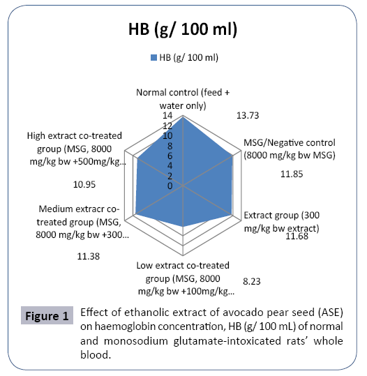
Figure 1: Effect of ethanolic extract of avocado pear seed (ASE) on haemoglobin concentration, HB (g/ 100 mL) of normal and monosodium glutamate-intoxicated rats’ whole blood.
Discussion
Avocado pear seed, obtained after consuming the avocado fruit flesh and usually discarded as waste could contain important minerals and be useful in mitigating monosodium glutamate intoxication. Thus, this study determined some minerals content of avocado pear seed flour and assessed the effect of the ethanolic extract of the avocado pear seed flour on the hematological indices of normal and monosodium glutamate-intoxicated rats via standard protocols. Potassium content in the avocado pear seed flour was higher (p<0.05) than the other determined minerals followed by phosphorous, sodium, calcium, and magnesium while the least was zinc followed by iron (Table 1), suggesting that avocado pear seed could be a rich source for particularly potassium, phosphorous, sodium and calcium which are of biochemical importance and, some, with specialized pumps. In particular, potassium plays a vital role in the maintenance of acid base balance and normal nerve stimulation and function [17] and in concert with sodium is critical to sodium-potassium pump. The potassium content (43.73%) reported in this study was higher than (0.41 %) reported by Parksoy et al. [18] for watermelon seed though quite lower than the value (350 %) reported by Akpabio [19] for almond seed. Magnesium content (10.23%) was higher than (6.40 %) reported by Nwofia et al. [20] for Carica papaya seed while calcium content reported in this study (21.12 %) was higher compared with the value (1.53 %) reported by Parksoy et al. [18] but comparable with the value (18.5 %) reported by Uzama et al. [21] for watermelon (Citrullus lanatus) seed. In particular, calcium is essential for bone health [22]. The phosphorus content (28.06%) could be compared with (31.33 ± 6.11 %) reported by Arukwe et al. [23] for Persea americana seed and higher than that (10%) reported by Akpabio [19] for almond seed and (0.74%) reported by Parksoy et al. [18] for Citrullus lanatus seed. Phosphorous is involved in bone growth, kidney function, cell growth and maintenance of acid-alkaline balance in human [24], suggesting that avocado pear seed could be a good phosphorous nutrient source in such conditions. Generally, high sodium resulting in high sodium:potassium ratio could worsen high blood pressure in human. However, sodium content of avocado peer seed flour (22.11 ± 0.01) vis a vis that of potassium as reported in this study could favour higher potassium:sodium ratio. The iron (1.04%) and zinc (2.04%) content reported in this study though slightly higher could be compared with the values (0.31%) and (0.09%) respectively reported by Arukwe et al. [23] also for Persea americana seed but the iron content compared with (1.22%) reported by Uzama et al. [21] while the zinc content was higher than the value (0.5%) reported by Uzama et al. [20]. Iron is involved in haem formation while zinc plays a vital role in wound healings. Generally, the respective minerals value was lower than the corresponding value obtained for watermelon rind and seed [25] while sodium, iron and zinc content in the sample were lower than the corresponding value reported by Egbuonu and Oriji [11] for mango seed kernel. The result suggests that avocado pear seed flour could not be good source for iron and zinc, though these (iron and zinc) are required only in trace amount in animal physiology. Thus, the preponderance of these minerals and at the concentration in the avocado pear seed flour may be nutritionally important warranting further investigations, for instance to screen out any possible toxic compound in the avocado pear seed or to assess the influence on important physiological functions in animal models.
In this study, the hematological effect of the ethanolic extract of the avocado pear seed flour was assessed in normal and monosodium glutamate-intoxicated rats. MSG intoxication according to Mariyamma et al. [7] caused a significant (p<0.05) reduction in HB, RBC and WBC but did not alter the PCV counts in the rats as compared to control, indicating and confirming adverse response on the hematological function of MSGintoxicated rats. Rats exposed to the ethanolic extract of the sample had a significant (p<0.05) reduction in HB, RBC and WBC but an increase (p<0.05) in the PCV counts compared to the control rats, suggesting sample extract-induced alteration of the rats hematological parameters and bio-functions. Exposing rats to intoxicating dose of MSG together with increasing concentration of the ethanolic extract of avocado pear seed elicited a dose dependent response on the hematological parameters that was similar, but marked, compared to that in the group exposed to either MSG or the sample extract alone, probably indicating that MSG and the ethanolic extract of avocado pear seed flour together elicited synergistic adverse response in the rats hematological indices and, perhaps, biofunctions. The hematology results obtained in this study for MSG intoxication could be compared with the effect (reduction), but not with the values reported by Mbah and Egbuonu [13], owing perhaps to the different methods used in the various study determination of the hematological parameters. The packed cell volume range observed in this study (21.50%-37.00%) was within the range (25%-45%) as reported by Oleforuh-Okoleh et al. [26] though for broiler chicks. This suggests unaltered (as observed in this study) or none adverse response of MSG and the sample extract either alone or in combination on the PCV count of the rats, in apparent agreement with earlier reported MSG-induced effect on rats’ hematology [27]. However, the observation on the WBCs of the rats especially in those concomitantly exposed to the extract and intoxicating dose of MSG was particularly worrisome considering the implication of apparent significant adverse influence related altered immune response in the rats [28-30]. Further investigations are warranted and recommended before harnessing the preponderant minerals in avocado pear seed reported in this study.
Conclusion
Thus, avocado pear seed usually discarded as a waste has appreciable concentration of the determined minerals, notably potassium, phosphorous, sodium and calcium. However, the ethanolic extract of avocado pear seed significantly reduced heamatological parameter counts of the rats and exacerbated the monosodium glutamate-induced effect in the rat’s hematology. Further studies are therefore warranted before the exploitation of the appreciable mineral contents of the avocado pear seed by animals.
References
- Adeyemi OO, Okpo SO, Ogunti OO (2002) Analgesic and anti-inflammatory effects of the aqueous extract of leaves of Perseaamericana Mill (Lauraceae). Fitoterapia 73: 375-380.
- Ojewole JA, Amabeoku GJ (2006) Anticonvulsant effect of Perseaamericana Mill (Lauraceae) (Avocado) leaf aqueous extract in mice. Phytotherapy Research 20: 696-700.
- Anaka ON, Ozolua RI, Okpo SO (2009) Effects of the aqueous seed extract of Perseaamericana mil (Lauraceae) on the blood pressure of spraguedawley rats. African Journal of Pharmacy and Pharmacology 3: 485-490.
- Alhassan AJ, Sule MS, Atiku MK, Wudil AM, Abubakar H, et al. (2012) Effects of aqueous avocado pear (Perseaamericana) seed extract on alloxan induced diabetes rats. Greener Journal of Medical Sciences 2: 005-011.
- Ilegbedion IG, Onyije FM, Chibuike OO (2013) Infiltration of inflammatory cells in the ovary following oral administration of monosodium glutamate. Bangladesh Journal of Medical Science 12: 413-418.
- Egbuonu ACC, Osakwe ON (2011) Effects of high monosodium glutamate on some serum markers of lipid status in male Wistar rats. Journal of Medicine and Medical Sciences 2: 653-656.
- Mariyamma T, Sujatha KS, George S (2009) Protective effect of Piper longum Linn on monosodium glutamate-induced oxidative stress in rats. Ind J Exp Biol 47: 186-192.
- Farombi EO, Abarikwu SO, Adedara IA, Oyeyemi MO (2007) Curcumin and kolaviron ameliorate di-n-butylphthalate-induced testicular damage in rats. Basic Clin Pharmacol Toxicol 100: 43-48.
- Egbuonu ACC, Ezeokonkwo CA, Ejikeme PM, Obidoa O, Ezeanyika LUS (2010) Some biochemical effects of sub-acute oral administration of L-arginine on monosodium glutamate-fed Wistar rats 2: Serum alkaline phosphatase, total acid phosphatase and aspartate aminotransferase activities. Asian Journal of Biochemistry 5: 89-95.
- Atasie OC, Egbuonu ACC (2017) Quality Assessment of Waterside River, Ogbor Hill, Aba 1: Effect of Three-Point Samples on Some Hematological Parameters of Wistar Rats. Int J Hydro 1: 00013.
- Egbuonu ACC, Oriji SO (2017) Pulverized Mangiferaindica (mango) seed kernel mitigated monosodium glutamate-intoxicated rats’ kidney histology and bio-functions. Journal of Nutritional Health and Food Science 5: 1-7.
- Mbah UO, Egbuonu ACC (2017a) Ethanolic extract of Solanummelongena Linn fruit mitigated monosodium glutamate-induced oxidative stress. International Journal of Biochemistry Research & Review 18: 1-8:
- Mbah UO, Egbuonu ACC (2017b) Ameliorative potentials of eggplant (Solanummelongena Linn) fruit ethanolic extract on monosodium glutamate-intoxicated rats' lipid profile, haematology and heart histology. International Journal of Biochemistry Research and Review 18: 1-10.
- Egbuonu ACC, Ekwuribe GA (2017) Pulverized Mangiferaindica (mango) seed-kernel modulated serum lipid profile in monosodium glutamate-challenged rats. Journal of Applied Biotechnology 5: 72-87.
- Lehane DP, Werner M (1973) Automated Microdetermination of Inorganic phosphorus in serum and urine by the ammonium molybdovanadate method. Annals of Clinical Laboratory Science 3: 448-453.
- Ochei J, Kolhatkar A (2008) Medical Laboratory Science: Theory and Practice. Tata McGraw Hill Publishing Co. Ltd, New York, USA, ISBN-13:978-0074632239.
- Kala BK, Mohan VR (2010) Nutritional and antinutritional potential of three accessions of itching bean (Mucunapruriens(L.) DC var. pruriens): An underutilized tribal pulse. International Journal of Food Science and Nutrition 61: 497-511.
- Parksoy MA, Aydin C, Turkmen O, Seymen M (2010) Modeling of some physical properties of watermelon (Citrulluslanatus (Thunb.) Mansf.)seeds depending on moisture contents and mineral compositions. Pak J Bot 42: 2775-2783.
- Akpabio UD (2012) Evalution of proximate composition, mineral element and anti-nutrients in almond seeds.AdvApplSci Res 3: 2247-2252.
- Nwofia GE, Ojimelukwe P, Eji C (2012) Chemical composition of leaves, fruit pulp and seeds in some Carica papaya (L) morphotypes. Int J Med Aromatic Plants 2: 200-206.
- Uzama D, Bwai DM, Magu J, KabirMGi, Oloninefa SD (2015) Proximate, mineral composition, phytochemical constituents and characterization of oil of Citrulluslanatus seeds. American Journal of Bioscience and Bioengineering 3: 72-75.
- Okwu DE, Emenike IN (2007) Nutritive value and mineral content of different varieties of citrus fruits. J Food Technol 5: 105-108.
- Arukwe U, Amadi BA, Duru MKC, Agomuo EN, Adindu EA, et al. (2012) Chemical composition of Perseaamericana leaf, fruit and seed. IJRRAS.
- Fallon S, Enig MG (2001) Nourishing Traditions. The Cookbook that Challenges Policitally Correct Nutrition and the Diet Dictocrats. Revised (2nd Edn) pp: 40-45.
- Egbuonu ACC (2015) Comparative assessment of some mineral, amino acid and vitamin compositions of watermelon (Citrulluslanatus) rind and seed. Asian Journal of Biochemistry 10: 230-236.
- Oleforuh-Okoleh VU, Olorunleke SO, Nte IJ (2015) Comparative response of bitter leaf (Vernoniaamygdalina) infusion administration on performance, haematology and serum biochemistry of broiler chicks. Asian J Anim Sci 9: 217-224.
- Kolawole OT (2013) Assessment of the effects of monosodium glutamate on some biochemical and hematological parameters in adult Wistar rats. American Journal of Bio Science1: 11-15.
- Maluly HDB, Areas MA, Borelli P, Reyes FGR (2013) Evaluation of biochemical, hematological and histological parameters in non-diabetic and diabetic Wistar rats fed monosodium glutamate. Food and Nutrition Sciences 4: 66-76.
- Egbuonu ACC, Ogbu AE, Ijeh II, Ezeanyika LUS (2015) Sub-chronic esculetin (6,7-dihydroxy-coumarin)-induced alteration in some haematological and serum parameters in normal male Wistar rats. Asian Journal of Biochemistry 10: 306-311.
- Ogunyemi IO, Abiola MT, Ojokuku S, Odesanmi OS (2015) Haematological effect of ethanolic extract of Uvariachamaeon monosodium glutamate (MSG)-induced toxicity in sprague-dawley rats Annals of Biological Research 6: 17-22.