M V Raghavendra Rao
Avalon University School of Medicine, USA
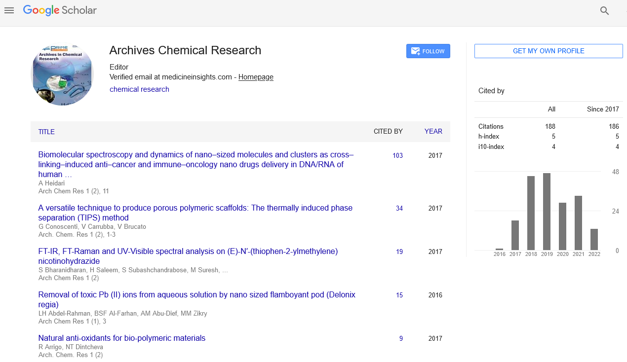
Posters & Accepted Abstracts: Arch Chem Res
Electrophoresis is the movement of charged particles (ions) in an electric field resulting in their migration towards the oppositely charged electrode. Molecules with a net positive charge (cation) move towards the negative cathode while those with negative charge (anion) migrate towards positive anode. It is a widely used analysis technique for the separation of biological molecules such as plasma proteins, lipoproteins and immunoglobulins. Traditionally, two classes of blood proteins are considered: serum albumin and globulin. They are generally equal in proportion, but albumin as a molecule is much smaller and lightly, negatively-charged, leading to an accumulation of albumin on the electrophoretic gel. Abnormal bands (spikes) are seen in monoclonal gammopathy of undetermined significance of multiple myeloma. The globulins are classified by their banding pattern (with their main representatives): 1. the alpha (α) band consists of two parts, 1 and 2: α1 - α1-antitrypsin, α1-acid glycoprotein; α2 - haptoglobin, α2-macroglobulin, α2-antiplasmin, ceruloplasmin.2.the beta (β) band -transferrin, LDL, complement. 3. the gamma (γ) band - immunoglobulin (IgA, IgD, IgE, IgG and IgM). Paraproteins (in multiple myeloma) usually appear in this band. Normal present medical procedure involves determination of numerous proteins in plasma including hormones and enzymes, some of them also determined by electrophoresis. However, gel electrophoresis is mainly a research tool, also when the subject is blood proteins. We are commonly using the following dyes/ stains. Acridine orange, C.I. 46005, very high purity. A grade of acridine orange of exceptionally high purity, suitable for quantitative work helps in detection of nucleic acids separated by gel electrophoresis. Alcian Blue 8GX, C.I. 74240 used in electrophoresis for detecting glycoproteins and other research needs. Bromophenol Blue, 24995 Bromophenol Blue is useful as an acid base indicator, a color marker and a dye. Suitable as a color marker in agarose gel electrophoresis and polyacrylamide gel electrophoresis and commonly used as a dye to stain proteins in slide. Coomassie® Blue 03707 protein stain for SDS gels used in dye binding techniques for protein quantification. Ethidium Bromide Cat. # 04033 intercalates double-stranded nucleic acids; frameshift mutagen; fluorescent stain for nucleic acids in electrophoresis used for DNA quantitation. Pyronin Y, C.I. 45005, Certified Cat. #18614 used in combination with methyl green for the selective and differential staining of nucleic acids. These are costly and sometimes non available. Keeping these things in view, we used cheaper, viable, available, and reliable, Lasonia inermis leaves.and Jamun,Java plum (Syzygium cumini) fruits. We obtained similar bands (Spikes) on gel electrophoresis. Botanical name- Lawsonia inermis.
Email:reachdrmvrrao@gmail.com
 Spanish
  Chinese
  Russian
  German
  French
  Japanese
  Portuguese
  Hindi
Hindi