Keywords
Amylases; Animal Experimentation; Melatonin; Pancreatitis, Acute Necrotizing
INTRODUCTION
Acute pancreatitis is a disease which develops with perivascular infiltration and inflammation characterized by lipid necrosis, polymorphonuclear leukocyte infiltration, hemorrhage, acinar cell necrosis and tissue edema in the pancreas.
Acute pancreatitis is still a serious disease as its diagnosis and treatment is difficult [1, 2]. It is usually accompanied by mild and restricted clinical conditions; however, necrotizing acute pancreatitis may sometimes result in a serious outcome. Despite current intensive care techniques, the development of nutritional support applications, fluid-electrolyte replacement and mechanic ventilation facilities, the mortality rate may reach 10-20% [1].
Studies published in the literature concerning acute pancreatitis demonstrate that, in the first 24 hours, many morphological changes, such as lipid necrosis in pancreatic tissue, hemorrhage and inflammation occurred in test subjects with occluded pancreatic ducts [3]. In the earlier and later stages of acute pancreatitis, proteolytic enzymes become active after the extravasation of pancreatic secretions into the interstitial space. Inflammation becomes more severe because of edema in the tissue, disruption of the microcirculation and cell ischemia; thus, toxic mediators start to accumulate in the tissue [4].
Two enzymes (amylase and lipase) are released from the acinar cells during acute pancreatitis and their concentration in the serum is used to confirm diagnosis. Serum amylase concentrations exceeding three times the normal upper limit support the diagnosis of acute pancreatitis. The specificity of the diagnostic value an increased amylase level is approximately 95%. It is therefore widely used in the diagnosis of acute pancreatitis and the assessment of its severity as it is considered an important indicator of the disease [4, 5, 6].
Melatonin is a hormone which affects the general immune system and cellular immunity both directly and indirectly. Melatonin, which is synthesized within pinealocytes in the pineal gland and secreted rhythmically in accordance with daylight, can overcome all the morphophysiological barriers in the human body and can reach the intercellular area in all organs and cell nuclei easily due to its lipophilic and hydrophilic features [7]. Melatonin is secreted by the intestines as well as by the pineal gland; however, 90% of the melatonin in the body is secreted by the pineal gland [7, 8]. The number of studies carried out regarding the effect of melatonin on amylase values in test subjects who have experimental acute pancreatitis and a high level of amylase is quite limited. The aim of this study was to address the effects of exogenous melatonin on the amylase level in acute pancreatitis. For this purpose, over the period of one week, melatonin (20 mg/kg/day) was injected intraperitoneally into rats with experimentally acute pancreatitis which had been developed through ligation of the pancreatic duct; the differences and changes which occurred in the serum amylase levels were examined in detail and the results were compared statistically with those of the control group.
MATERIALS AND METHODS
In this experimental study, 20 adult Winstar Albino male rats (average weight 200-250 g) were used. The test subjects were divided into two groups of 10 rats each. In the 10 control rats, only experimental acute pancreatitis was caused and they received no other treatment. In the 10 rats of the experimental group, in addition to having acute pancreatitis caused experimentally, intraperitoneal melatonin (20 mg/kg/ day) was injected every day for one week. At the end of one week, blood samples were taken from the test subjects and the test subjects were then sacrificed.
The surgical and medical procedures performed on the experimental animals were carried out under general anesthesia and standard sterile conditions. General anesthesia was administered with 20 mg/kg Ketamin HCl plus 5 mg/kg Xylazine HCl (i.m.) and prophylactic antibiotics were used in all animals. Acute pancreatitis was provoked through surgical ligation of the common pancreatic duct by a 2 cm midline laparotomy. The pancreas was explored and the procedure finalized with duct ligation using 5/0 polypropylene suture.
In the test subjects, 24 hours after the acute pancreatitis was developed, melatonin (Sigma, St. Louis, MO, U.S.A.) 20 mg/kg/day was injected intraperitoneally for one week everyday between 06:00 and 08:00 pm. During the study, one test animal in each group died due to technical complications and problems related to the dose of anesthesia; thus, the study was carried out with 9 test subjects in each group. At the end of the 7th day, 2 mL blood samples were taken from the subjects by means of intracardiac puncture, and were stored in plain biochemistry tubes without anticoagulants (Becton Dickinson, Franklin Lakes, NJ, U.S.A.).
Twenty minutes after withdrawal, the blood samples were centrifuged at 3,500 g for 15 minutes and the serum collected through centrifuging was diluted with isotonic NaCl and the amylase levels were measured with an auto analyzer. Abbott Architect C8000(Chicago, IL, U.S.A.) was used as an amylase kit and the values were recorded in IU/mL.
ETHICS
This experimental animal study was conducted in accordance with the principles and procedures outlined in the “NIH Guide for the care and use of animals” and carried out with ethical approval (Decision number; 2008/116) obtained from the Animal Care Ethics Committee of Abant Izzet Baysal University Medical School.
During the study, all the experimental animals were kept at normal room temperature (22°C) in a 12-h light and 12-h dark cycle and were fed with standard pellet rat food (210 kcal/100 g/day) and tap water.
STATISTICS
Statistical analysis was carried out using the SPSS-11 for Windows program (SPSS Inc., Chicago, IL, U.S.A.). Amylase levels, shown as mean ± standard deviation (SD), were analyzed by means of the Student’s t-test. A two-tailed P value of less than 0.001 was considered as statistically significant.
RESULTS
Before starting the experimental studies, the pancreatic tissue of all the test subjects in both groups was determined to be healthy and the structure of the ducts was normal after laparotomy. Normal samples of pancreatic tissue were taken from a few of the test subjects and examined for comparison and histopathological assessment; however, these test subjects were not used in the experimental study (Figure 1).
Figure 1. a. The macroscopic appearance of a normal pancreas and
duct structure in rats. b. Normal pancreas tissue in rats at light
microscopy (H&E x40).
When re-laparotomy was performed at the end of the experimental study, findings of dilatation in the pancreatic duct as well as edema and inflammation in the pancreas were observed macroscopically (Figure 2a); after taking a blood sample by intracardiac puncture to measure the serum amylase level, the samples of pancreatic tissue were taken for histopathological tests. In the microscopic investigation of the pancreas tissue, polymorphonuclear cell infiltration, edema, inflammation and ductal dilatation were observed (Figure 2b).
Figure 2. a. The macroscopic appearance of acute pancreatitis
caused by duct ligation. There is very clear extreme dilatation in the
choledochus and edema in the pancreatic tissue. b. Histopathological
findings of acute pancreatitis in test subjects (H&E x40).
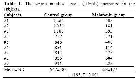
The serum amylase levels at the end of the 7th day of the experimental study were found to have a minimum of 717 IU/mL and a maximum of 1,262 IU/mL in the rats of the control group (Table 1). The average serum amylase was 947±182 IU/mL. On the other hand, it was found that, in the melatonin group, the amylase levels ranged between 116 and 684 IU/mL, and the average value was determined to be 358±177 IU/mL. The difference between the amylase levels of the control and melatonin groups was statistically significant (t=6.95; P<0.001).
DISCUSSION
Figure 1 shows the consistency of the macroscopic and microscopic appearance before acute pancreatitis with the normal pancreatic tissue. The observation of duct dilatation along with edema in the pancreas macroscopically after experimental acute pancreatitis was developed is given in Figure 2. The microscopic determination of the histopathological findings of acute pancreatitis proves that acute pancreatitis was successfully developed after the ligation of the pancreatic duct.
In the subjects of the control group, the fact that the serum amylase level at the end of the 7th day varied between 717 and 1,262 IU/mL (mean±SD: 947±182 IU/mL) was considered to be a biochemical indicator showing that experimental acute pancreatitis had been developed successfully. The increase in amylase level (358±177 IU/mL) in the group which was given exogenous melatonin was less than half of the levels in the control group. According to this finding, it can be concluded that using exogenous melatonin prevents amylase values from increasing more than necessary or decreases a serum amylase level which was already too high
Melatonin is a hormone which can be found in body fluids such as cerebrospinal fluid, saliva and bile; it is not stored and is rapidly released into the general circulation immediately after being synthesized by the pineal gland. The concentration of melatonin in these body fluids is almost 2-3 times higher than its concentration in serum [9]. The night-time secretion of melatonin takes place in the pineal gland while it is secreted by gastrointestinal system mucosa during the day, especially after the intake of food.
Melatonin does not undergo auto-oxidation, redox cycling or reactions producing hydroxyl radicals. Therefore, in contrast to other antioxidants, it does not have a toxic effect even if it is used in very high doses (300 mg/day) and for a long time [10].
Melatonin, which can overcome all body barriers and the placenta, can easily be effective for all intracellular components. Thus, melatonin can protect the cell wall, organelles and cell nucleus from damage by free radicals [8, 11]. Deactivation of melatonin occurs mainly in the liver by microsomal enzymes which metabolize melatonin into 6-hydroxymelatonin.
It has previously been shown that normal serum amylase levels in healthy rats on a standard diet are approximately 250 IU/mL, and 65 IU/mL after 12 h fasting [12]. Pancreatic enzyme activity in serum is changed with feeding and pancreatic functioning. The question in the current study was to evaluate whether or not the melatonin had any effect on serum amylase levels. The results of published studies carried out to investigate the effect of melatonin on amylase level differ from each other. Melatonin has been shown to decrease the level of amylase in some studies whereas it has increased it in others [2, 13, 14, 15, 16]. In the rats with a pancreaticobiliary fistula, intraperitoneal administration of melatonin significantly stimulated serum amylase concentrations. Despite the in vivo observations demonstrating that melatonin is a potent pancreatic secretagogue, none of above substances was able to directly affect amylase release from isolated pancreatic acini [14, 16]. A large number of experimental studies have been carried out to demonstrate the protective effect of different doses of melatonin on tissue damage caused by ischemia reperfusion injuries in the liver, kidney and intestines, in addition to burns, compressed injuries and certain medications [17, 18, 19].
Giving exogenous melatonin to healthy organisms resulted in a considerable increase in amylase activity [2, 13, 15]. It has been demonstrated that melatonin decreases lipid peroxydation and tissue edema in experimental acute pancreatitis and thus causes the amylase activity to decrease [13, 14]. As is seen, the results regarding the effects of melatonin on amylase levels vary to a great extent. In the current study, it was found that melatonin decreased the elevated level of amylase in rats with pancreatitis. In considering the results of experimental studies carried out previously and the data obtained in the current study, it can be concluded that injections of exogenous melatonin increase amylase levels in healthy organisms while they help to reduce elevated amylase levels due to acute pancreatitis.
Acknowledgements
The authors would like to thank Dr. Bulent Gunduz and Dr. Aysel Kukner for their collaboration and also Dr. Aysu Kiyan for using SPSS to carry out the statistical analyses
Conflict of interest
The authors have no potential conflicts of interest
References
- Alhan E, Kalyoncu NI, Kural BV, Erçin C. Effects of melatonin on acute necrotizing pancreatitis in rats. Z Gastroenterol 2004; 42:967-72. [PMID 15455265]
- Eşrefoğlu M, Gül M, Ates B, Batçioğlu K, Selimoglu MA. Antioxidative effect of melatonin, ascorbic acid and N-acetylcysteineon caerulein-induced pancreatitis and associated liver injury in rats. World J Gastroenterol 2006; 12:259-64. [PMID 16482627]
- Lerch MM, Saluja AK, Dawra R, Ramaraò P, Saluja M, Steer ML. Acute necrotizing pancreatitis in the opossum: earliest morphological changes involve acinar cells. Gastroenterology 1992; 103:205-13. [PMID 1612327]
- Banks PA, Freeman ML; Practice Parameters Committee of the American College of Gastroenterology. Practice guidelines in acute pancreatitis. Am J Gastroenterol 2006; 101:2379-400. [PMID 17032204]
- Frossard JL, Steer ML, Pastor CM. Acute Pancreatitis. Lancet 2008; 371:143-52. [PMID 18191686]
- Matull WR, Pereira SP, O'Donohue JW. Biochemical markers ofacute pancreatitis. J ClinPathol 2006; 59:340-44. [PMID 16567468]
- Konturek SJ, Konturek PC, Brzozowski T, Bubenik GA. Role of melatonin in upper gastrointestinal tract. J PhysiolPharmacol 2007; 58(Suppl 6):23-52. [PMID 18212399]
- Konturek SJ, Konturek PC, Brzozowska I, Pawlik M, SliwowskiZ, Czesnikiewicz-Guzik M, et al. Localization and biological activities of melatonin in intact and diseased gastrointestinal tract (GIT). J PhysiolPharmacol 2007; 58:381-405. [PMID 17928638]
- Tan D, Manchester LC, Reiter RJ, Qi W, Hanes MA, Farley NJ. High physiological levels of melatonin in the bile of mammals. Life Sci 1999; 65:2523-9. [PMID 10622237]
- Reiter RJ. Interactions of the pineal hormone melatonin with oxygen-centered free radicals: a brief review. Braz J Med Biol Res 1993; 26:1141-55. [PMID 8136717]
- Bülbüller N, Dogru O, Umaç H, Gürsu F, Akpolat N. The effects of melatonin and pentoxiphylline on L-arginine induced acute pancreatitis. UlusTravmaAcilCerrahiDerg 2005; 11:108-14. [PMID 15877240]
- Cloutier M, Gingras D, Bendayan M. Internalization and transcytosis of pancreatic enzymes by the intestinal mucosa. J HistochemCytochem 2006; 54:781-94. [PMID 16517974]
- Jaworek J, Konturek SJ, Tomaszewska R, Leja-Szpak A, Bonior J, Nawrot K, et al. The circadian rhythm of melatonin modulates the severity of caerulein-induced pancreatitis in the rat. J Pineal Res 2004; 37:161-70. [PMID 15357660]
- Jaworek J, Nawrot-Porabka K, Leja-Szpak A, Bonior J, Szklarczyk J, Kot M, et al. Melatonin as modulator of pancreatic enzyme secretion and pancreatoprotector. J PhysiolPharmacol 2007; 58(Suppl 6):65-80. [PMID 18212401]
- Szabolcs A, Reiter RJ, Letoha T, Hegyi P, Papai G, Varga I, etal. Effect of melatonin on the severity of L-arginine-induced experimental acute pancreatitis in rats. World J Gastroenterol 2006; 12:251-8. [PMID 16482626]
- Jaworek J, Nawrot K, Konturek SJ, Leja-Szpak A, Thor P, Pawlik WW. Melatonin and its precursor, L-tryptophan: influence on pancreatic amylase secretion in vivo and in vitro. J Pineal Res 2004; 36:155-64. [PMID 15009505]
- Sener G, Sehirli AO, Satiroglu H, Keyer-Uysal M, CYegen B. Melatonin improves oxidative organ damage in a rat model of thermal injury. Burns 2002; 28:419-25. [PMID 12163279]
- Jahovic N, Cevik H, Sehirli AO, Yegen BC, Sener G. Melatonin prevents methotrexate-induced hepatorenal oxidative injury in rats. J Pineal Res 2003; 34:282-7. [PMID 12662351]
- Kaçmaz A, User EY, Sehirli AO, Tilki M, Ozkan S, Sener G. Protective effect of melatonin against ischemia/reperfusion-induced oxidative remote organ injury in the rat. Surg Today 2005; 35:744- 50. [PMID 16133669]


