Keywords
Exercise, Glucagon, Insulin, Pancreas, Pancreatic Polypeptide, Somatostatin
Abbreviations CHE: control heavy exercise; CS: control sedentary; DHE: diabetic heavy exercise; DS: diabetic sedentary; STZ: streptozotocin; ZDF: Zucker diabetic fatty
INTRODUCTION
Diabetes mellitus is a serious medical problem. The worldwide prevalence of diabetes is rising dramatically. The total number of people with diabetes is projected to rise from 171 million in 2000 to 366 million in 2030 [1]. Cardiovascular complications are the major cause of morbidity and mortality in diabetic patients [2, 3, 4, 5]. Exercise training has long been utilized as an adjunct to pharmacotherapy in the management of diabetes. In terms of pancreatic function, experiments in humans and animals have variously demonstrated that exercise improves insulin resistance, increases insulin sensitivity, increases pancreatic beta-cell mass and generally enhances betacell function and insulinotropic action, especially in type 2 diabetes [6, 7, 8, 9, 10, 11, 12]. Reports on the effect of exercise on the glucose-related metabolic parameters in type 1 diabetes remain controversial, especially in animal models of diabetes [10, 13]. Moreover, the cellular basis of the physiological and clinical effects of exercise training in type 1 diabetes is poorly understood. It is well known that insulin, glucagon, somatostatin and pancreatic polypeptide work in tandem to maintain normoglycemia.
This study investigates the effects of a heavy exercise program on the distribution of the pancreatic hormones insulin, glucagon, somatostatin and pancreatic polypeptide in the streptozotocin (STZ)-induced diabetic rat, a model of type 1 diabetes.
METHODS
Animal Model and Exercise Protocol
Diabetes was induced in male Wistar rats (243.3±1.6 g) with a single intraperitoneal injection of STZ (60 mg/kg body weight, Sigma, St. Louis, USA) dissolved in a citrate buffer. Weight-matched control rats received citrate buffer alone. Animals were divided into 4 groups: 10 control sedentary (CS), 10 diabetic sedentary (DS), 10 control heavy exercise (CHE) and 10 diabetic heavy exercise (DHE) rats. One week after STZ treatment, the CHE and DHE rats started an exercise program which involved 5 60-min sessions per week on a treadmill (Model 800, IITC Life Science, Woodland Hills, CA, U.S.A.) for a period of 12 weeks for the animals in each of the exercise groups. The entire experiment was completed in 12 to 23 weeks. Exercising rats ran at a speed of 18 m/min and on a running belt at a 5% incline. Every exercise session began with a 10 min warm-up when the speed was increased progressively from 0 to 18 m/min. Each lane of the 5 lane treadmill had a shock grid located at one end of the running belt. Mild electrical shocks were used sparingly to motivate the animals to run. Experiments were staggered to ensure equal levels of exercise between different groups of animals. All animals were maintained on the same normal laboratory rodent chow and water ad libitum. Body weight and blood glucose (One Touch BasicPlus; Lifescan, Johnson & Johnson, Langhorne, PA, U.S.A.) were measured at the time of sacrifice. The pancreata were removed within 12 hours after exercise training.
Immunohistochemistry
After sacrificing the rats, the pancreata were rapidly removed from the CS, CHE, DS and DHE rats. Isolated pancreata were trimmed free of adherent fat and connective tissue, and were cut into small pieces (2 mm3) and fixed overnight in freshly prepared Zamboni’s fixative. The tissue samples were later dehydrated in graded concentrations of ethanol, cleared in xylene and subsequently embedded in paraffin wax at 55°C. Sections of 6 μm thickness were cut on a microtome (Shandon AS325, Cheshire, UK). The sections were deparaffinized, transferred into absolute ethanol and processed for immunohistochemistry using established Avidin Biotin Complex methods [14, 15]. Briefly, the sections were then incubated for 30 min in 0.3% hydrogen peroxide solution in methanol to block endogenous peroxidase activity and later treated with a blocking reagent for 30 min before incubation in antibodies against insulin, glucagon, somatostatin and pancreatic polypeptide for 24 h at 4°C. The sections were then washed and incubated for 30 min with prediluted biotinylated anti-rabbit IgG (Sigma, Poole, Dorset, UK) for 30 min, before incubation in streptavidin peroxidase conjugate for 45 min. The peroxidase activity was revealed by incubating the
specimens for 3 min in 3,3-diaminobenzidine tetrahydrochloride containing 0.03% hydrogen peroxide in PBS. The slides were later washed and counterstained with hematoxylin for 30 s before mounting in Cytoseal 60 (Stephens Scientific, Riverdale, NJ, U.S.A.).
The antisera to insulin, glucagon were supplied diluted (Dako, Copenhagen, Denmark). Somatostatin and pancreatic polypeptide were each used at a dilution of 1:2,000. No specific immunostaining was observed in pancreatic tissue when primary antisera were omitted.
Morphometric Analysis of Pancreatic Islet Cells
Sections of pancreata were examined with a Carl Zeiss Scientific microscope (Göttingen, Germany) and photographed with a digital camera attached to the microscope. Slides, containing sections of the pancreas, were prepared from all animals. Twenty slides were selected at random from the different animals and 5 sections were labeled for insulin, 5 for glucagon, 5 for somatostatin and 5 for pancreatic polypeptide. Twenty digital photographs were taken of each slide for subsequent offline analysis. The total number of cells in the islets of the CS, DS, CHE and DHE rats were counted using the Axiovision Microimaging System® (Carl Zeiss, Göttingen, Germany). In addition, insulin, glucagon, somatostatin and pancreatic polypeptidepositive cells within a given islet were also counted.
ETHICS
Approval for this project was obtained from the Faculty of Medicine and Health Sciences Ethics Committee, United Arab Emirates University. All animals received care according to the criteria outlined in the “Guide for the Care and Use of Laboratory Animals (1996)” prepared by the National Academy of Sciences.
STATISTICS
Data were expressed as mean and standard error of the mean (SEM). Statistical comparisons were made using one-way ANOVA with the Bonferroni post hoc adjustment for multiple comparisons. Two-tailed P values less than 0.05 were considered significant. Data were analyzed by means of the SPSS (SPSS Inc., Chicago, IL, U.S.A.; version 15.0 for Windows) statistical package.
RESULTS
General Characteristics
Table 1 shows the data observed in the four groups of rats. Body weight, at time of sacrifice, was significantly (P<0.001) lower in DS (266±10 g) as compared to CS (426±13 g) rats. Body weight was not significantly (P=1.000) altered by heavy exercise in either the diabetic (264±14 g) or the control (412±9 g) rats.
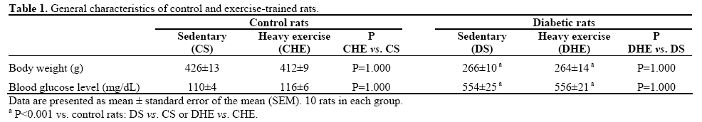
Blood glucose was significantly (P<0.001) elevated in DS (554±25 mg/dL) as compared to CS (110±4 mg/dL) rats. Blood glucose was not additionally altered (P=1.000) by heavy exercise in either the diabetic (556±21 mg/dL) or the control (116±6 mg/dL) rats.
Insulin
Typical micrographs showing the distribution of insulin immunolabeling in the islets of the CS, CHE, DS and DHE rats are depicted in Figure 1. The percentage distribution of insulin-positive cells from the CS, CHE, DS and DHE rats is shown in Figure 2. The percentage distribution of insulin-positive cells was significantly (P<0.001) lower in DS (24.2±2.3%, n=83) as compared to CS (87.5±2.0%, n=82) rats (Figure 2). Insulin-labeling of the islet cells was not additionally altered (P=1.000) by heavy exercise in either the diabetic (24.3±3.0%, n=84) or the control (88.0±1.4%, n=95) rats.
Figure 1. Light micrographs showing the pattern of distribution of
insulin-immunoreactive cells (arrow) in the pancreatic islets of
control sedentary (CS), control heavy exercise (CHE), diabetic
sedentary (DS) and diabetic heavy exercise (DHE) rats.
(Magnification: x200).
Figure 2. Graphs showing the pattern of distribution of insulinpositive
islets. Data are mean±SEM; the total number of islets
evaluated was 82, 95, 83 and 84 in control sedentary (CS), control
heavy exercise (CHE), diabetic sedentary (DS) and diabetic heavy
exercise (DHE) groups, respectively.
Glucagon
Typical micrographs demonstrating the distribution of glucagon in the islets of the CS, CHE, DS and DHE rats are shown in Figure 3. The percentage distribution of glucagon-positive cells from the CS, CHE, DS and DHE rats is shown in Figure 4. The percentage distribution of glucagon-positive cells was significantly (P=0.018) higher in DS (44.0±1.7%, n=84) as compared to CS (34.7±2.1%, n=68) rats. The glucagon labeling of islet cells was not additionally altered by heavy exercise in either the diabetic (50.7±2.1%, n=75; P=0.165) or the control (33.3±2.3%, n=84; P=1.000) rats.
Figure 3. Light micrographs showing the pattern of distribution of
glucagon-immunoreactive cells (arrow) in the pancreatic islets of
control sedentary (CS), control heavy exercise (CHE), diabetic
sedentary (DS) and diabetic heavy exercise (DHE) rats.
(Magnification: x200).
Figure 4. Graphs showing the pattern of distribution of glucagonpositive
islets. Data are mean±SEM; the total number of islets
evaluated was 68, 84, 84 and 75 in control sedentary (CS), control
heavy exercise (CHE), diabetic sedentary (DS) and diabetic heavy
exercise (DHE) groups, respectively.
Somatostatin
Typical micrographs showing the distribution of somatostatin in the islets of the CS, CHE, DS and DHE rats are shown in Figure 5. The percentage distribution of somatostatin-positive cells from the CS, CHE, DS and DHE rats is shown in Figure 6. The percentage distribution of somatostatin-positive cells was not significantly (P=0.263) altered in DS (28.2±2.0%, n=77) as compared to CS (21.9±2.7%, n=73) rats. Somatostatin labeling of the islet cells was not significantly (P=1.000) altered by heavy exercise in either the diabetic (32.3±2.0%, n=74) or the control (19.4±1.7%, n=97) rats.
Figure 5. Light micrographs showing the pattern of distribution of
somatostatin-immunoreactive cells (arrow) in the pancreatic islets of
control sedentary (CS), control heavy exercise (CHE), diabetic
sedentary (DS) and diabetic heavy exercise (DHE) rats.
(Magnification: x200).
Figure 6. Graphs showing the pattern of distribution of somatostatinpositive
islets. Data are mean±SEM; the total number of islets
evaluated was 73, 97, 77 and 74 in control sedentary (CS), control
heavy exercise (CHE), diabetic sedentary (DS) and diabetic heavy
exercise (DHE) groups, respectively.
Pancreatic Polypeptide
Typical micrographs showing the distribution of pancreatic polypeptide in the islets of the CS, CHE, DS and DHE rats are shown in Figure 7. The percentage distribution of pancreatic polypeptide-positive cells from the CS, CHE, DS and DHE rats is shown in Figure 8. The percentage distribution of pancreatic polypeptide-positive cells was significantly (P=0.014) increased in DS (20.8±1.6%, n=79) as compared to CS (12.7±1.8%, n=72) rats. Polypeptide labeling of the islet cells was not additionally altered by heavy exercise in either the diabetic (19.9±2.1%, n=62; P=1.000) or the control (16.2±1.4%, n=96; P=0.987) rats.
Figure 7. Light micrographs showing the pattern of distribution of
pancreatic polypeptide-immunoreactive cells (arrow) in the
pancreatic islets of control sedentary (CS), control heavy exercise
(CHE), diabetic sedentary (DS) and diabetic heavy exercise (DHE)
rats. (Magnification: x200).
Figure 8. Graphs showing the pattern of distribution of pancreatic
polypeptide-positive islets. Data are mean±SEM; the total number of
islets evaluated was 72, 96, 79 and 62 in control sedentary (CS),
control heavy exercise (CHE), diabetic sedentary (DS) and diabetic
heavy exercise (DHE) groups, respectively.
DISCUSSION
The effects of heavy exercise on the distribution of pancreatic hormones in the STZ-induced diabetic rat were investigated. The exercise program commenced one week after the administration of STZ and continued for 12 weeks. The main findings of the study were that: i) the percentage of insulin-positive cells was lower in the islets of the diabetic rats as compared to the controls; ii) the percentage of glucagon-positive and pancreatic polypeptide-positive cells was higher in islets from the diabetic rats as compared to the controls; iii) the percentage of somatostatin-positive cells was similar in islets from the diabetic rats as compared to controls.
The study utilized a heavy exercise regime consisting of enforced running for one h/day, 5 days/week, for 12 weeks at a speed of 18 m/min at a belt gradient of 5%. Earlier studies in both normal and diabetic rats have demonstrated a close correlation between running speed and oxygen uptake suggesting that running speed can be used to assess training intensity [16]. It has also been shown in STZ-induced diabetic rats that, for treadmill speeds from 3 to 19 m/min, oxygen consumption increases progressively as a function of running speed, thus the relationship between the two can be expressed by a simple linear equation. Based only on running speed, i.e., without taking elevation into account, O2 uptake would be equal to approximately 80% of VO2 max [17]. For comparison, this would translate into approximately hard, strenuous running [18]. Thus, the duration and the intensity of exercise applied in the current study are comparable to previous studies which have demonstrated the effects of exercise on the cardiovascular system [19].
As expected, the body weight of the DS rats was significantly lower (62%) as compared to the CS rats and body weight was not additionally altered by heavy exercise in either the diabetic or the control rats. Our observation that body mass in the control rats and in the diabetic rats was not significantly influenced by exercise is consistent with data from some previous studies carried out on normal Sprague-Dawley rats trained on a treadmill for 14 weeks [20] and in STZinduced diabetic rats trained on a treadmill using a progressive protocol set at 27 m/min for 1 h/day, 5 days/week for a total of 8 weeks [21]. Clearly, these exercise regimens and the animal models differed from our study. The disparity in exercise regimen and animal model may contribute to the difference in the results obtained. In other studies, where Zucker diabetic fatty (ZDF) rats, a model of type 2 diabetes, were subjected to a swimming regimen of 60 min/day, 3 days/week for a total of 12 weeks, body weight was not significantly different from the control and the exercise-trained ZDF rats [22].
Blood glucose of the DS rats was significantly higher (5-fold higher) as compared to the CS rats and blood glucose was not additionally altered by heavy exercise in either the diabetic or the control rats. Previous studies have demonstrated exercise-induced improvements in glucose metabolism including myocardial glucose oxidation and glycolysis, improved blood glucose levels and reduced ATP levels in diabetic rats [23, 24, 25]. It was somewhat surprising to find that heavy exercise did not appear to have any obvious effects on blood glucose. The disparity between these results and ours may be due to differences in the methodology used by the investigators. For example, Hall et al. [24] trained normal and diabetic Sprague-Dawley rats at 20 m/min for 60 min for 5 days/week for a total of 10 weeks. Mokhtar et al. [25] observed a slight but not significant decrease in blood glucose level in exercise-trained diabetic rats as compared to controls. In their study, they used 50 mg/kg body weight of STZ to induce diabetes in contrast to 60 mg/kg body weight of STZ in our study. This may contribute to the differences observed between their studies and ours. Another study which showed a beneficial effect on plasma glucose level was that of Tansey et al. [26]. They demonstrated that type 1 diabetic patients between 10 and 17 years of age had a significant reduction in blood glucose levels after 75 min of moderate exercise on a treadmill after school physical activity. It is therefore difficult to directly compare the results of Tansey et al. with ours due to the long duration of the exercise and different subjects.
However, it should be noted that some previous studies have also failed to demonstrate any significant effect of exercise on glycemic control or insulin levels in diabetic rats [13]. STZ-induced diabetic Sprague- Dawley rats trained on a treadmill using a progressive protocol set at 27 m/min, for 1 h/day, 6 days/week at a 10% inclination for a total of 10 weeks did not show any changes in blood glucose level [27].
Pancreatic islets are neuroendocrine organs which control blood glucose homeostasis. The precise interplay of beta, alpha, delta and pancreatic polypeptide cell populations results in a fine-tuned and balanced release of insulin, glucagon, somatostatin and pancreatic polypeptide hormones [28], and the patterns of distribution of the pancreatic islets within the pancreas vary from species to species [29]. The percentage of insulin-positive cells decreased and glucagon-positive cells increased in islets from diabetic rats as compared to controls. A previous study reported an increase in plasma glucagon in diabetic rats; this would be consistent with an increase in glucagonpositive cells in diabetic rat islets [30]. The decrease in insulin-positive cells can, for the most part, be attributed to the selective diabetogenic action of STZ on beta cells [31] and this, together with an increase in glucagon-positive cells and perhaps an increased release of glucagon, can explain the hyperglycemia observed in the diabetic rats.
Heavy exercise had no significant effect on insulin and glucagon immunolabeling of islet cells. The fact that exercise did not affect insulin cells is consistent with a previous study which showed that pancreatic insulin concentration was unaltered with training in diabetic rats [32]. General intense and/or prolonged exercise is normally associated with a decline in plasma insulin and an increase in glucagon [33]. Another study which employed different intensities of exercise prior to the administration of STZ demonstrated that exercise, especially moderate exercise, has a protective effect on pancreatic beta-cells. Islet cell degeneration and weak insulin immunohistochemical staining were observed in STZ-induced diabetic rats and there was an increased intensity of staining for insulin and preservation of beta-cell numbers in the exerciseapplied diabetic rats [10]. Costun et al. [10] subjected normal and STZ (50 mg/kg body weight)-induced diabetic Wistar rats to 15 min of swimming daily for 12 weeks. Again, the major differences between their study and ours do not allow for a direct comparison. Somatostatin acts as a paracrine agent to inhibit both insulin and glucagon levels and therefore, to modulate their output [34, 35]. The percentage of somatostatinpositive cells was not significantly altered in DS as compared to CS rats and was not additionally altered by heavy exercise in either the diabetic or the control rats.
Pancreatic polypeptide may function as an important feedback inhibitor of pancreatic secretion after a meal [36]. The percentage of pancreatic polypeptide-positive cells was significantly increased in diabetic rats as compared to controls and was not additionally altered by heavy exercise in either the diabetic or the control rats. Previous experiments have demonstrated increases in plasma pancreatic polypeptide with exercise, with aging and in diabetic patients [36, 37]. STZ-induced diabetic rats gain less body weight as compared to controls; this may partly be attributed to the anorexic effects of pancreatic polypeptide which are exacerbated with exercise [38, 39, 40]. The percentage of the pancreatic polypeptide cells counted was slightly more elevated than normal [14]. The majority of the sections used for pancreatic polypeptide-positive cell morphometry were retrieved from the tail end of the pancreas which contains a relatively larger number of pancreatic polypeptide-positive cells.
The reasons for the absence of significant alterations in body weight, blood glucose and distribution of pancreatic hormones are not clear but may be due to several factors including: 1) the intensity of the exercise may not be appropriate for this type of animal model. Several studies have shown that moderate intensity exercise is more beneficial to achieving better glycemic control and regeneration of the pancreatic beta cells [10] as compared to heavy exercise. In fact, it has been shown that heavy exercise induces oxidative stress and may be harmful to an already compromised body [41, 42]; 2) the duration of the exercise regimen used in this study may not have been long enough to induce significant changes in the parameters measured; 3) the loss of insulin-producing pancreatic beta cells in STZ (60 mg/kg body weight)-treated rats may result in few or no beta cells left to stimulate; hence, no reduction in glucose level will be observed.
The role of exercise in controlling body weight is hardly needed in type 1 diabetes since most subjects would have normal or lean weight as compared to those with type 2 diabetes where most patients would be overweight or obese. Moreover, exercise training
may induce unwarranted complications in type 1 diabetes, such as hypoglycemia [43]. Since the question of insulin resistance does not arise in type 1 diabetes, exercise training may not be as beneficial in type 1 as it is in type 2 diabetes where enhancement of insulin sensitivity is important in reducing insulin resistance.
In conclusion, alterations in the distribution of pancreatic hormones in STZ-induced diabetic rats are not normalized with heavy exercise.
Conflict of interest The authors have no potential conflicts of interest
References
- Wild S, Roglic G, Green A, Sicree R, King H. Global prevalence of diabetes: estimates for the year 2000 and projections for 2030. Diabetes Care 2004; 27(5):1047-1053. [PMID 15111519]
- Winer N, Sowers JR. Epidemiology of diabetes. J Clin Pharmacol 2004; 44(4):397-405. [PMID 15051748]
- Goldberg RB. Cardiovascular disease in patients who have diabetes. Cardiol Clin 2003; 21(3):399-413, vii. [PMID 14621454]
- Candido R, Srivastava P, Cooper ME, Burrell LM. Diabetes mellitus: a cardiovascular disease.Curr Opin Investig Drugs 2003; 4(9):1088-1094. [PMID 14582453]
- Julien J. Cardiac complications in non-insulin-dependent diabetes mellitus. J Diabetes Complications 1997; 11:123-130. [PMID 9101398]
- Bloem CJ, Chang AM. Short-term exercise improves beta-cell function and insulin resistance in older people with impaired glucose tolerance. J Clin Endocrinol Metab 2008; 93(2):387-392. [PMID 18000089]
- Park S, Hong SM, Lee JE, Sung SR. Exercise improves glucose homeostasis that has been impaired by a high-fat diet by potentiating pancreatic beta-cell function and mass through IRS2 in diabetic rats. J Appl Physiol 2007; 103(5):1764-1771. [PMID 17761790]
- Choi SB, Jang JS, Hong SM, Jun DW, Park S. Exercise and dexamethasone oppositely modulate beta-cell function and survival via independent pathways in 90% pancreatectomized rats. J Endocrinol 2006; 190(2):471-482. [PMID 16899580]
- Dela F, vonLinstow ME, Mikines KJ, Galbo H. Physical training may enhance beta-cell function in type 2 diabetes. Am J Physiol Endocrinol Metab 2004; 287(5):E1024-E1031. [PMID 15251867]
- Coskun O, Ocakci A, Bayraktaroglu T, Kanter M. Exercise training prevents and protects streptozotocin-induced oxidative stress and beta-cell damage in rat pancreas. Tohoku J Exp Med 2004; 203(3):145-154. [PMID 15240923]
- Zonderland ML, DubbeldamS, Erkelens DW, van Haeften TW. Lower beta-cell secretion in physically active first-degree relatives of type 2 diabetes patients. Metabolism 2000; 49(7):833-838. [PMID 10909991]
- Park S, Hong SM, Sung SR. Exendin-4 and exercise promotes beta-cell function and mass through IRS2 induction in islets of diabetic rats. Life Sci 2008; 82(9-10):503-511. [PMID 18237751]
- Mokhtar N, Rousseau-Migneron S, Tancrede G, Nadeau A. Partial correction of impaired creatinekinase activity in diabetic rat heart by physical training. Metabolism 1992; 41(9):1004-1008. [PMID 1334210]
- Adeghate E, Donath T. Morphometric and immunohistochemical study on the endocrine cells of pancreatic transplants. Exp Clin Endocrinol 1991; 98(3):193-199. [PMID 1685711]
- Adeghate E, Howarth FC, Rashed H, Saeed T, Gbewonyo A.The effect of a fat-enriched diet on the pattern and distribution of pancreatic islet cells in the C57BL/6J mice. Ann N Y Acad Sci 1084, 361-370. 2006. [PMID 17151315]
- Hoydal MA, Wisloff U, Kemi OJ, Ellingsen O. Running speed and maximal oxygen uptake in rats and mice: practical implications for exercise training. Eur J Cardiovasc Prev Rehabil 2007; 14(6):753- 760. [PMID 18043295]
- Rodrigues B, Figueroa DM, Mostarda CT, Heeren MV, IrigoyenMC, De Angelis K. Maximal exercise test is a useful method for physical capacity and oxygen consumption determination in streptozotocin-diabetic rats. Cardiovasc Diabetol 2007; 6:38.:38. [PMID 18078520]
- Fletcher GF, Balady GJ, Amsterdam EA, Chaitman B, Eckel R, Fleg J, Froelicher VF, Leon AS, et al. Exercise standards for testing and training: a statement for healthcare professionals from the American Heart Association. Circulation 2001; 104(14):1694-1740. [PMID 11581152]
- Harthmann AD, De Angelis K, Costa LP, Senador D, SchaanBD, Krieger EM, Irigoyen MC. Exercise training improves arterial baro- and chemoreflex in control and diabetic rats. Auton Neurosci 2007; 133(2):115-120. [PMID 17196889]
- Natali AJ, Turner DL, Harrison SM, White E. Regional effects of voluntary exercise on cell size and contraction-frequency responses in rat cardiac myocytes. J Exp Biol 2001; 204(Pt 6):1191- 1199. [PMID 11222134]
- DeBlieux PM, Barbee RW, McDonough KH, Shepherd RE. Exercise training improves cardiac performance in diabetic rats. ProcSocExp Biol Med 1993; 203(2):209-213. [PMID 8502662]
- de Lemos ET, Reis F, Baptista S, Pinto R, Sepodes B, Vala H, Rocha-Pereira P, Silva AS, Teixeira F. Exercise training is associated with improved levels of C-reactive protein and adiponectin in ZDF (type 2) diabetic rats. Med Sci Monit 2007; 13(8): BR168-174. [PMID 17660720]
- Broderick TL, Poirier P, Gillis M. Exercise training restores abnormal myocardial glucose utilization and cardiac function in diabetes. Diabetes Metab Res Rev 2005; 21(1):44-50. [PMID 15386820]
- Hall JL, Sexton WL, Stanley WC. Exercise training attenuates the reduction in myocardial GLUT-4 in diabetic rats. J Appl Physiol1995; 78(1):76-81. [PMID 7713847]
- Mokhtar N, Rousseau-Migneron S, Tancrede G, Nadeau A. Physical training attenuates phosphocreatine and long-chain acyl- CoA alterations in diabetic rat heart. J Appl Physiol 1993; 74(4):1785-1790. [PMID 8514697]
- Tansey MJ, Tsalikian E, Beck RW, Mauras N, Buckingham BA, Weinzimer SA, Janz KF, et al. The effects of aerobic exercise on glucose and counterregulatory hormone concentrations in children with type 1 diabetes. Diabetes Care 2006; 29(1):20-25. [PMID16373890]
- Paulson DJ, Mathews R, Bowman J, Zhao J. Metabolic effectsof treadmill exercise training on the diabetic heart. J Appl Physiol 1992; 73(1):265-271. [PMID 1506379]
- Soria B, Andreu E, Berna G, Fuentes E, Gil A, Leon-Quinto T, Martin F, et al. Engineering pancreatic islets. Pflugers Arch 2000; 440(1):1-18. [PMID 10863992]
- Wieczorek G, Pospischil A, Perentes E. A comparative immunohistochemical study of pancreatic islets in laboratory animals (rats, dogs, minipigs, nonhuman primates). Exp Toxicol Pathol 1998; 50(3):151-172. [PMID 9681646]
- Ponery AS, Adeghate E. Distribution of NPY and SP and their effects on glucagon secretion from the in vitro normal and diabetic pancreatic tissues. Peptides 2000; 21: 1503-1509. [PMID 11068097]
- Szkudelski T. The mechanism of alloxan and streptozotocinaction in B cells of the rat pancreas. Physiol Res 2001; 50(6):537- 546. [PMID 11829314]
- Noble JD, Farrell PA. Effect of exercise training on the onset of type I diabetes in the BB/Worrat. Med Sci Sports Exerc 1994; 26(9):1130-1134. [PMID 7808247]
- Shilo S, Sotsky M, ShamoonH. Islet hormonal regulation of glucose turnover during exercise in type 1 diabetes. J Clin Endocrinol Metab 1990; 70(1):162-172. [PMID 1967178]
- Hauge-Evans AC, King AJ, Carmingnac D, Richardson CC, Robinson ICAF, Low MJ, Christie MR, Persaud SJ, Jones PM. Somatostatin secreted by islet delta cells fullfils roles as a paracrine regulator of islet function. Diabetes 2009;58(2): 403-411. [PMID 18984743]
- Ludvigsen E, Olsson R, Stridsberg M, Janson ET, Sandler S. Expression and distribution of somatostatin receptors subtypes in the pancreatic islet of mice and rats. J HistochemCytochem 2004; 52(3): 391-400. [PMID 14966206]
- Lonovics J, Devitt P, Watson LC, Rayford PL, Thompson JC. Pancreatic polypeptide. A review. Arch Surg 1981; 116(10):1256- 1264. [PMID 7025798]
- Martins C, Morgan L, Bloom SR, Robertson M. Effets of exercise on gut peptides, energy intake and appetite. J Endocrinol2007; 193(2): 251-258. [PMID 17470516]
- Ueno N, Asakawa A, Satoh Y, Inui A. Increased circulating cholecystokinin contributes to anorexia and anxiety behavior in mice overexpressing pancreatic polypeptide. RegulPept 2007; 141(1-3):8- 11. [PMID 17306894]
- Kojima S, Ueno N, Asakawa A, Sagiyama K, Naruo T, Mizuno S, Inui A. A role for pancreatic polypeptide in feeding and body weight regulation. Peptides 2007; 28(2):459-463. [PMID 17207558]
- Chaudhri O, Small C, Bloom S. Gastrointestinal hormones regulating appetite. Philos Trans R SocLond B BiolSci 2006; 361(1471):1187-1209. [PMID 16815798]
- Saraceni C, Broderick TL. Cardiac and metabolic consequencesof aerobic exercise training in experimental diabetes. Curr Diabetes Rev 2007; 3(1):75-84. [PMID 18220658]
- Fisher-wellman K, Bloomer RJ. Acute exercise and oxidative stress: a 30 year history. Dynamic Med 2009;8:1 [PMID 19144121]
- Sandoval DA, Guy DLA, Richardson MA, Ertl AC, Davis SN. Effect of low and moderate antecedent exercise on counterregulatoryresponse subsequent hypoglycemia in type 1 diabetes. Diabetes 2004; 53: 1798-1806. [PMID 15220204]


