Keywords
Animal; Genes, ras; Injections, Subcutaneous; Mesocricetus; Neoplasm Metastasis; Neoplastic Processes; Pancreatic Neoplasms; Transplantation, Homologous
Abbreviations
SCI: subcutaneous cell implantation; STI: subcutaneous tissue implantation
INTRODUCTION
The process of metastasis in pancreatic cancer is not clear. Subcutaneously implanted nude mice models have been widely used in in-vivo research. However, the response to the therapy may sometimes be a false positive. The immunological rejection response to the graft may give a false impression that the drug works [1]. The homologous implantation model appears to be suitable for in-vivo experiments because of the low rejection rate.
Nitrosamine-induced pancreatic cancer in Syrian golden hamsters resembles that of humans immunologically, biologically and morphologically [2]. Thus, subcutaneously implanted tumor models in syngeneic golden hamsters appear suitable for use in in-vivo experiments for the biological study of pancreatic cancer. However, there have been no studies done to see if cell implantation and tissue implantation show different metastatic behaviors in this model.
The purpose of the present study was to clarify the behavioral differences of subcutaneously inoculated pancreatic cancer cells versus subcutaneously implanted pancreatic cancer tissue with respect to the process of metastasis depending on the method of tumor implantation.
MATERIALS AND METHODS
Cells
A cultured cell line derived from a pancreatic cancer induced by N-nitrosobis (2- hydroxypropyl) amine (BHP) in Syrian golden hamsters, HaP-T1, established by Saito et al. [3], was used in this study. The cell culture was maintained in Eagle's Minimum Essential Medium, containing glutamine, non-essential aminoacids and NaHCO3, as described previously [3], through serial passages. This cell line shows a mutation from GGT to GAT in codon 12 of the K-ras gene [4].
Animals
Thirty-five Syrian golden hamsters of both sexes from 8 to 26 weeks of age were used.
Preparation of Suspension of Cell Lines
Subconfluent cultures were washed once with PBS, and harvested with trypsin 0.25% and EDTA 0.02%. After checking cell viability with the trypan blue-dye exclusion test, the cells were counted and adjusted to 2x10⁶ cells/mL, using cold serum-free culture medium. They were kept cold until use.
Preparation of Tumor Grafts
0.1 ml of tumor cell suspensions was injected into the subcutis of 4 animals. After one month, the tumors were resected aseptically, and cut into pieces of approximately 1 mm3. They were maintained in a cold serum-free medium until use. Part of the resected tumors was taken for histopathological study.
Experimental Design
The animals were divided into two randomized groups, subcutaneous cell inoculation (SCI), and subcutaneous tissue implantation (STI). All implantations in both groups were performed on the back of the animal. The growth of the tumor and the body weight were monitored weekly. After one month, tumorectomy was performed to avoid death of the hamsters due to tumor necrosis or invasion of the deep tissues. Thus, we could study the process of metastasis during the early stage of tumor growth. The resected tumor specimens were studied histopathologically. The animals were followed-up until death. After death, necropsy was performed.
Subcutaneous Implantation
SCI (n=15) animals were anesthetized with diethyl-ether inhalation. After asepsy, 0.1 mL of cell suspension was inoculated once, subcutaneously, using a 29-Gauge needle. STI (n=20) hamsters were anesthetized with diethyl-ether inhalation. After asepsy, a hole was opened in the cutis using a scalpel, and then one piece of the tumor was implanted in the subcutis. The hole was closed using a 4.0 nylon suture (Keisei Co., Tokyo, Japan).
Resection of the Implanted Tumor
After one month, hamsters were anesthetized with diethyl-ether inhalation and sodium pentobarbital 5 mg/kg body weight, intraperitoneally. They were placed in a sterile field. Asepsy was carried out. The skin was opened with a scalpel 2 mm from the developed tumor to avoid rupture of the capsule, and possible spreading of the tumor cells. The dissection was made "circumferencially" around the tumor. Thick vessels were linked using 4.0 nylon sutures. After the linkage, the tumor was isolated in the lower part using Kelly, and was sectioned. Therefore, the resected specimen consisted of tumor and the upper skin. Next, the hole was closed with simple sutures using 4.0 nylon.
Follow-up of the Animals
The hamsters were followed-up weekly, in order to observe local recurrence of the tumor, palpable lymph nodes and general condition, i.e. if they become moribund or not. After death, the animals were necropsied in order to confirm the presence or absence of metastasis. The liver, lungs and pancreas were removed and fixed in formalin and some parts were frozen in liquid nitrogen for DNA analysis.
Histopathological Examinations
The resected tumor and necropsied tissues were stained with hematoxilin-eosin and alcian-blue/periodic acid Schiff.
Detection of K-ras Point Mutation at Codon 12
DNA extraction and detection of the K-ras gene were made according to the previous described PCR/RFLP method [4]. When mutation is present, the sample shows two bands, a mutant and a wild type. DNA extracted from HaP-T1, and from the liver of a 12-week-old hamster without tumor, were used as positive and negative controls, respectively.
ETHICS
The Syrian golden hamsters (GN strain), used in the present study, were purchased from the Nippon Institute for Biological Science (Oume, Japan), and they were maintained in the Laboratory Animal Center of our University, in a 12h/12h light/dark cycle, fed standard rations and water ad libitum. The use of these animals was approved by the Animal Studies Committee of our University.
STATISTICAL ANALYSIS
Results were shown as mean values ±SD. Survival time was compared between the groups by means of the Mann-Whitney test, while the Fisher exact test was used to compare the incidence of metastasis. Histopathology and PCR/RFLP analysis for the detection of lung metastases were compared by means of the Mc-Nemar test. Statistical evaluations were performed by means of the SPSS/PC+ package running on a personal computer. A two-tailed P value less than 0.05 was accepted as statistically significant [5].
RESULTS
Success rate of Implantation, Appearance of Resected Tumors and Survival Time
All 35 animals developed tumors at the site of implantation, palpable after one week. At the time of resection, the diameter of the tumor ranged between 18 and 22 mm. Five hamsters in the STI group, but none in the SCI group, showed adhesion to deep tissue. The skin covering the tumor was adhered in all animals of both groups. All tumors showed a wellcircumscribed surrounding capsule. Some necrosis was found in the center of all tumors in the SCI group, and in the center and periphery of all tumors in the STI group. All resected specimens were confirmed histologically as moderately differentiated adenocarcinoma. However, tumors of the STI group consisted of more connective tissue, as shown in Figure 1. Thus, the success rate of implantation was 100%. The mean survival time in the SCI group was 151±17.5 days (range: 120-156), and in the STI group was 137±12.9 days (range: 130-170); the survival time was not significantly different between the two groups (P=0.200) (Table 1).
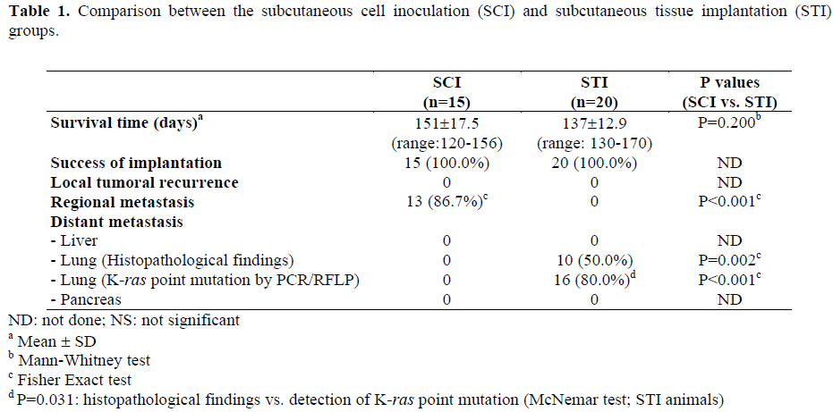
Figure 1. A subcutaneously implanted hamster. A. Appearance before tumor resection. B. Panoramic view after
resection. C. Resected specimen without the covering skin. D. Histopathologic view showing a moderately
differentiated adenocarcinoma (H&E, 200x).
Follow-up, Local Recurrence and Findings in the Necropsy
In the follow-up, neither the SCI nor the STI group showed local recurrence at the site of implantation. Thirteen SCI hamsters (86.7%) developed right axillary lymph node metastasis (Table 1), which was palpable between 4 and 12 weeks after the resection. This lymph node in the early stage was movable. After approximately 2 weeks, it became adhered to deep tissue and grew to 6 cm in diameter. When the lymph node reached this size, the animals died. In the necropsy, there was no local recurrence, or macroscopic internal-organ metastases. The metastatic axillary lymph node showed central necrosis (Figure 2). All lymph nodes were confirmed as metastasis at the histopathological and molecular level. The lungs, liver and pancreas did not show any metastases. The remaining two animals of this group without axillary lymph node metastasis did not have any metastases in their internal organs. The cause of death was unknown.
Figure 2. A case from the SCI group. A. Appearance
of the right lymph node. B. Necropsied lymph node
specimen cut sagitally. C. Histopathologic view of the
lymph node (H&E, 100x).
The STI animals did not show any palpable lymph nodes. They became moribund about 17 to 21 weeks after tumorectomy, and quickly lost weight (about 10 g/day during the last 3 days of life). The occurrence of axillary lymph node metastases was significantly lower than that observed in the SCI group (P<0.001). After death, when necropsy was performed, 10 of 20 animals (50.0%) showed metastases in the lungs at the histopathological level, which was significantly higher when compared with SCI group (P=0.002). On the other hand, the incidence of lung metastases found by PCR/RFLP analysis (Figure 3) was 80.0%. This figure was significantly higher in comparison with the SCI group as well as in comparison with the histopathological findings observed in the same animals (P=0.031). No metastases were found in the liver and pancreas. The remaining 4 animals in this group did not show any macroscopic or microscopic metastatic sites.
Figure 3. An ethidium-bromide stained agarose gel
electrophoresis of PCR/RFLP analysis. (1: mutant
band; 2: wild band; mk: marker; pc: positive control
(HaP-T1 cell line); li: liver; lu: lung; pa: pancreas; rt:
subcutaneously-resected tumor (STI); nc: normal liver
tissue.
DISCUSSION
With the purpose of studying the response to the drugs in-vivo, many studies prefer to use the subcutaneous transplantable tumor models because the method is simple and monitoring of the results is easy. Since the majority of these experiments use human cell lines, nude mice have been widely used. However, in most of these studies, the animals have not been studied over a long period because of the immunological response which increases as the animal gets older [6]. Klein and Bevan [7] stated that it could be due to age-related reconstitution of the nude mouse immune system by endogenous production of interleukin-2. Thus, monitoring of the local response to the therapy obscures the validity of long-term treatment studies since tumor reduction may be due to histocompatibility of the tumor rejection rather than specific antitumor activity. Therefore, we used a homologous model of subcutaneous implantation.
Kyriaziz et al. [8] subcutaneously injected human carcinoma of the larynx and human colon carcinoma cell lines into nude mice. After observing for 6 months, they noted regional lymph node metastases, capsule infiltration and invasion of lymphatic vessels. However, they did not resect the local tumor, as was done in the present study. In our preliminary findings, when the local tumor was resected within 21 days, no metastases were found in either group at 6 months follow-up (data not shown).
Kyriaziz et al. [9] implanted pieces of various human tumors, including pancreatic cancer, in the anterior part of the lateral thoracic region in nude mice. Most of the implanted tumors metastasized to regional and mediastinal lymph nodes and the lungs. The authors stated that the lung metastases occurred through lymphatic and hematogenous routes.
There appear to be two patterns of arrest of tumor cells at target sites; one is based on anatomic and physiologic factors, and the other is based on selectivity [1]. In the pathogenesis of the metastasis cascade, the process consists of a long series of sequential interrelated steps. Thin-walled venules, like lymphatic channels, offer very little resistance to penetration by tumor cells and provide the most common pathways for tumor cell entrance into circulation [1, 10]. Thus, cells entering the lymphatics could have been sequestered via the draining lymph node in the SCI group. Metastatic cells of STI group could have entered the venous circulation, via the metastatic cascade, and encountered their first barrier in the lung, as Nicholson and Poste reported [1]. This could be explained by the non-specific trapping of metastatic tumor cells, which often occurs in the first organ encountered by the circulating cells. Greene and Harvey [11] stated that the localization of a metastatic colony to a particular organ might depend on the formation of an initial bond between the tumor cells and the adhesive molecules on the luminal side of the vascular endothelium of that organ. However, the reasons why lymphatic dissemination was absent in the STI group should be clarified in the future.
Growth rates, invasiveness and metastatic behavior of the transplanted tumors differ depending on the route of implantation [2, 12]. Although the subcutaneous route of implantation was similar in the present experiments, the histology of the STI resected tumors showed more developed architecture of the interstitial tissue. Therefore, the tumoral architecture, including vascularization, may be an important factor in the process of the spread of metastatic cells. Moreover, in the STI model, it should be taken into account that the implanted tumor tissue was derived from the tumor cell line inoculated subcutaneously in Syrian golden hamsters. The different pattern of metastasis might be due to the fact that a second step of implantation could modify the cell biology, thus favoring hematogenous spread.
In human pancreatic cancer, the first target organ of hematogeneous metastases is the liver because the tumoral cells usually spread via the portal vein [13]. We reported a high liver metastatic rate when orthotopic implantation of pancreatic tumor tissue was performed [4]. Thus, other routes of tumor implantation, i.e. intrapancreatic or intraperitoneal, could favor tumor spread to the liver, more closely resembling to the human pattern. Lung metastases found in the present experiments may be a consequence of non-specific trapping of metastatic tumor cells, as stated before.
Ductal adenocarcinoma, the most common form of pancreatic cancer in humans, is associated with activation of the K-ras oncogene in approximately 90% of cases [14]. This has been shown in other nitrosamine pancreatic cancer induced models to take place an elevated high rate [9, 15, 16, 17]. The present cell line has had the point mutation confirmed [4]. Moreover, the early diagnosis of pancreatic cancer in humans may be carried out by the detection of the K-ras point mutation. In the present study, the incidence of lung metastases found by PCR/RFLP analysis was greater than the histopathological findings (80% vs. 50%) and was statistically significant as well. Thus, detection of the K-ras point mutation could identify the micrometastasis, which could not be found using histopathologic examinations alone. Therefore, the sensitivity of the detection of the K-ras point mutation might be greater than that of histology.
The cause of death of the animals not showing any metastases was unknown. However, we believe that it was related to the metastasis phenomena, considering that healthy animals have a two to three year lifespan. We think that these animals could have had metastases in the brain, bone or bone marrow, sites, which were not observed at necropsy.
In conclusion, homologous SCI and STI models showed different patterns of metastasis. The event of metastasis appeared to occur between 21 and 30 days after subcutaneous implantation. The SCI model combined with tumorectomy may be used as a lymphatic metastasis model, (simple to monitor) and the STI model combined with tumorectomy may be used as a hematogenic metastatic model of the lung. However, these findings are preliminary and further studies are required to clarify the mechanisms of metastasis. In the future, the different patterns of metastases after implantation of tumor cells and tumor tissue should be evaluated using other cell lines.
Acknowledgements
The authors would like to thank Mr. Yoshihiro Kuwabara, the Laboratory Animal Center, and the Molecular Research Genetics Center of our University, for their support.
References
- Nicolson GL, Poste G. Tumor implantation and invasion at metastatic sites. Int Rev Exp Pathol 1983; 25:77-181.
- Pour PM, Wilson RB. Experimental tumors of the pancreas. In: Moossa AR, ed. Tumor of Pancreas. 1st ed. Baltimore: Waverly Press, 1980: 37-158.
- Saito S, Nishimura N, Kubota Y, Yamazaki K, Shibuya T, Sasaki H. Establishment and characterization of a cultured cell line derived from nitrosamineinduced pancreatic ductal adenocarcinoma in Syrian golden hamsters. Gastroenterol Jpn 1988; 23:183-94. [88255761]
- Morioka CY, Saito S, Ohzawa K, Watanabe A. Homologous orthotopic implantation models of pancreatic ductal cancer in Syrian golden hamsters: which is better for metastasis research-cell implantation or tissue implantation? Pancreas 2000; 20:152-7. [20170203]
- Altman DG. Practical statistics for medical research. 1st ed. New York: Chapman and Hall, CRC, 1999.
- Hanna N, Fidler IJ. Expression of metastatic potential of allogenic and xenogeneic neoplasms in young nude mice. Cancer Res 1981; 41:438-44. [81088203]
- Klein JR, Bevan MJ. Secretion of immune interferon and generation of cytotoxic T cell activity in nude mice are dependent on interleukin 2: age-associated endogenous production of interleukin 2 in nude mice. J Immunol 1983; 130:1780-3. [83162365]
- Kyriazis AP, DiPersio L, Michael GJ, Pesce AJ, Stinnett JD. Growth patterns and metastatic behavior of human tumors growing in athymic mice. Cancer Res 1978; 38:3186- 90. [79001572]
- Kyriaziz AP, Kyriaziz AA, McCombs WB 3rd, Kereiakes JA. Biological behavior of human malignant tumors grown in the nude mouse. Cancer Res 1981; 41:3995-4000. [82025384]
- Fidler IJ. Origin and biology of cancer metastasis. Cytometry 1989; 10:673-80. [90059577]
- Greene HSN, Harvey EK. The relationship between the dissemination of tumor cells and the distribution of metastases. Cancer Res 1964; 24:799-811.
- Hart IR. "Seed and soil" revisited mechanisms of site specific metastasis. Cancer Metastasis Rev 1982; 1:5-16. [83284869]
- Fidler IJ. Critical factors in the biology of human cancer metastasis: twenty-eight G.H.A. Clowes memorial award lecture. Cancer Res 1990; 6130-8. [90381682]
- Sakorafas GH, Tsiotou AG, Tsiotos GG. Molecular biology of pancreatic cancer; oncogenes, tumour suppressor genes, growth factors, and their receptors from a clinical perspective. Cancer Treat Rev 2000; 26:29- 52. [20127810]
- Erill N, Cuatrecasas M, Sancho FJ, Farre A, Pour PM, Lluis F, Capella G. K-ras and p53 mutations in hamster pancreatic ductal adenocarcinomas and cell lines. Am J Pathol 1996; 149:1333-9. [97017067]
- Sugio K, Gazdar AF, Albores-Saavedra J, Kokkinakis DM. High yields of K-ras mutations in intraductal papillary mucinous tumors and invasive adenocarcinomas induced by N-nitroso(2-hydroxypropyl)(2- oxopropyl)amine in the pancreas of female Syrian hamsters. Carcinogenesis 1996; 17:303-9. [96225904]
- Konishi Y, Tsutsumi M, Tsujiuchi T. Mechanistic analysis of pancreatic ductal carcinogenesis in hamsters. Pancreas 1998; 16:300-6. [98208451]


