Keywords
Pancreas; Pancreatectomy; Pancreatic Fistula
Abbreviations
NBCA: n-butyl cyanoacrylate; PC: pseudocyst; PPPD: pylorus preserving pancreaticoduodenectomy
INTRODUCTION
Pancreatic fistulas are a heterogeneous group of disorders in which both disruption of the pancreatic duct and leakage of amylase and lipase-rich fluid to the skin are present. The most common causes include pancreatic inflammation (acute or chronic pancreatitis), pancreatic resection and trauma [1]. Traditionally, they have been classified as high output (secretion of drain fluid greater than 200 mL/day) or low output (less than 200 mL/day), although the utility of this classification remains unclear [2, 3]. Conventional treatment strategy consists in establishing adequate drainage and fistula control, aggressively treating sepsis, providing nutritional support and reducing pancreatic exocrine secretions with total parenteral nutrition or octreotide. The combination of external drainage and medical therapy results in fistula closure in approximately 85% of patients [4]. If closure fails to occur with medical therapy, surgical treatment is necessary [5].
The use of percutaneous injection of a sclerosing substance, prolamine, into the pancreatic duct is reported for the management of three cases of chronic pancreatic fistula following pancreatic resection.
PATIENTS AND METHODS
From 1994 to 2004, 150 pancreatic resections were performed in our Institute; the overall mortality rate was 3.3% (5 cases), while morbidity was of 33.3% (50 cases). Among surgical complications, pancreatic fistulas were the most frequent and were observed in 22 cases (14.7%). Pancreatic fistula was defined as a drainage of more than 50 mL of amylase-rich fluid (more than three times the normal upper limit present in serum) on or after postoperative day 10, or pancreatic anastomotic disruption demonstrated radiographically [6]. They were successfully treated with medical or radiological therapy in 20 cases (90.9%); only in 2 cases (9.1%) was surgical treatment carried out. In 3 out of 20 cases (15.0%) of pancreatic fistulas treated conservatively, a sclerosing substance, prolamine (Ethibloc-Ethicon, Norderstedt, Germany), was used (Table 1).

The patients were 2 males and 1 female 26, 48, and 58 years of age (mean 44 years), respectively, who had undergone pancreatic resection: 2 pylorus-preserving pancreaticoduodenectomies (PPPD) for ductal adenocarcinoma of the pancreatic head and one total excision of a body-tail cystic lesion. In the PPPDs, legation of the Wirsung duct and suture of the pancreatic stump was performed because of a friable pancreatic parenchyma and an undilated Wirsung duct. All cases were treated with octreotide in the postoperative period (0.2 mg/day tid) for at least seven days. The postoperative course was uneventful and they were discharged after 8, 15, and 16 days, respectively.
In the follow-up period, a large pancreatic sterile collection, with diameters of 9, 10, and 12 cm, respectively, developed after 2, 3, and 4 months, respectively after the pancreatic resection. In all cases, the first treatment was percutaneous CT-guided drainage of the pseudocyst which resolved the lesion (Figure 1) but it created an external iatrogenic fistula.
Figure 1. Abdominal CT scan shows a large
pseudocyst of the head of the pancreas which is treated
with percutaneous CT-guided drainage. (Case 1)
In fact, output from the drainage was 150, 200, and 220 mL/days, respectively, of amylase-rich fluid which persisted for a long time: 2, 3 and 4 months, respectively. In this period, octreotide (0.2 mg/day tid) was administered to each patient. In the case treated with total excision of a cystic mass located in the body-tail, an endoscopic sphincterotomy was also performed, but without success.
A CT scan with barium injection via drainage catheter allowed to show disruption of the pancreatic duct, and the distal Wirsung duct (Figure 2). Persistent, intractable pancreatic fistulas were treated as follows: 1) a CT scan with barium injection via drainage catheter which showed disruption of the pancreatic duct, and the distal Wirsung duct (Figure 2); the catheter was not in the duct but just outside of it; 2) Wirsung duct embolization with 7.5 mL of sclerosing substance, prolamine, injected, via the catheter, under CT scan guidance (Figure 3). Three days later, an abdominal CT scan was performed to establish whether or not drainage catheter could be removed (Figure 4). The first abdominal ultrasound was performed after one month and the ultrasound examination was performed subsequently every 3 months until 12 months after the treatment.
Figure 2. Abdominal CT scan showing the fistula and
the Wirsung duct after injection of barium via drainage
catheter. (Case 1)
Figure 3. Abdominal CT scan showing the Wirsung
duct after the injection of 7.5 mL of the sclerosing
substance, prolamine, via drainage catheter. (Case 1)
Figure 4. Abdominal CT scan performed three days
after treatment with prolamine shows a complete
resolution of the pseudocyst and a Wirsung duct
occluded by the sclerosing substance (Case 1)
RESULTS
The results of this method are summarized in Tables 2 and 3. In cases 1 and 2 the fistula output decreased rapidly: from 220 to 10 mL/day in case 1 and from 150 to 20 mL/day in case 2. There were no clinical signs of pancreatitis and no serum amylase level increase after the procedure and the drainage catheter was removed after 4 days. An abdominal ultrasound examination, performed 1 month later, did not show any pseudocysts or other collections. The patients had postinjection diarrhea and required enzyme replacement. They have not experienced recurrence of the pancreatic juice fistula or reduction of glucose tolerance 12 months after the treatment.

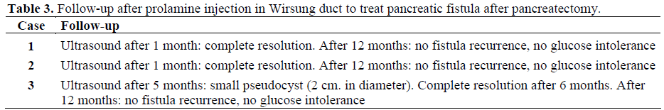
In case 3, the fistula output remains high after treatment (100 mL/day). There were no complications in this case, but the patient had post-injection diarrhea and required enzyme replacement. In contrast with cases 1 and 2, the drainage catheter was removed after 60 days because of the persistent high volume fistula output (superior than 50 mL/day). An abdominal ultrasound examination showed a significant decrease in the collection after 5 months and a complete resolution after 6 months. Twelve months after the treatment, there were no recurrences of a pancreatic fistula or reduction of glucose tolerance.
DISCUSSION
The safety of pancreatic resections has increased in recent years but, although the mortality rate has decreased tgo less than 5% in high volume centers [7, 8, 9, 10], morbidity is still high ranging from 10 to 47% [7, 8, 10]. Among surgical complications pancreatic fistulas represent the most common and clinically relevant; in high volume centers, their rate is in the range of 3 to 13% [7, 9] and the consequences are life-threatening (i.e., bleeding, pancreatic fluid collection and abscesses) with a prolonged hospital stay for specialized treatment and increased costs.
Computed tomography is widely available for the assessment of such complications. Fistulography may help in this setting by showing the communication with the main pancreatic duct.. Endoscopic retrograde cholangiopancreatography (ERCP) has lost much of its contribution to the staging of pancreatic fistulas since the development of secretin-enhanced magnetic resonance pancreatography (S-MRCP). This procedure gives a planar reconstruction of the pancreatic duct and, after secretin injection, the preferential direction of the pancreatic flow (through the fistula). Moreover, it allows us to precisely locate the disruption without any risk of infection due to the external injection of contrast [11].
The goal of fistula treatment is to achieve its closure in the shortest time possible. Several therapeutic modalities should be considered: conservative measures, radiological, endoscopic and surgical treatment. According to previous reports [12], the initial treatment for a pancreatic fistula is conservative and it varies with the severity of the leak. Moreover, it has been well-established that approximately 85% of pancreatic fistulas heal with conservative therapy alone [13] and that the time for closure is from 4 to 18 weeks [13, 14]. Additional therapies have been advocated by some authors in an attempt to speed closure of the leak [15] and to avoid surgical treatment. Endoscopic stenting of the pancreatic duct allows favorable results for incomplete main pancreatic duct disruption [16]. Transpapillary stents can bypass the high resistance of the sphincter of Oddi, ductal strictures and calculi, thereby reducing intraductal pressure and the driving force behind the fistula. Endoscopic pancreatic sphincterotomy should be considered the treatment of choice in patients with a pancreatic fistula after body-tail resection. Percutaneous embolization of the pancreatic duct with different sclerosing substances is a new method for resolving pancreatic fistulas as soon as possible. Embolization of the pancreatic duct was first described by Little et al. in 1977 [17]. They reported the technique of intraoperative pancreatic duct embolization for the prevention or treatment of a pancreatic fistula. Embolization of the duct in the pancreatic remnant during pancreaticoduodenectomy for chronic pancreatitis has been shown by Gall et al. [18] to reduce the frequency of postoperative recurrent pancreatitis by preventing leakage at the pancreaticojejunostomy anastomosis. In several series, this approach has been shown to preserve the endocrine function of the pancreas while it caused exocrine atrophy [19]. The percutaneous approach to embolization, using a prolamine solution, was first described in 1997 by Buecker et al. [14] in a 54-year-old woman in order to treat a pseudocystoductal fistula following necrosectomy for acute pancreatitis. Subsequently, a few cases of percutaneous embolization of the pancreatic duct have been described but they regarded only single case reports. Some different tissue glues were used for embolization: fibrin glue [20], neoprene, n-butyl cyanoacrylate (NBCA) [19] and prolamine solution [14, 19]. Prolamine solution was the first sclerosing substance proposed in the treatment of persistent pancreatic fistulas as an alternative to surgery [14]. The injection of the prolamine solution into the pancreatic duct causes its occlusion with a decrease in the serum enzyme levels while the endocrine function remains the same [21]. Moreover, this agent can be opacified and observed with a CT scan. The relative disadvantages of this agent for percutaneous use include a high degree of viscosity and complete absorption with recanalization within 14 days [19].
Prolamine injection is usually performed after the unsuccessful insertion of a percutaneous drainage catheter, used to treat pancreatic collections, with or without proven fistulas, between the pancreatic duct and the pseudocyst which are more difficult to treat and need longer drainage time. The question of when to stop unsuccessful catheter drainage is not yet clear. Buecher et al. [14] performed percutaneous embolization 6 weeks after catheter drainage insertion because of a persistent pancreatic fistula with 200 mL fluid daily and Findeiss et al. [19] performed percutaneous embolization after 11 months in a pancreatic fistula having an output of more than 200 mL daily. In our experience, the patients’ drainage catheters yielded 220, 150, and 200 mL fluid, respectively, daily after 2, 3, and 3 months, respectively. In unsuccessful cases, Hirota et al. [15] suggested repeating the embolization at an interval of 1-2 weeks.
Independently of the correct timing and the number of embolizations, the results were good with complete resolution of the pancreatic fistula and no increase in serum glucose levels [14, 15, 18].
In our experience, percutaneous embolization was performed only once and the treatment allowed the complete closure of the pancreatic fistulas shortly thereafter. Moreover, we report a series of three patients with successful outcomes while, until now, only single case reports have, for the most part, been seen.
Chronic pancreatic fistulas following pancreatic resection is a difficult problem often requiring operative intervention due to anatomical causes. However, in selected cases, new techniques such as prolamine ductal injection offer a less invasive option with good results. This is a simple and safe technique which should follow percutaneous catheter drainage of a pseudocyst. It consists of: 1) a CT scan with barium injection via a drainage catheter to show the duct disruption and the whole Wirsung duct; 2) Wirsung duct embolization with 7.5 mL of prolamine, injected, via the catheter, under CT scan guidance. This treatment avoids surgery allowing a shorter recovery time and a lower risk of morbidity.
References
- Kozarek RA, Traverso LW. Pancreatic fistulas: etilogy, consequences and treatment. Gastroenterologist 1996; 4:238-44. [PMID 8957097]
- Martin FM, Rossi RL, Munson JL, ReMine SG, Braasch JW. Management of pancreatic fistulas. Arch Surg 1989; 124:571-3. [PMID 2712699]
- Zinner MJ, Baker RR, Cameron JL. Pancreatic cutaneous fistulas. SurgGynecolObstet 1974; 138:710-2. [PMID 4823372]
- Munoz-Bongrand N, Sauvanet A, Denys A, Sibert A, Vilgrain V, Belghiti J. Conservative management of pancreatic fistula after pancreaticoduodenectomy with pancreaticogastrostomy. J Am CollSurg 2004; 199:198-203. [PMID 15275873]
- Alexakis N, Sutton R, Neoptolemos JP. Surgical treatment of pancreatic fistula. Dig Surg 2004; 21:262- 74. [PMID 15308865]
- Yeo CJ, Cameron JL, Maher MM, Sauter PK, Zahurak ML, Talamini MA, Lillemoe KD, Pitt HA. A prospective randomized trial of pancreaticojejunostomy after pancreaticoduodenectomy. Ann Surg 1995; 222:580- 92. [PMID 7574936]
- Bassi C, Butturini G, Molinari E, Mascetta G, Salvia R, Falconi M, et al. Pancreatic fistola rate after pancreatic resection. Dig Surg 2004; 21:54-9. [PMID 14707394]
- Buchler MW, Friess H, Wagner M, Kulli C, Wagener V, Z'Graggen K. Pancreatic fistula after pancreatic head resection. Br J Surg 2000; 87:883-9. [PMID 10931023]
- Rau C, Candinas D, Gloor B. Technique of pancreatic anastomosis. Swiss Surg 2003; 9:135-9. [PMID 12815835]
- Knaebel HP, Diener MK, Wente MN, Buchler MW, Seiler CM. Systematic review and meta-analysis of technique for closure of the pancreatic remnant afterdistal pancreatectomy. Br J Surg 2005; 92:539-46.[PMID 15852419]
- Fukukura Y, Fujiyoshi F, Sasaki M, Nakajo M. Pancreatic duct: morphologic evaluation with MR cholangiopancreatography after secretin stimulation. Radiology 2002; 222:674-80. [PMID 11867784]
- Hashimoto N, Yasuda C, Ohyanagi H. Pancreatic fistula after pancreatic head resection; incidence, significance and management. Hepatogastroenterology 2003; 50:1658-60. [PMID 14571810]
- Howard TJ, Stonerock CE, Sarkar J, Lehman GA, Sherman S, Wiebke EA, et al. Contemporary treatment strategies for external pancreatic fistulas. Surgery 1998; 124:627-33. [PMID 9780981]
- Buecker A, Keulers P, Guenther RW. Successful closure and embolization of a fistula between the pancreatic duct and a pseudocyst using ethibloc. CardiovascInterventRadiol 1997; 20:394-96. [PMID 9271654]
- Hirota M, Kamekawa K, Tashima T, Mizumoto M, Ohara C, Beppu T, et al. Percutaneous embolization of the distal pancreatic duct to treat intractable pancreatic juice fistula. Pancreas 2001; 22:214-6. [PMID 11249080]
- Le Moine O, Matos C, Closset J, Deviere J. Endoscopic management of pancreatic fistola after pancreatic and other abdominal surgery. Best Pract Res Clin Gastroenterol 2004; 18:957-75. [PMID 15494289]
- Little JM, Lauer C, Hogg J. Pancreatic duct obstruction with an acrylate glue: a new method for producing pancreatic exocrine atrophy. Surgery 1977 ;81:243-9. [PMID 841462]
- Gall FP, Zirngibl H, Gebhardt C, Schneider MU. Duodenal pancreatectomy with occlusion of the pancreatic duct. Hepatogastroenterology 1990; 37:290- 4. [PMID 2373461]
- Findeiss LK, Brandabur J, Traverso LW, Robinson DH. Percutaneous embolization of the pancreatic duct with cyanoacylate tissue adesive in disconnected duct sindrome. J VascIntervRadiol 2003;14:107-11. [PMID 12525595]
- 20. Haber GB. Tissue glue for pancreatic fistula. Gastrointest Endosc 2004; 59:122-5. [PMID 15044890]
- Cavuoti OP, Moody FG, Martinez G. Role of pancreatic duct occlusion with prolamine (Ethibloc) in necrotizing pancreatitis. Surgery 1988;103:361-6. [PMID 2449741]


