Michaela B Rehman, Elisa Larrieu-Ardilouze and Sébastien Levesque*
CHU de Poitiers, 2 Rue de la milétrie, France
*Corresponding Author:
Sébastien Levesque
DMC Cardiologie, CHU Poitiers, 2 rue de la milétrie 86000 Poitiers, France.
Tel: 0549444628
E-mail: selevesq@yahoo.fr
Received Date: March 17, 2016 Accepted Date: March 24, 2016 Published Date: March 29, 2016
Citation: Rehman MB, Larrieu-Ardilouze E, Levesque S. Recanalized Image of a Thrombotic Occlusion: A Lotus Root- Like Panda Face Appearance by Optical Coherence Tomography. Interv Cardiol J 2015, 2:1. DOI: 10.21767/2471-8157.100015
In OCT, images of recanalized thrombotic occlusion have a lotus root-like appearance. This is due to septa that divide the lumen into multiple channels that communicate together and converge into a single lumen in the proximal and distal sites of the occlusion. We report a case of a recanalized mid left anterior descending (LAD) occlusion confirmed by OCT, where the lotus root appearance has a delightful panda face.
Keywords
Lotus root, Coronary artery, Optical coherence tomography
Optical Coherence Tomography (OCT) uses polarization properties to differentiate tissue characteristics (calcified, fibrous or lipid-rich plaques) inside coronary arteries. In OCT, images of recanalized thrombotic occlusion have a lotus root-like appearance.
There is little data on this relatively new semiological entity, which has only been described in OCT.
There are no series, only case-reports [1,2]. The lotus-root like appearance is due to septa that divide the lumen into multiple channels that communicate together and converge into a single lumen in the proximal and distal sites of the occlusion. In angiography, recanalization of a thrombotic occlusion often has a “braid-like” aspect but this can be litigious, as it is not specific for recanalization.
We report a case of a recanalized mid left anterior descending (LAD) occlusion confirmed by OCT, where the lotus root appearance has a delightful panda face. A 51-year-old woman with essential hypertension, dyslipidemia and active smoking was admitted to our hospital for chest pain over the last three weeks. Her ECG was in sinus rhythm with Q waves in the anterior leads. Transthoracic echocardiography showed a kinetic apex and anterior wall and a 50% left ventricular ejection fraction. Coronary angiography (Figure 1A) revealed a recanalized thrombotic mid left anterior descending artery (LAD) occlusion confirmed by OCT (Figure 1B), where the lotus root appearance had a delightful panda face! The occlusion was treated with a drug eluting stent with an excellent angiographic result.
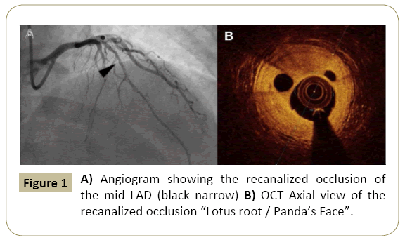
Figure 1: A) Angiogram showing the recanalized occlusion of the mid LAD (black narrow) B) OCT Axial view of the recanalized occlusion “Lotus root / Panda’s Face”.
It is amusing to show how serious pathology can smile as us through this adorable face.
Figures at a glance
References
- Kato M, Dote K, Sasaki S (2011) Recanalized image of thrombotic occlusion with coronary plaque rupture: a lotus root-like appearance by optical coherence tomography.Canadian Journal of Cardiology27: 871.
- Sakurai S, Takashima H, Waseda K, Ando H, Kurita A, et al. (2014) Multiple recanalized images of thrombotic occlusion 19 years after percutaneous coronary intervention: Insights from optical coherence tomography and intravascular ultrasound.International journal of cardiology172: 480-481.

