Samina Khokher*
Chief Consultant Surgeon, Head of Surgical Special Unit, Services Hospital, Lahore, Pakistan
Rafia Shahzad
Radiologist, INMOL Hospital, Lahore, Pakistan
Samia Shahbaz
House Officer, Surgical Special Unit, Services Hospital, Pakistan
Corresponding Author:
Samina Khokher
MBBS, FCPS, PhD, Chief Consultant Surgeon, Head of Surgical Special Unit, Services Hospital, Lahore, Pakistan
Tel: 0343 2532430
E-mail: drsamkhokher@yahoo.com
Submitted date: May 30, 2016; Accepted date: June 21, 2016; Published date: June 28, 2016
Keywords
Pseudoangiomatous; Stromal hyperplasia; Breast; Giant tumor
Introduction
A broad spectrum of primary tumors and tumor like lesions arising from the stromal tissue of the breast has been described. Because of their diverse morphological appearance and biological behavior, many of these lesions present as a diagnostic and therapeutic challenge. Pseudoangiomatous Hyperplasia (PASH) is a non-neoplastic proliferation of the myofibroblastic cells in the mammary stroma and it was first described in 1986.1 Extra-mammary PASH has only been reported in the mammary like glands in the anogenital region of women.2 PASH is usually reported as an incidental finding on histopathological examination of benign breast lesions and presentation as a giant tumor is rare. Large palpable tumor in the breast is a matter of great concern for the patient because it causes deformity, discomfort and mental distress for the fear of mastectomy or incurability. We report two cases of giant tumorous PASH. Informed consent of the patients was taken for taking photographs and for the publication.
Case Report
Case 1
A 32 years old multiparous woman, presented to the Breast Clinic of Services hospital, Lahore in June 2014 with history of a lump in her left breast for the last three years. Initially lump grew in size gradually but later there was rapid growth leading to painful swelling of the whole left breast. On examination, the left breast was about twice the size of right and had a palpable mass of 16 cm diameter in it. The overlying skin and nipple areola complex were normal and axillary lymph nodes were not palpable. Sonomammography revealed a large well circumscribed homogeneously hypoechoic solid mass measuring 15 × 9.5 cm in left breast compressing upon surrounding parenchyma. Core Needle Biopsy (CNB) was difficult as the mass was extremely hard and it revealed hyalinized fibrous tissue with no epithelial component on histopathology. Repeat biopsy was done which showed paucicellular material with dense hyalinized stroma with no evidence of malignancy. After the general workup a 15.5 × 15.5 × 8 cm well circumscribed mass was excised through circumferential incision in the upper outer quadrant of left breast. The histopathology report showed the tumor having epithelial and stromal components. The stroma was composed of spindle shaped cells having elongated nuclei with inconspicuous nuclei and eosinophilic cytoplasm exhibiting extensive sclerosis and small slit like spaces lined by flattened epithelial cells exhibiting strong positivity for CD 34 immunostain. The diagnosis was benign breast lump with features of PASH. The patient is tumor free on one year’s follow up with excellent cosmetic outcome and breast symmetry (Figure 1).
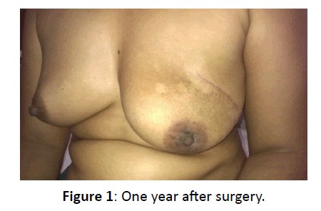
Figure 1: One year after surgery.
Case 2
A 15 years old girl presented to the Breast Clinic of INMOL hospital, Lahore in September 2015, with history of a lump in her left breast for the last two years. Initially it increased in size gradually but later rapidly, causing dragging sensations in the breast and obvious asymmetry. On examination the left breast was ptotic and larger than the right. There were prominent veins and larger sized nipple areola complex. The suprasternal notch to nipple distance was 17 cm on the right and 33 cm on the left. There was a well-defined mobile palpable mass of about 12 cm diameter in it. Sonomammography revealed a large circumscribed mass measuring 11 × 8.4 cm occupying whole of the left breast with barely appreciable normal breast parenchyma. Mass had heterogeneous echogenecity with scanty flow on Doppler evaluation (Figure 2). CNB revealed breast parenchyma with marked stromal fibrosis suggestive of PASH. After the general workup a mass, 11 × 11 × 9 cm in dimensions was excised through circumferential incision centered at 3 O’clock on left breast (Figure 3). The histopathology report was consistent with the diagnosis of PASH. Six months after the surgery, there was wrinkled skin adjacent to the operation scar mark and marked improvement in the breast symmetry (Figure 4). The suprasternal notch to nipple distance was 17 cm on the right and 19 cm on the left.
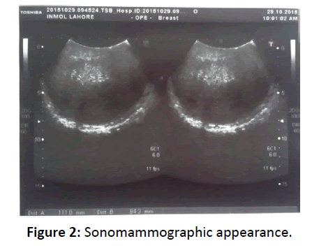
Figure 2: Sonomammographic appearance.
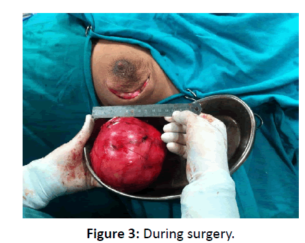
Figure 3: During surgery.
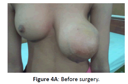
Figure 4A: Before surgery.
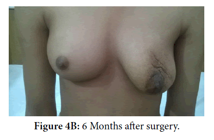
Figure 4B: 6 Months after surgery.
Discussion
PASH is an uncommon form of benign overgrowth of mesenchymal tissue in the breast which was first described in 1986.1 It is a benign angioma like proliferation of the myofibroblastic tissue in the mammary stroma which resembles but is not actually consistent with the angiomatous proliferation of low grade angiosarcoma. This distinction is important as PASH follows a benign course while angiosarcoma has the clinical course of a malignant tumor and therefore bears entirely different biological behavior and therapeutic implications.
A palpable breast mass is the commonest presentation of all benign and malignant lesions in the breast. A broad spectrum of benign and malignant tumors arising from the epithelial and stromal tissues in the breast has been described. Diagnostic evaluation by triple assessment is the key to diagnosis and appropriate management. Clinical presentation of PASH as a giant tumor is extremely rare3,4 and it is usually reported as an incidental finding on the histopathological examination of benign breast lesions. Both the cases reported by us had huge sized tumors in the breast (15 cm in case 1 and 11 cm in case 2) while the average diameter reported for PASH is 5 to 6 cm ranging between 0.6 and 12 cm.5,6 Both the cases reported by us had slow growth initially but later had rapid growth and associated pain in the breast. The local women population has low level of knowledge about breast health and typically they report late with large neglected malignant masses.7 The same attitude has been observed for benign breast masses as both these cases reported when the whole breast was involved and there was obvious asymmetry and dragging pain in the breast.
The imaging features of PASH are nonspecific and do not lead to a confident diagnosis. At Ultrasound, the lesions are seen as well-defined solid masses. Sometimes these masses are hypoechoic while hyperechogenicity or heterogeneous echogenicity has also been described. The sound attenuation characteristics vary from posterior enhancement to mild posterior shadowing. These lesions may contain numerous lacelike reticular areas with scattered cystic changes and biopsy is necessary to confirm the diagnosis.8,9 Lesions in both of our cases were well circumscribed having homogeneous hypoechogenicity in case one and heterogeneous echogenicity in the second case. Both of the cases reported by us were young and as per guidelines did not have mammography.
The treatment of PASH depends upon the clinical presentation as no additional treatment is recommended for incidental diagnosis of PASH and treatments ranging from observation to the surgical tragedy of bilateral mastectomies have been reported for tumorous PASH.10,11 A case of an adolescent girl at the age of 12 years has been reported who underwent bilateral mastectomy for the treatment of this benign disease.10 Both of the cases reported by us were managed without mastectomy and had good cosmetic outcome. Further improvement can be achieved by plastic reconstruction if required after scar maturation and natural molding has completed.
Conclusion
PASH presenting as a giant tumor is very rare. Two cases have been reported. Surgical excision and breast conservation provides relief from the deformity, discomfort and mental distress. Earlier excision of rapidly growing PASH is mandatory to avoid breast asymmetry. Any residual asymmetry can be dealt with by plastic reconstruction if required after scar maturation.
References
- Vuitch MF, Rosen PP, Erlandson RA. Pseudoangiomatous hyperplasia of mammary stroma. Hum Pathol 1986; 17: 185-191.
- Kazakov DV, Bisceglia M, Mukensnabi P, Michal M. Pseudoangiomatous stromal hyperplasia in leions involving anogenital mammary-like glands. Am J Surg Pathol 2005; 29: 1243-1246.
- Teh HS, Chiang SH, Leung JWT, Tan SM, Mancer JFK. Rapidly enlarging tumoral pseudoangiomatous stromal hyperplasia in a 15 year old patient. Distinguishing sonographic and magnetic resonance imaging findings and correlation with histologic findings. J Ultrasound Med 2007; 26: 1101-1116.
- Kutluturk K, Usta S, Unal B, Karadag N, Akatli AN. Pseudoangiomatous hyperplasia of the breast presenting as a giant breast tumor: A case report. J Breast Health 2015; 11: 39-41.
- Castro CY, Whitman GJ, Sahin AA. Pseudoangiomatous hyperplasia of the breast. Am J Clin Oncol 2002; 25: 213-216.
- Virk RK, Khan A. Pseudoangiomatous hyperplasia: An overview. Arch Pathol Lab Med 2010; 134: 1070-1074.
- Khokher S, Qureshi MU, Riaz M, Akhtar N, Saleem A. Clinicopathologic profile of breast cancer patients in Pakistan: Ten years data of a local cancer hospital. Asian Pac J Cancer Prev 2012 13: 693-698.
- Irshad A, Ackerman SJ, Pope TL, Moses CK, Rumboldt T, et al. Rare breast lesions: Correlation of imaging and histologic features with WHO classification. RadioGraphics 2008; 28: 1399-1414.
- Jones KN, Glaze Brook KN, Reynolds C. Pseudoangiomatous stromal hyperplasia: Imaging findings with pathologic and clinical correlation. AJR 2010; 195: 1036-1042.
- Singh KA, Lewis MM, Runge RL, Carlson GW. Pseudoangiomatous stromal hyperplasia. A case for bilateral mastectomy in a 12 year old girl. Breast J 2007; 13: 603-606.
- Dai H, Connor C, Cui W, Gatewood J, Fan F. Bilateral diffuse pseudoangiomatous stromal hyperplasia: A case of bilateral mastectomy in a 28 year old women. Case Rep Pathol 2014.

