Keywords
Cell Adhesion; Cytokines; Endothelium; Pancreatic Neoplasms; Surgery
Abbreviations
MEC: microvascular endothelial cell; HUVEC: human umbilical vein endothelial cell
INTRODUCTION
In the western world, pancreatic cancer leads to approximately 150,000 deaths each year, making it one of the five leading causes of cancer-related deaths [1]. Potential curative resection is still the only option that offers a chance for cure in pancreatic cancer patients, but this can only be performed in about 10- 15% of pancreatic cancer patients [2]. the prognosis for these patients is still poor, since more than 80% die within 5 years after surgery. Recurrences are found locally, in the intra-abdominal cavity (peritoneal and hepatic) and to a lesser extent at extraabdominal sites [3].
Hematogenous metastasis develops from circulating tumor cells. These cells can be detected in 60-80% of pancreatic cancer patients and this percentage increases during surgical procedures [4, 5, 6].
Operative trauma in the tissue itself may favor development of tumor recurrence. An association between surgical trauma and tumor recurrence has been supported by previous in vivo and in vitro studies [7, 8, 9, 10, 11, 12]. These studies showed that surgical trauma brings about enhanced locoregional tumor recurrence by the inflammatory reaction it provokes. The inflammatory reaction is not confined locally, but spreads out systemically as well, yet to a somewhat lesser degree [13, 14, 15, 16, 17]. Indeed, in several in vivo studies, surgical trauma enhanced tumor recurrence at distant sites as well [18, 12].
The inflammatory reaction provoked by surgical trauma leads to the activation of leukocytes and monocytes with the release of pro-inflammatory cytokines and reactive oxygen species. Thus, it has been found that, after major abdominal surgery, the proinflammatory cytokines interleukin-6 (IL-6), interleukin-1beta (IL-1beta) and tumor necrosis factor-alpha (TNF-alpha) in the peripheral blood are elevated [13, 14, 15, 16, 17].
The adhesion of circulating tumor cells to the microvascular endothelium of organs at distant sites - like the liver and the lungs - is an important step in blood-borne metastasis. In previous in vitro experiments we showed that pro-inflammatory cytokines are capable of enhancing the adhesion of human colon carcinoma cells to the microvascular endothelium, most likely by the up-regulation of adhesion molecules to the endothelium [11].
In this study, we investigated the influence of the pro-inflammatory cytokines IL-1beta, TNF-alpha and IL-6 on the adhesion of human pancreatic carcinoma cells to microvascular endothelial cells. Therefore, a reproducible human in vitro model was developed. Moreover, the expression of cell adhesion molecules on both endothelial and tumor cells was assessed as well as the influence of blocking antibodies to cell adhesion molecules in tumor cell adhesion assays.
MATERIALS AND METHODS
Cells
Human microvascular endothelial cells (MECs) at passage 4 were purchased from Cambrex (Verviers, Belgium) and maintained in EGM-2-MV Bullet kit according to the manufacturer’s instructions at 37°C, 95% relative humidity and 5% CO2. Confluent monolayers were passaged by trypsin/EDTA (0.025%/0.01%) and cells were used up to passage 8.
The human pancreatic carcinoma cell lines PanC1, MiaPaCa and BxPC3 were also grown in an EGM-2-MV Bullet kit in order to create similar culture conditions to the endothelial cells, which was necessary for the experiments. The tumor cells were maintained by serial passage after trypsinization using trypsin/EDTA (0.05% 0.02%) (all, except penicillin, obtained from Gibco, Breda, the Netherlands; penicillin from Yamanouchi, Leiderdorp, the Netherlands). Before the adhesion assay, tumor cells were harvested using trypsin and maintained in suspension culture for two hours to regenerate cellsurface proteins.
Adhesion Assay
To quantify tumor cell adhesion to MECs, a standardized cell adhesion assay was developed according to the methods of Catterall et al . [19]. Briefly, endothelial monolayers were established in 96 well microtiter plates (Perkin Elmer, Groningen, the Netherlands). To do this, confluent cells were trypsinized and 2x10⁴ endothelial cells were added to each well.
The plates were incubated at 37°C, 95% relative humidity, 5% CO2 and the medium was replaced daily by fresh medium. The MECs reached confluence in 3 to 4 days as determined by light microscopy.
To determine the effect of cytokines on tumor cell adhesion, endothelial monolayers were pre-incubated with 0.1 or 10 ng/mL recombinant human IL-1beta, TNF-alpha and IL-6 (R&D Systems, Uithoorn, The Netherlands) for 4 or 12 hours. Untreated monolayers served as controls. Not only was the effect of the endothelial pre-incubation investigated but the effect of tumor cell preincubation as well. Therefore, BxPC3 cells were pre-incubated with 10 ng/mL IL-1beta for 12 hours before the adhesion assay.
To quantify tumor cell adhesion, the tumor cells were labeled with calcein-AM (Molecular Probes, Leiden, the Netherlands) for 45 minutes, washed 3 times and added to the wells (3x10⁴ per well). The plates were centrifuged for 1 minute at 80 g in a Heraeus centrifuge (Etten Leur, the Netherlands) and incubated at 37°C for 1 hour. Thereafter, the wells were washed twice with medium to remove non-adherent tumor cells. The remaining fluorescence per well was measured on a Perkin Elmer (Gouda, the Netherlands) plate reader using 485 nm excitation and 530 nm emission filters.
Immunocytochemistry
Endothelial and tumor cells were prepared for staining by cytospin preparation, fixed in acetone for 10 minutes and stored at -20°C until used.
The cytospins were incubated for 30 minutes at room temperature with the following primary antibodies: mouse anti-human monoclonal antibodies to E-selectin (R&D Systems, Uithoorn, The Netherlands), ICAM- 1, VCAM-1 (Dako Cytomation Heverlee, Belgium), sialyl Lewis a (sLea), sialyl Lewis x (sLex) (Sanbio, Uden, the Netherlands), lymphocyte function-associated antigen-1 (LFA-1) (alphaLbeta2) and very late activation antigen-4 (VLA-4) (alpha4beta1) (Becton Dickinson, Alphen a/d Rijn, the Netherlands). Negative controls were incubated with PBS. As secondary antibodies, biotinylated goat anti-mouse antibodies were used followed by incubation with streptavidin-biotinylated alkalin-phosphatase complex. Substrate development was carried out with new fuchsin 4%. The cytospins were counterstained with hematoxylin.
The expression of cell adhesion molecules was quantified by two different observers using semi-quantitative scoring system ranging from no expression (-), weakly positive (±) to positive expression (+).
Enzyme Immuno Assay (EIA)
Endothelial cells and tumor cells were grown to confluence as described for the adhesion assays in 96-well flat-bottomed multititer plates (Becton Dickinson, Alphen a/d Rijn, the Netherlands). The endothelial cells were pre-incubated with either cell culture media alone or with media containing IL-1beta, TNF-alpha or IL-6 (0.1 and 10 ng/mL). The tumor cells were pre-incubated with either cell culture media alone or with media containing IL-1beta or TNF-alpha (10 ng/mL). Following this pre-incubation, the cells were washed with phosphate buffered saline (room temperature, pH 7.4), fixed in ethanol/methanol for 45 minutes and then washed again. Subsequently, the wells were incubated for 10 minutes with 1% goat serum to block unspecific binding sites. Mouse monoclonal antibody to E-selectin, ICAM-1, VCAM-1 (R&D Systems, Uithoorn, The Netherlands) sLea, sLex (Sanbio, Uden, the Netherlands), LFA-1 (alphaLbeta2) or VLA-4 (alpha4beta1) (Becton Dickinson, Alphen a/d Rijn, the Netherlands) was added for 1 hour, followed by the addition of a second antibody, biotinylated goat anti-mouse antibody (Sigma Zwijndrecht, the Netherlands) in a dilution of 1:250. Increased sensitivity was obtained using the ExtrAvidin- Peroxidase system (Sigma, Zwijndrecht, the Netherlands). Adding 2,2'-azino-bis(3- ethylbenzothiazoline-6-sulfonic acid) diammonium salt in citrate-phosphate buffer with urea hydrogen peroxide-developed substrate. Incubation of the endothelial cells without the primary antibody served as a negative control. As a positive control, the ExtrAvidin-Peroxidase (Sigma, Zwijndrecht, the Netherlands) system was added followed by substrate development without washing away the peroxidase. After 40 minutes, the reaction was stopped with sodium fluoride and photometrical evaluation was performed with computer-controlled ELISA reader at a lambda equal to 405 nm.
Function Blocking Assay
Endothelial monolayers were pre-incubated with 10 ng/mL IL-1beta for 12 hours. One hour before the adhesion assay was performed, 50 �g/mL of function blocking monoclonal mouse antibodies to human Eselectin (R&D Systems, Uithoorn, The Netherlands) were then added to the endothelial monolayers. The same inhibition assays were carried out with monoclonal mouse antibodies to human ICAM-1 (25 �g/mL) and VCAM-1 (60 �g/mL) (R&D Systems, Uithoorn, The Netherlands) (the concentration of the antibody was in accordance with the instructions of the manufacturer).
STATISTICS
Data are reported as mean±SD values. The data were analyzed using one- or two-way analysis of variance (ANOVA) according to the experimental design applied. The simple and the repeated contrasts were applied to ANOVA in order to compare the various experiments with the control experiment as well as adjacent categories, respectively. The effects within specific categories were evaluated using nested designs. The SPSS version 13.0 for Windows was used to analyze the data. Two-tailed P values less than 0.05 were considered to be statistically significant. Experiments (n=6) were performed at least twice with comparable results while quadriplate wells were used in the EIA assays.
RESULTS
Validation of Assay
Labeling tumor cells with calcein-AM did not decrease their viability (greater than 95% using trypan blue). To determine the stability of calcein labeling, the fluorescence of the labeled cells and of the supernatant of the labeled cells was measured. The fluorescence of the labeled cells remained constant for at least 60 minutes indicating retention of the dye within the cells (data not shown). This result was also seen in the adhesion assay, where maximal tumor cell adhesion was reached after a one hour incubation followed by a decrease at longer incubation times (data not shown). Therefore, for all subsequent experiments, the incubation time was 1 hour. Dilution series with labeled tumor cells on MEC monolayers showed a linear correlation between cell number and measured fluorescence (data not shown) which was used as a standard to calibrate the fluorescence measured. In this way, the amount of adhered tumor cells in the experimental wells could be determined.
Adhesion to Endothelial Cells
In all assays, PanC1 cells adhered to untreated MEC in a percentage ranging from 10 to 20%, i.e. the basal or control adhesion. The basal adhesion of BxPC3 ranged from 20 to 30% in all assays and that of MiaPaCa ranged from 5 to 20%
Pre-incubation of MEC with IL-1beta or TNF-alpha, but not with IL-6 resulted in timeand concentration-dependent-enhanced tumor cell adhesion (Figure 1). Four-hour preincubation with 0.1 ng/mL IL-1beta resulted in a significant enhancement of BxPC3 only (113% vs. control, P=0.016), while 4-hour pre-incubation with 10 ng/mL IL-1beta resulted in a significant enhancement for MiaPaCa (144% vs. control, P=0.011) and BxPC3 (127% vs. control, P<0.001). After a 4-hour pre-incubation of MEC with TNFalpha, adhesion for all 3 cell lines was significantly enhanced (PanC1: P=0.004 and P=0.010 for 0.1 and 10 ng/mL, respectively; MiaPaCa and BxPC3: P<0.001 at each concentration). As far as 4-hour preincubation with IL-6 was concerned, only a slight enhancement adhesion of MiaPaCa (135% vs. control, P=0.039) was obtained with 10 ng/mL IL-6. A twelve-hour preincubation with IL-6 did not show any significant enhancement of adhesion. The enhancement of adhesion for all cell lines was significant after 12 hours of pre-incubation with 0.1 ng/mL of IL-1beta and the adhesion for all cell lines significantly increased by increasing the IL-1beta concentration to 10 ng/mL; for PanC1, adhesion reached 159% vs. control (P<0.001), for MiaPaCa it was 204% (P<0.001) and for BxPC3 it was 127% (P<0.001). TNF-alpha resulted in enhanced adhesion for all cell lines at 0.1 ng/mL as well as at 10 ng/mL, but a significant increase with the concentration was observed only for BxPC3; for PanC1 it was 155% (P<0.001) and 144%(P<0.001), for MiaPaCa 178% (P=0.001) and 203% (P<0.001) and for BxPC3 144% (P<0.001) and 137% (P<0.001) vs. control at 01. and 10 ng/mL TNF-alpha, respectively.
Figure 1. Adhesion of PanC1, MiaPaCa and BxPC3 to MECs after 4 and 12 hours of pre-incubation of MECs with 0.1
or 10 ng/mL IL-1beta, TNF-alpha or IL-6. Mean±SD values are shown. Generally, n=6 samples were evaluated for
each experiment. (Two-way ANOVA). Black P values refer to the comparison with the control group (untreated
MECs): simple contrast was applied within the two time evaluations by a nested design. Red P values refer to the
comparison between 0.1 and 10 ng/mL; repeated contrast was applied within the two time evaluations using a nested
design.
A time-dependent increase in adhesion was observed with pre-incubation with 10 ng/mL IL-1beta for all three cell lines (P<0.001) as well as at the two concentrations (0.1 ng/mL: P=0.003; 10 ng/mL: P=0.023) of TNF-alpha for PanC1 cells only while a significant decrease in adhesion was observed for the BxPC3 cell line from 4- to 12-hour preincubation with 10 ng/mL IL-6 (P=0.001).
Pre-incubation with higher concentrations of the pro-inflammatory cytokines (50 and 100 ng/mL) did not result in a more pronounced enhancement of adhesion (data not shown).
The Role of Cell Adhesion Molecules
It is known that both sialyl Lewis a (sLea) and sialyl Lewis x (sLex) are ligands for Eselectin; LFA-1 (alphaLbeta2) is a ligand for ICAM-1, and VLA-4 (alpha4beta1) for VCAM-1.
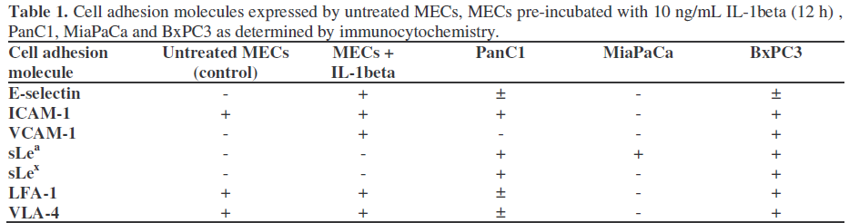
Table 1 shows the expression of cell adhesion molecules on untreated MECs, MECs preincubated with 10 ng/mL IL-1beta for 12 hours, as well as on PanC1, MiaPaCa and BxPC3. Untreated MECs express ICAM-1, as well as LFA-1 and VLA-4, whereas Eselectin and VCAM-1 are not expressed. Both these adhesion molecules are up-regulated by exposure of MECs to IL-1beta. BxPC3 cells express all tested adhesion molecules, PanC1 cells do not express VCAM-1 only, whereas MiaPaCa cells selectively express sLea.
This difference in the adhesion molecule pattern is displayed in the percentage of adherent cells to MECs, which is highest for BxPC3 and lowest for MiaPaCa (data not shown).
We further evaluated the endothelial cell adhesion molecules by EIA (a semiquantitative assay) (Figure 2). The staining intensity (in optical density units measured at 405 nm) of E-selectin expression on unstimulated MECs was 0.2. Following preincubation with 0.1 and 10 ng/mL IL-1beta for 12 hours the staining intensities reached 0.19 (P=0.739) and 0.50 (P<0.001), respectively. Pre-incubation with 0.1 and 10 ng/mL TNF-alpha significantly up-regulated E-selectin expression to a staining intensity of 0.47 and 0.59, respectively (both P<0.001). IL-6 did not up-regulate E-selectin expression. ICAM-1 expression was upregulated from 0.2 to 0.27 (P<0.001) and to 0.40 (P<0.001) by pre-incubation with 0.1 and 10 ng/mL IL-1beta, respectively. Preincubation with 0.1 and 10 ng/mL TNF-alpha up-regulated expression to 0.48 (P<0.001) and to 0.47 (P<0.001), respectively whereas once again IL-6 did not influence expression. A slight but significant enhancement in the expression of VCAM-1 was induced in all but one experiment (the pre-incubation with 0.1 ng/mL IL-6; P=0.062); maximal induction was reached with 10 ng/mL TNF-alpha (from 0.23 to 0.31; P=0.001).
Figure 2. Anti-E-selectin, anti-ICAM-1 and anti-
VCAM-1 expression on MECs pre-incubated for 12
hours with 0.1 and 10 ng/mL TNF-alpha, IL-1beta or
IL-6 assessed by EIA. Bars represent the mean±SD
absorbance values (OD 405 nm). Generally, n=4 wells
were evaluated for each experiment. (One-way
ANOVA). Black P values refer to the comparison with
the control group (untreated MECs); simple contrast
was applied. Red P values refer to the comparison
between 0.1 and 10 ng/mL; repeated contrast was
applied.
Next, we focused in more detail on interactions in inhibition assays between tumor cells and endothelial cells by using monoclonal antibodies against adhesion molecules on MECs. None of the function blocking antibodies tested significantly influenced basal adhesion apart from a slight decrease in PanC1 cells with anti-E-selectin (Figure 3). Pre-incubation with IL-1beta significantly (P<0.001) enhanced adhesion of all three cell lines both in the absence or the presence of all the antibodies tested. Anti-Eselectin was not capable of inhibiting the enhanced adhesion of all 3 tumor cell lines to IL-1beta pre-incubated MECs while slight increases of adhesion were observed in PanC1 cells with anti-ICAM-1 and anti-VCAM-1 antibodies. No significant modifications were found in MiaPaCa cells, while slight decreases of adhesion were obtained in BxPC3 with anti-ICAM-1 (with and without the addition of anti-E-selectin) and anti- VCAM-1 antibodies. (Figure 3). Furthermore, pre-incubation of MECs with TNF-alpha instead of IL-1beta gave similar results (data not shown). The cell adhesion molecules sLea, sLex, LFA-1 and VLA-4 are probably not the ligands on MECs responsible for increased tumor cell adhesion after exposure to the proinflammatory cytokines. First of all, sLea and sLex are neither expressed on MECs or on activated MECs and its counterpart E-selectin is not expressed on MiaPaCa and only weakly expressed on PanC1 and BxPC3 (Table 1). Furthermore, although both LFA-1 and VLA- 4 are expressed on MECs and are slightly upregulated after exposure to pro-inflammatory cytokines (data not shown), their counterparts are not expressed on MiaPaCa. However, the adhesion of MiaPaCa clearly increased to cytokine pre-incubated MECs and, therefore, LFA-1 and VLA-4 are unlikely to be the adhesion molecules on MEC which induce enhanced adhesion.
Figure 3. Adhesion of PanC1, MiaPaCa and BxPC3 to
MECs. Prior to tumor cell adhesion, the MECs were
pre-incubated for 12 hours with 10 ng/mL IL-1beta
and/or for 1 hour with anti-E-selectin (E-sel), anti-
ICAM-1 (ICAM-1), both anti-E-selectin and anti-
ICAM-1 (E-sel+ICAM-1) or anti-VCAM-1 (VCAM-
1). Mean±SD values are shown. Generally, n=6
samples were evaluated for each experiment. (One-way
ANOVA). Black P values refer to the comparisons
with values observed without added antibodies (either
untreated MAC or IL-1beta pre-incubated MECs): the
simple contrast was applied. Red P values refer to the
comparisons between untreated MAC and IL-1beta
pre-incubated MEC; repeated contrast was applied.
Exposure of Tumor Cells
Since not only the endothelium but the circulating tumor cells as well are exposed to factors released during surgery, the influence of pro-inflammatory cytokines on adhesion molecule expression of the tumor cells was investigated subsequently by EIA (Figure 4). For PanC-1 the expression of VLA-4 shows a slight but significant decrease after IL-1beta pre-incubation (P=0.003). The expression of LFA-1 on MiaPaCa after IL-1beta preincubation is also slightly decreased (P=0.016). On BxPC3 however, a slight but significant increase in ICAM-1 (P<0.001) and LFA-1 (P=0.049) was observed. Comparable results were obtained after pre-incubation of the tumor cells with TNF-alpha (data not shown).
Figure 4. Adhesion molecule expression on PanC1,
MiaPaCa and BxPC3 obtained with EIA. Orange bars
represent basal expression; green bars represent
expression after 12 hours pre-incubation with 10
ng/mL IL-1beta. Bars represent the mean±SD
absorbance values (OD 405 nm). Generally, n=4 wells
were evaluated for each experiment. (Two-way
ANOVA; the effect of the pre-incubation with IL-1beta
was evaluated within the various categories using a
nested design).
Therefore, the influence of exposing BxPC3 to IL-1beta on tumor cell adhesion was investigated (Figure 5). The overall analysis showed that pre-incubation with IL-1beta significantly enhanced adhesion (P<0.001) while BxPC3 did not (P=0.241). The effect of incubation with BxPC3 was not significantly related to the presence or absence of preincubation with IL-1beta (interaction term of two-way ANOVA: P=0.096). In particular, the effect of pre-incubation with BxPC3 was not significant either in control MECs (P=0.050) or in IL-1beta pre-incubated cells (P=0.706) while the effect of pre-incubation with IL-1beta was equally significant in control (P<0.001) and pre-incubated (P<0.001) BxPC3 cells.
Figure 5. The effect of the pre-incubation of MECs
with 10 ng/mL IL-1beta and/or BxPC3. Bars represent
the mean±SD absorbance values (OD 405 nm).
Generally, n=6 samples were evaluated for each
experiment. (Two-way ANOVA: the effects of preincubation
with IL-1beta and BxPC3 were evaluated
within the various categories using a nested design).
Black P values refer to the comparisons with control
MECs (without IL-1beta pre-incubation). Red P values
refer to the comparisons between control BxPC3 and
pre-incubated BxPC3 cells.
DISCUSSION
Previous in vivo studies suggested a tumorpromoting effect from surgical trauma, not only locally, but systemically as well. This theory was further explored in a human in vitro model, showing that pro-inflammatory cytokines, released during surgical trauma, enhance the adhesion of human colon carcinoma cells to mesothelial cells and microvascular endothelial cells [11]. Therefore, surgical trauma may enhance tumor recurrence loco-regionally as well as at distant sites.
In this human in vitro study, we demonstrated that the pro-inflammatory cytokines IL-1beta and TNF-alpha significantly enhance the adhesion of 3 human pancreatic carcinoma cell lines to the microvascular endothelium. IL-6, which is distinctly elevated during surgical trauma and is an activator of several inflammatory mechanisms, did not influence interactions between tumors and endothelial cells.
In previous studies [11], we excluded the possibility that enhanced adhesion after cytokine exposure was caused by enhanced growth of endothelial cells resulting in a higher number of MEC and, consequently, more binding receptors since proliferation assays did not show enhanced growth of the exposed MECs as compared to normal MECs during the 12 hour pre-incubation period.
Interactions between tumor cells and endothelial cells are accomplished by adhesion molecules. The results of the EIA showed that the endothelial adhesion molecules E-selectin and ICAM-1 are significantly up-regulated after pre-incubation with TNF-alpha or IL-1beta. The question then arises as to whether the increased tumor cell binding is dependent on the up-regulation of E-selectin or ICAM-1. VCAM-1 is not likely to be the adhesion molecule responsible since no significant enhancement in MEC expression was found after cytokine preincubation, except for a slight increase after pre-incubation with 10 ng/mL TNF-alpha. In previous studies, the adhesion of several pancreatic carcinoma cell lines to human umbilical vein endothelial cells (HUVECs), which are macrovascular cells, could be inhibited by antibodies against the E-selectin adhesion molecule [20, 21]. In these studies no inhibition in adhesion to activated endothelium was observed using monoclonal antibodies against E-selectin. The absence of inhibition could not be attributed to the malfunctioning of antibodies or technical problems since we brought about strong inhibition in the adhesion of HT29 colon carcinoma cells to activated HUVECs using the same protocol and antibodies [11]. However, in that particular study, we were not able to block the adhesion of HT29 cells to MECs. Therefore, it was concluded that another adhesion molecule or a complex of adhesion molecules was responsible for HT29 cell interactions with MECs. Again, in this study no inhibition could be reached using Eselectin antibodies in the adhesion of tumor cells to MECs. Even for MiaPaCa, with limited adhesion options according to immunohistochemistry results concerning the adhesion molecules tested, i.e. only binding through E-selectin on activated MECs with sLea on MiaPaCa seemed possible, no inhibition was observed using the E-selectin antibody, suggesting that another adhesion molecule or a complex of adhesion molecules seems responsible for the enhanced adhesion to MEC.
Antibodies against other major adhesion molecules on MECs, ICAM-1 and VCAM-1, did not lead to a reduction in tumor cell adhesion to activated MECs either. Thus, both colon carcinoma cells and pancreatic carcinoma cells seem to exhibit different adhesion patterns to macrovascular endothelial cells as compared to microvascular endothelial cells. In contrast to the E-selectin dependent adhesion to HUVECs, adhesion to MECs is E-selectin independent and formed by another novel adhesion molecule or a complex of adhesion molecules consisting of more than E-selectin and ICAM-1.
In the systemic inflammatory response during and after surgery, the circulating tumor cells - like endothelial cells - are exposed to proinflammatory cytokines as well. Therefore, the influence of pro-inflammatory cytokines regarding adhesion molecule expression on the tumor cells was evaluated as well. Only ICAM-1 expression on BxPC3 showed a slight enhancement after exposure to IL-1beta and TNF-alpha; the expression of the other adhesion molecules was not affected. These rather moderate changes in adhesion molecule expression after IL-1beta or TNF-alpha exposure might be caused by the already high basal adhesion molecule expression on tumor tissue as compared to normal tissue [22]. Since only BxPC3 showed a difference after IL-1beta or TNF-alpha exposure, we performed adhesion assays with this cell line. However, pre-incubation of BxPC3 with IL- 1beta gave comparable results when compared to untreated BxPC3; therefore, in this study, no effect of tumor cell exposure to pro-inflammatory cytokines was detected. It is interesting that tumor cells are capable of producing pro-inflammatory cytokines themselves and that these tumoral cytokines may influence interactions with the microvascular endothelium [23].
In conclusion, pro-inflammatory cytokines derived from surgical trauma enhance the adhesion of these 3 tumor cell lines to MECs. In addition, these cytokines increase the expression level of several adhesion molecules on the cell surface of MECs. The inability of function blocking antibodies to Eselectin, ICAM-1 and VCAM-1 to block increased tumor cell adhesion to MECs, suggests that other adhesion molecules or even a complex of adhesion molecules determine this enhanced tumor cell adhesion by the pro-inflammatory cytokines which were evaluated.
Although surgery is the cornerstone in the treatment of pancreatic cancer and the only chance of cure, it may bring about more successful tumor cell implantation locally and in distant organs resulting in an increased risk of metastasis formation. Further studies, including in vivo studies, are required to unravel the precise mechanisms by which surgery enhances the development of distant metastases. Ultimately, this may lead to new treatment modalities targeting the development of post-operative tumor development.
References
- Jemal A, Thomas A, Murray T, Thun M. Cancer statistics, 2002. CA Cancer J Clin 2002; 52:23-47. [PMID 11814064]
- Beger HG, Rau B, Gansauge F, Poch B, Link KH. Treatment of pancreatic cancer: challenge of the facts. World J Surg 2003; 27:1075-84. [PMID 12925907]
- Griffin JF, Smalley SR, Jewell W, Paradelo JC, Reymond RD, Hassanein RE, Evans RG. Patterns of failure after curative resection of pancreatic carcinoma. Cancer 1990; 66:56-61. [PMID 2354408]
- Bilchik A, Miyashiro M, Kelley M, Kuo C, Fujiwara Y, Nakamori S, et al. Molecular detection of metastatic pancreatic carcinoma cells using a multimarker reverse transcriptase-polymerase chain reaction assay. Cancer 2000; 88:1037-44. [PMID 10699892]
- Miyazono F, Takao S, Natsugoe S, Uchikura K, Kijima F, Aridome K, et al. Molecular detection of circulating cancer cells during surgery in patients with biliary-pancreatic cancer. Am J Surg 1999; 177:475-9. [PMID 10414697]
- Z'graggen K, Centeno BA, Fernandez-del Castillo C, Jimenez RE, Werner J, Warshaw AL. Biological implications of tumor cells in blood and bone marrow of pancreatic cancer patients. Surgery 2001; 129:537- 46. [PMID 11331445]
- van den Tol PM, van Rossen EE, van Eijck CH, Bonthuis F, Marquet RL, Jeekel H. Reduction of peritoneal trauma by using nonsurgical gauze leads to less implantation metastasis of spilled tumor cells. Ann Surg 1998; 227:242-8. [PMID 9488523]
- Bouvy ND, Marquet RL, Jeekel J, Bonjer HJ. Laparoscopic surgery is associated with less tumour growth stimulation than conventional surgery: an experimental study. Br J Surg 1997; 84:358-61. [PMID 9117307]
- van Rossen ME, Hofland LJ, van den Tol MP, van Koetsveld PM, Jeekel J, Marquet RL, van Eijck CH. Effect of inflammatory cytokines and growth factors on tumour cell adhesion to the peritoneum. J Pathol 2001; 193:530-7. [PMID 11276014]
- Eggermont AM, Steller EP, Sugarbaker PH. Laparotomy enhances intraperitoneal tumor growth and abrogates the antitumor effects of interleukin-2 and lymphokine-activated killer cells. Surgery 1987; 102:71-8. [PMID 3495896]
- ten Kate M, Hofland LJ, van Grevenstein WM, van Koetsveld PV, Jeekel J, van Eijck CH. Influence of proinflammatory cytokines on the adhesion of human colon carcinoma cells to lung microvascular endothelium. Int J Cancer 2004; 112:943-50. [PMID 15386356]
- Shiromizu A, Suematsu T, Yamaguchi K, Shiraishi N, Adachi Y, Kitano S. Effect of laparotomy and laparoscopy on the establishment of lung metastasis in a murine model. Surgery 2000; 128:799- 805. [PMID 11056443]
- Aosasa S, Ono S, Mochizuki H, Tsujimoto H, Osada S, Takayama E, et al. Activation of monocytes and endothelial cells depends on the severity of surgical stress. World J Surg 2000; 24:10-6. [PMID 10594196]
- Badia JM, Whawell SA, Scott-Coombes DM, Abel PD, Williamson RC, Thompson JN. Peritoneal and systemic cytokine response to laparotomy. Br J Surg 1996; 83:347-8. [PMID 8665188]
- Baigrie RJ, Lamont PM, Kwiatkowski D, Dallman MJ, Morris PJ. Systemic cytokine response after major surgery. Br J Surg 1992; 79:757-60. [PMID 1393463]
- Ure BM, Niewold TA, Bax NMA, Ham M, van der Zee DC, Essen GJ. Peritoneal, systemic, and distant organ inflammatory responses are reduced by a laparoscopic approach and carbon dioxide versus air. SurgEndosc 2002; 16:836-42. [PMID 11997833]
- Vittimberga FJ Jr, Foley DP, Meyers WC, Callery MP. Laparoscopic surgery and the systemic immune response. Ann Surg 1998; 227:326-34. [PMID 9527054]
- Raa ST, Oosterling SJ, van der Kaaij NP, van den Tol MP, Beelen RH, Meijer S, et al. Surgery promotes implantation of disseminated tumor cells, but does not increase growth of tumor cell clusters. J SurgOncol 2005; 92:124-9. [PMID 16231370]
- Catterall JB, Gardner MJ, Jones LM, Thompson GA, Turner GA. A precise, rapid and sensitive in vitro assay to measure the adhesion of ovarian tumour cells to peritoneal mesothelial cells. Cancer Lett 1994; 87:199-203. [PMID 7812941]
- Iwai K, Ishikura H, Kaji M, Sugiura H, Ishizu A, Takahashi C, et al. Importance of E-selectin (ELAM-1) and sialylLewis(a) in the adhesion of pancreatic carcinoma cells to activated endothelium. Int J Cancer 1993; 54:972-7. [PMID 7687590]
- Nozawa F, Hirota M, Okabe A, Shibata M, Iwamura T, Haga Y, Ogawa M. Tumor necrosis factor alpha acts on cultured human vascular endothelial cells to increase the adhesion of pancreatic cancer cells. Pancreas 2000; 21:392-8. [PMID 11075994]
- Tempia-Caliera AA, Horvath LZ, Zimmermann A, Tihanyi TT, Korc M, Friess H, Buchler MW. Adhesion molecules in human pancreatic cancer. J SurgOncol 2002; 79:93-100. [PMID 11815996]
- Kaji M, Ishikura H, Kishimoto T, Omi M, Ishizu A, Kimura C, et al. E-selectin expression induced by pancreas-carcinoma-derived interleukin-1 alpha results in enhanced adhesion of pancreas-carcinoma cells to endothelial cells. Int J Cancer 1995; 60:712-7. [PMID 7532161]


