Keywords
Malaria in pregnancy; Diagnosis; RDT; Performance
Malaria is a life-threatening protozoan disease caused by malaria parasite belonging to the genus Plasmodium. Malaria caused by Plasmodium falciparum which is referred to as falciparum malaria, formerly known as malignant tertian malaria is the most widely spread and pathogenic of the human species, with untreated infections causing severe disease and death, particularly in young children, pregnant women and nonimmune adults [1].
Nearly half of the world's population was at risk of malaria in 2016 and malaria is prevalent in poorer tropical areas of Africa where most malaria cases and deaths occur, while regions of South-East Asia, Eastern Mediterranean, Western Pacific, and the Americas are also at risk [2]. In 2016, 91 countries had ongoing malaria transmission with 216 million cases of malaria, up from 211 million cases in 2015, and the estimated number of malaria deaths did not change at 445,000 [2]. More than 80% of the deaths worldwide occur in Sub-Sahara African [2]. In Nigeria, there is an estimated 142,054,940 at risk, 20,156,313 cases and 122,800 deaths, estimated for year 2016 [2].
Malaria infection during pregnancy is particularly troublesome throughout the world. Studies have reported that non-immune pregnant women are at high risk of malaria which can result in high rate of miscarriage and cause over 10% of maternal deaths, severe anaemia, and impaired foetal growth. An estimated 3 million suffered complications from low birth weight as a result of maternal malaria infection during pregnancy. There are 75,000-2,00,000 (5%-12%) of low birth weights directly due to malaria infections and 35% of preventable low birth weights [3-5]. In Nigeria, one of the most common complications of malaria in pregnancy is anaemia and it has a negative impact on the outcome of pregnancy [6,7].
The clinical diagnosis, otherwise called prognosis of malaria, which is more widely used based on the symptoms of malaria is rather non-satisfactory. This is because the symptoms are very “non-specific” and overlaps with those of other febrile illnesses [8]. Accurate diagnosis and prompt treatment of pregnancyassociated malaria (PAM) is essential to avert adverse pregnancy outcomes and so the WHO Test, Treat, Track Global Initiative becomes even more imperative in pregnancy [9,10]. Wrong diagnoses may lead to presumptive medication and hence many patients may leave the health facility without the right treatment. The consequences of malaria in pregnancy range from maternal anaemia, low birth weight, spontaneous abortion, still birth among others [5,11,12]. Therefore, treatment of these infections may prevent potential risks of adverse pregnancy outcome [13].
Several methods to diagnose malaria exist, each with a certain degree of accuracy. Microscopy has been the gold standard for malaria diagnosis but it is compromised by poor infrastructure and the need for individuals with expertise in microscopy who are not readily available in many health facilities in malaria endemic countries [14]. Microscopy was reported to detect about 75% of malaria infections in high transmission areas whereas in low transmission areas, this method was reported to miss up to 88% of infections [15]. Furthermore, microscopy was reported not to be sensitive, which might have been due to lack of good quality reagents, well maintained microscopes and it is timeconsuming [16-20].
Previous studies conducted in many malaria endemic countries reveal better sensitivity of rapid diagnostic tests (RDTs) as compared to microscopy [21-24]. This study was designed to determine the prevalence of malaria in febrile pregnant women in the areas of study and to evaluate the performance of two rapid diagnostic methods compared with gold standard of malaria microscopy.
Materials and Methods
The study was conducted at two sites, Ijede General Hospital and Ikorodu General Hospital, both in Ikorodu Local Government Area of Lagos state. The study site is located within Latitudes 6°37'N to 6°45'N and Longitudes 3°3'E to 3°5'E. Ijede is 8 km from the semi urban town of Ikorodu which is in turn 20 km from Lagos metropolis. Ijede community has a homogeneous population of about 10,000 inhabitants. The inhabitants are peasant farmers and fishermen. The community has a good access road, pipe borne water and relatively stable electric power supply because of its proximity to Egbin thermal station, a major National Electricity grid. Ikorodu on the other hand is more urban with many schools and hospitals. It has a population of about 7,00,000 people who are mostly traders.
Study design
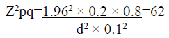
Sample size determination: Assuming a prevalence of 20% for malaria in pregnancy, for an error rate of 10% and a confidence interval of 95%, the sample size needed is determined by:
Where,
Z=z-score at 95% confidence interval=1.96
P=prevalence rate (20%)
q=Failure rate (80%)
d=error rate (10%)
Sixty-two participants will be required for this study.
Study population
The study population comprises of pregnant women attending antenatal clinic at Ijede General Hospital and Ikorodu General Hospital. The inclusion criteria were those women with fever or history of fever in 24-48 h preceding presentation at the hospital and those that signed or thumbprint the consent form. Patients with hyper parasitemia and signs of severe malaria were excluded.
Ethical approval
The study was conducted as a sub-study of another study “Characterization of molecular markers associated with Plasmodium falciparum resistance to antimalarial drugs and evaluation of PCR methods for parasite density estimation in rural and semi-urban site in southwest Nigeria” for which ethical approval was obtained from the Nigerian Institute of Medical Research Institutional Review Board. All work was performed according to the guidelines for human experimentation in clinical research and Helsinki declaration.
Data and sample collection
Questionnaires were designed to collect demographic data, history of drug use, participation in IPT, ITN possession, information on gestational age, history of fertile illness. 0.5 ml venous blood sample was collected from the peripheral vein in each participant by a trained laboratory Scientist or technician into a microtainer labelled EDTA bottles. The bottles were inverted several times to dissolve the anticoagulant and so prevent blood clotting. Left over blood samples were used to make thick and thin blood films and perform the two RDT immediately.
Preparation of Thick and Thin Blood Films Slides were labeled with sample number, Patient’s Initials and date. The slides were put on a level surface on top of a template to guide the size of the thick smear made and approximate positions of the thick and thin blood films that would be made. A drop of blood was placed onto the middle of the slide for the thin blood film and three larger drops about 1 cm from that for the thin film was spotted for the thick film. Using another slide as a spreader, the small drop of blood was touched with the spreader and allowed to run along its edge. The spreader was then firmly pushed in one swift stroke along the slide at an angle of 450 making sure it was in even contact with the surface of the slide. Using the corner of the spreader the drops of blood were spread to make an even, thick film about 1 cm in diameter. The films were allowed to dry for at least 15 min in a flat level position away from dust protected from flies.
Fixing and staining of films
Thin films were fixed by allowing a drop of methanol to run off the thin film away from the thick film for 10-20 s avoiding the fumes of the methanol reaching the thick films. They were placed vertically on racks with the thick film on the upper end away from the base of the drying rack, the methanol running dry away from it. The slides were allowed to air dry until the next day, placed in the oven for 15 min at 500°C. Slides were then immersed in 3% Giemsa stain prepared by diluting Giemsa with buffered water pH 7.2, in troughs, stained for 30 min. The slides were removed from staining trough and rinsed gently under the running tap and allowed to dry for 15 min before examination under the microscope.
Microscopic examination of blood films and estimation of parasitemia
A drop of immersion oil was dropped on the thick blood film and examined using the 100x oil immersion lenses on a binocular light microscope with 10x ocular objectives so that the total magnification was 1000x. Two hand tally counters were used, one to count parasites and one to count the fields. A third counter was necessary if gametocytes were seen or more than one species of parasite was present. For a slid to be declared negative 200 high powered fields are read. If parasites are seen, then:
If >100/HPF, then 2 representative fields are read. If moderate levels are present, then 200 parasites are counted, and the number of fields noted. If low levels are present, i.e., <1/HPF, then 200 fields are read. On slides with low levels of parasitaemia (i.e., the readers examine many fields before coming across a parasite), I asked another qualified slide reader to verify immediately that a parasite has been seen. If they cannot agree then the opinion of another qualified reader was sought.
The results are reported as the number of parasites per HPF or number of parasites per 200 HPF. The parasite density per μl is calculated by multiplying the number per HPF by 500, based on assumptions that 5-8 μl of blood is used in making a thick blood films and that 0.002 μl of blood is in a HPF, i.e., 10x eye piece, 100x objectives [9].
Parasite lactate dehydrogenase and aldolase kits
The OptiMAL dipstick test with lot number 46110.36.01and expiry date November 2009 was used in this study. It consisted of individually packed dipstick kits, detecting parasite pLDH specific for P. falciparum in one capture site and pan-pLDH detecting all four Plasmodium species in a separate capture line. The test device consists of two tubes, in which the dipstick stands for 10 min each, so results are read after 20 min. Washing buffer was provided for each individual kit in a mini plastic tube and a capillary pipette also.
Each kit is opened just before it is used. One drop of wash buffer, provided was put in the first well and four drops in the second well just before tests were carried out. Blood sampling (8-12 μl) was done with the plastic capillary pipette provided up to the indicated mark. The dipstick is then inserted into the first well such that contents of the well migrate up the nitrocellulose membrane of the dipstick. The presence of pLDH is revealed using monoclonal antibodies directed against isoforms of the enzyme but there is no cross-reaction with human LDH.
Dr. Greg’s aldolase kit had lot number 080620 Mal and expiry date 09/2011 was used in this study (EU). It consists of individually packed cassette with components that will perform the test per individual, i.e., alcohol swap, dropper and lancet, test cassette and wash buffer. The kit is designed to be used for combined P. falciparum/non-P. falciparum (P. vivax, P. ovale, P. malariae) diagnosis, targeting principally two antigens simultaneously just as in the Optimal kit.
Each kit was opened just before use. Using the dropper provided, two to three drops of blood was squeezed into the sample well of the cassette. After about 30 s, 2-3 drops of diluents was squeezed also into the sample well and the result read after 20 min. Test line one captures antigen specific to P. falciparum. Test line two captures antigen specific to non P. falciparum. The third line is for control.
For both the Optimal IT and Dr Greg’s kit, results were interpreted in a similar manner. No lines were visible in the result window before use. After the test, results were interpreted as:
Control line alone visible was read as negative
Control line and Test line one visible was read as positive for non-P. falciparum.
Control line and Test line one and two visible was read as for Positive for P. falciparum and or non-P. falciparum.
Preparation of qPCR DNA standards
Ready-made DNA from laboratory cultures of 3D7 grown to 8-11% parasitemia and centrifuged in percol gradient to isolate ring stage parasites, the stage of life cycle normally detected in peripheral blood, were used to make 10-fold serial dilutions to 1:106 with uninfected whole blood. Parasite densities were determined by counting parasites against 50,000 RBCs in thin blood films of 1:100 dilutions. It was confirmed by counting 500/hpf of duplicate thick films and converted to parasites per μl by counting 500 hpf of duplicate thick films using the actual RBC count of 3.9 × 10⁶ cells/ul. Parasite densities for the rest of the dilution series were extrapolated from the values obtained from the 1:100 dilution.
DNA extraction and nested qPCR
DNA was extracted from whole blood using the CORBETT X-TRACTOR GENETM ROBOT - (Montreal Biotech Inc.) in accordance with the manufacturer’s protocol which is completely automated as stated below. 50 μl of sterile PBS was added into each well of the lysis block. 150 μl blood samples was added according to earlier prepared plate plans and mixed by pipetting up and down 3x. Having dispensed reagent volumes of 118 ml DX wash (DXW) into a 170 ml reagent tub loaded into position A1 of reagent block R1, 60 ml of DX final wash (DXF) into another 170 ml reagent tub and loaded in position B1 of the reagent block R1, 60 ml of DX binding (DXB) solution containing 0.6 g of DX Binding additive into a 270 ml reagent tub and loaded in position C1, all reagents were covered with appropriate lids. A run was then started by clicking on the Corbet icon, switching on the CORBETT X-TRACTOR GENETM ROBOT, double clicking the Robotics 4 icon, selecting cancel on the vacuum extractor wizard window, selecting the blood extraction protocol from the program window and then clicking on jump to the end. Protocol is reviewed, closing the pop-up window to return to extraction protocol. Sample wells were selected at position B1, entering sample IDs at appropriate rows in the pop up window. 10 ml of DX liquid digest (DXL) was put in a 15 ml falcon tube, 1 ml DX digest enzyme added and gently mixed by inverting 10x taking care to avoid foaming. This was poured into a 17 ml reagent tub and installed at position C1. Elution buffer is loaded just before the elution step on prompt by the machine. Full boxes of filter tips are put in place and the run started by clicking control and selecting start. Elution plates are removed and eluents transferred into pre labeled 96 well PCR plates maintaining the same orientation. Samples were stored at 20° until required for PCR.
Quantitative PCR was used to determine presence of malaria parasites in the samples and the quantities in which they were present. The Opticon 2 Real-Time PCR Detection System using the Monitor software (version 3.1; BioRad Laboratories). The TaqMan assay which uses a fluorogenic probe to detect a specific PCR product was used.
Results
Patient characteristics
A total of 113 pregnant women comprising 90 from Ijede 23 from Ikorodu were enrolled in this study. The average age of the participants was 29.96 ± 4.67. The number of pregnant women enrolled during the study period included 17% in first trimester, 48% in the second trimester and 35% in the third trimester. There were 32 primigravida, 32 secundigravida and 49 multigravidas. According to their gravidity, the primigravidae had the highest proportion of parasitaemic participants with 25% (8/32) being positive with an average parasitemia of 1,845 ± 1881.13. Only 4 out of the 32 secundigravida (28%) were positive for peripheral parasitemia and they had a mean parasitemia of 11,530.2 ± 14194.96, while 20% of the multigravida (10/49) was positive with a mean parasitemia of 13,284.5 ± 18394.5.
Detection by microscopy, aldolase and PLDH
Twenty-three pregnant women were positive by microscopy giving a prevalence of 20.4%, while the prevalence by Aldolase and PLDH were 45.1% and 12.4%, respectively. The distribution of parasitemia among the women of different ages by microscopy and rapid diagnostic tests (Aldolase and PLDH) (Table 1). The pregnant women of age group 20-24 years (Table 1). Old were the most vulnerable to malaria infection with a prevalence of 54.5% and 22.7% by Dr. Grey’s Aldolase and PLDH tests respectively while microscopy showed women of age group 25-29 years as the most vulnerable with a prevalence of 25.7%. The table also shows that only Dr. Grey’s Aldolase test reveal women of age 40 years and above to have malaria infection while no infection was detected by microscopy and PLDH test in this group of women. The results showed that the primigravidae had the highest prevalence of parasitaemic (25%) while the secundigravidae had the lowest prevalence of 12.5%.
|
Age group
|
No. examined
|
Microscopy |
Aldolase |
Parasite Lactate dehydrogenase |
| Positive |
Negative |
Positive |
Negative |
Negative |
| Freq. |
% |
Freq. |
% |
Freq. |
% |
Freq. |
% |
Freq. |
% |
Freq. |
% |
| 20-24 |
22 |
5 |
22.7 |
17 |
77.3 |
12 |
54.5 |
10 |
45.5 |
5 |
22.7 |
17 |
77.3 |
| 25-29 |
35 |
9 |
25.7 |
26 |
74.3 |
17 |
48.6 |
18 |
51.4 |
4 |
11.4 |
31 |
88.6 |
| 30-34 |
33 |
6 |
18.2 |
27 |
81.8 |
12 |
36.4 |
21 |
63.6 |
4 |
12.1 |
29 |
87.9 |
| 35-39 |
17 |
3 |
17.6 |
14 |
82.4 |
8 |
47.1 |
9 |
52.9 |
1 |
5.9 |
16 |
94.1 |
| = 40 |
6 |
0 |
0.0 |
6 |
100.0 |
2 |
33.3 |
4 |
66.7 |
0 |
0.0 |
6 |
100.0 |
| Total |
113 |
23 |
20.4 |
90 |
79.6 |
51 |
45.1 |
62 |
54.9 |
14 |
12.4 |
99 |
87.6 |
Table 1: Prevalence of malaria infection based on different techniques in relation to age of pregnant women.
Comparison of rapid diagnostic tests with microscopy results
Overall, 54 (47.8%) samples were positive for malaria parasite antigen based on RDT (Dr. Grey’s Aldolase and PLDH) whereas 23 (20.4%) were positive by microscopy (Table 2). Of the 54 RDT positive samples, 51 (94.4%) were positive by Dr. Grey’s Aldolase while 14 (25.9%) were positive by PLDH. Of the 51 positive samples by Grey’s Aldolase, 17 (33.3%) samples were positive by microscopy and 34 (66.7%) samples were negative. 34 (66.7%) of the positive samples by Grey’s Aldolase were negative for both microscopy and pLDH. 3 (13.0%) of the 23 positive samples by microscopy were negative by RDTs (i.e., both Grey’s Aldolase and PLDH).
| Age group |
No. examined |
RDT
(Aldolase and Parasite Lactate dehydrogenase) |
Microscopy |
| Positive |
Negative |
Positive |
Negative |
| Freq. |
% |
Freq. |
% |
Freq. |
% |
Freq. |
% |
| 20-24 |
22 |
13 |
59.1 |
9 |
40.9 |
5 |
22.7 |
17 |
77.3 |
| 25-29 |
35 |
17 |
48.6 |
18 |
51.4 |
4 |
11.4 |
31 |
88.6 |
| 30-34 |
33 |
14 |
42.4 |
19 |
57.6 |
4 |
12.1 |
29 |
87.9 |
| 35-39 |
17 |
8 |
47.1 |
9 |
52.9 |
1 |
5.9 |
16 |
94.1 |
| = 40 |
6 |
2 |
33.3 |
4 |
66.7 |
0 |
0.0 |
6 |
100.0 |
| Total |
113 |
54 |
47.8 |
59 |
52.2 |
14 |
12.4 |
99 |
87.6 |
Table 2: Performance of different brands of rapid diagnostic tests in diagnosing pregnancy associated with malaria (PAM).
Sensitivity and specificity of diagnostic techniques
Based on microscopy as gold standard, the overall sensitivity of presumptive diagnosis based on Aldolase and pLDH were 73% and 64%, respectively. The corresponding specificity rates were 59% and 100% respectively. pLDH gives no false positive but a false negative rate of 36%. Aldolase gave a false positive rate of 41% and false negative rate of 27%.
Positive and negative predictive values
The positive predictive value (PPV) of pLDH was 100% and negative predictive value (NPV) was 92%. The PPV and NPV of Aldolase were 30% and 90%, respectively.
Discussion
The performance of RDTs (pLDH and Aldolase) in diagnosing pregnancy associated with malaria (PAM) was evaluated against microscopy among pregnant women in Lagos State, Southwest, Nigeria. Studies of the use of rapid diagnostic tests (RDTs) in pregnant women have been undertaken mainly for the purpose of diagnosing placental malaria at delivery [25-27]. The data presented in this study shows that RDTs are potentially useful tools in the diagnosis of malaria in this setting. The levels of sensitivity of RDTs ranged from 64-73% for P. falciparum, however World Health Organisation (WHO) stated that for RDTs to be a useful diagnostic tool, it must achieve sensitivity of greater than 95% [28].
In this study, aldolase test was better in sensitivity when compared to the pLDH test; its lack of specificity makes it difficult to compare both performances. For P. falciparum detection, aldolase test followed by microscopy appeared to be more sensitive than the pLDH test. Aldolase test had a better capacity to detect low density P. falciparum infections, but it gave a relatively high number of false positive results (41%) so that its specificity and positive predictive value were lower than that of pLDH. Previous studies have reported aldolase to be more sensitive due to its capacity to detect low parasitaemia [29].
The results of this study have also shown that microscopy may underestimate the real malaria burden during pregnancy. Similar finding was reported by Singer et al (2004). who likewise observed that that Polymerase Chain Reaction (PCR) detected more positive cases than RDTs whilst assessing only placental blood samples [27]. In this study, a small proportion of blood samples negative for both RDTs (pLDH and aldolase) and microscopy were checked by PCR (result not shown) and the analyses showed that only two more samples were positive with PCR. The use of microscopy in diagnosis of malarial infection in adult population that comprises pregnant women can pose some challenges due to placental sequestration of parasites thus reducing the sensitivity of microscopy.
The performance of RDTs (aldolase) in malaria diagnosis in this study is in agreement with previous studies conducted, showing that RDTs performed better than microscopy in malaria diagnosis under field conditions [21,30-33]. The performance of pLDH was well below optimum in sensitivity. It missed detection of low density parasitaemia that was detected by microscopy although the sensitivity became 83% for parasitaemia above 100 p/μl, this value is still less than the recommended performance of 95% at this threshold. Some studies have also reported reduced sensitivity of RDTs as a result of low parasitaemia, and this could explain the reason for the cases with low parasite densities missed by RDTs [34,35]. The use of RDTs act as an appropriate alternative diagnostic tool for malaria instead of relying on presumptive treatment based on clinical observations in areas with limited expert microscopy and laboratory infrastructure.
Conclusion
This study revealed that aldolase (RDTs) was more sensitive than pLDH but lacked specificity when microscopy is used as reference in diagnosing P. falciparum in pregnant women. Further research should be conducted into aldolase RDT based optimum use in low transmission settings.
References
- Miller LH, Good MF, Milon G (1994) Malaria pathogenesis. Science. 264:1878-1883.
- World Health Organization (2016) WHO World Malaria Report World Health Organization.
- World Health Organization (2017) WHO Malaria in pregnant women.
- Parise M, Nahlen B, Menendez C, Steketee R (2001) The burden of malaria in pregnancy in malaria-endemic areas. Am. J. Trop. Med. Hyg.64:28-35.
- Mockenhaupt FP, Rong B, Günther M, Beck S, Till H, et al. (2000) Submicroscopic P. falciparum infections in pregnancy in Ghana. Trop Med Int Heal. 94:477-483.
- Olayemi O, Aimakhu CO, Udoh ES (2003) Attitudes of patients to obstetric analgesia at the University College Hospital, Ibadan, Nigeria. J. Obstet. Gynaecol.23:38-40.
- Kwenti TE, Kwenti TDB (2016) Anaemia and its association with month and blood phenotype in blood donors in Fako division, Cameroon. BMC Hematol.16:29.
- Bartoloni A, Zammarchi L (2012) Clinical aspects of uncomplicated and severe malaria. Mediterr. J. Hematol. Infect. Dis.4:1.
- Mayor A, Serra-Casas E, Bardají A, Sanz S (2009) Sub-microscopic infections and long-term recrudescence of Plasmodium falciparum in Mozambican pregnant women. Malar J. 8:9.
- World Health Organization (2016) T3: Test. Treat. Track initiative, WHO.
- Adegnika AA, Verweij JJ, Agnandji ST, Chai SK (2006) Microscopic and sub-microscopic Plasmodium falciparum infection, but not inflammation caused by infection, is associated with low birth weight. Am. J. Trop. Med. Hyg. 75:798-803.
- Tagbor H, Bruce J, Browne E, Greenwood B, Chandramohan D, et al. (2008) Malaria in pregnancy in an area of stable and intense transmission: is it asymptomatic? Trop. Med. Int. Heal. 13:1016-1021.
- Mockenhaupt FP, Bedu-Addo G, von Gaertner C (2006) Detection and clinical manifestation of placental malaria in southern Ghana. Malar J. 5:119.
- Jelinek T, Grobusch MP, Nothdurft HD (1999) Sensitivity and specificity of dipstick tests for rapid diagnosis of malaria in nonimmune travellers. J. Clin. Microbiol. 37:175-179.
- Okell LC, Ghani AC, Lyons E, Drakeley CJ (2009) Submicroscopic infection in P. falciparum endemic populations: a systematic review and meta-analysis. J. Infect. Dis. 200:1509-1517.
- Reyburn H, Mbatia R, Drakeley C, Carneiro I (2004) Overdiagnosis of malaria in patients with severe febrile illness in Tanzania: a prospective study. BMJ. 329:1212.
- Bejon P, Andrews L, Hunt-Cooke A, Sanderson F (2006) Thick blood film examination for Plasmodium falciparum malaria has reduced sensitivity and underestimates parasite density. Mal. J. 5:104.
- Othnigué N, Wyss K, Tanner M, Genton B (2006) Urban malaria in the Sahel: prevalence and seasonality of presumptive malaria and parasitaemia at primary care level in Chad. Trop. Med Int. Heal. 11:204-210.
- Dhorda M, Piola P, Nyehangane D (2012) Performance of a Histidine-Rich Protein 2 rapid diagnostic Ttest, Paracheck Pf®, for detection of malaria infections in Ugandan Pregnant Women. Am. J. Trop. Med.Hyg. 86:93-95.
- Gerstl S, Dunkley S, Mukhtar A, De Smet M (2010) Assessment of two malaria rapid diagnostic tests in children under five years of age, with follow-up of false-positive pLDH test results, in a hyperendemic falciparum malaria area, Sierra Leone. Malaria Journal, Malar J. 9:28.
- Tham JM, Lee SH, Tan TM, Ting RC, Kara UA, et al. (1999) Detection and species determination of malaria parasites by PCR: comparison with microscopy and with ParaSight-F and ICT Malaria Pf Tests in a clinical environment. J. Clin. Microbiol. 37 1269-1273.
- Singh NEER, Saxena AJA, Awadhia SB, Shrivastava RITA, Singh MP, et al. (2005) Evaluation of a rapid diagnostic test for assessing the burden of malaria at delivery in India. Am. J. Trop. Med. Hyg.73.
- de Oliveira AM, Skarbinski J, Ouma PO, Kariuki S, Barnwell JW , et al. (1980) Performance of malaria rapid diagnostic tests as part of routine malaria case management in Kenya. Am. J. Trop. Med. Hyg. 80:470-474.
- Minja DTR Minja DTR, Schmiegelow C, Mmbando B, Boström S, Oesterholt M, et al. (2012) Reliability of rapid diagnostic tests in diagnosing pregnancy-associated malaria in north-eastern Tanzania. Malar. J. 11:211.
- Leke N, Grenier D, Goldner M, Mayrand D (1999) Effects of hydrogen peroxide on growth and selected properties of Porphyromonas gingivalis. FEMS Microbiol. Lett. 174:347-353.
- Mankhambo L, Kanjala M, Rudman S, Lema VM, Rogerson SJ, et al. (2002) Evaluation of the OptiMAL rapid antigen test and species-specific PCR to detect placental P. falciparum infection at delivery. J. Clin. Microbiol. 40:155-158.
- Singer LM, Newman RD, Diarra A, Moran AC, Huber CS, et al. (2004) Evaluation of a malaria rapid diagnostic test for assessing the burden of malaria during pregnancy. Am. J. Trop. Med. Hyg. 70:481-485.
- Molineaux L, Gramiccia G (1999) New Perspectives: Malaria Diagnosis. Report of joint WHO/USAID informal consultation.
- van den Broek I, Hill O, Gordillo F, Angarita B (2006) Evaluation of three rapid tests for diagnosis of P. falciparum and P. vivax malaria in Colombia. Am. J. Trop. Med. Hyg. 75:1209-1215.
- Tjitra E, Suprianto S, Dyer M, Currie BJ (1999) Field evaluation of the ICT Malaria P.f/P.v immunochromatographic test for detection of P. Falciparum and P.Vivax in patients with a presumptive clinical diagnosis of malaria in eastern Indonesia. J Clin Microbiol. 37:2412-2417.
- Batwala V, Magnussen P, Nuwaha F (2010) Are rapid diagnostic tests more accurate in diagnosis of Plasmodium falciparum malaria compared to microscopy at rural health centres? Malar. J. 9:349.
- Kamel MM, Attia SS, Emam GD, Al Sherbiny NAE (2016) The validity of rapid malaria test and microscopy in detecting malaria in a preelimination region of Egypt. Scientifica. 2016: 4048032.
- Ayogu E, Ukwe C, Nna E (2016) “Assessing the reliability of microscopy and rapid diagnostic tests in malaria diagnosis in areas with varying parasite density among older children and adult patients in Nigeria,” J. Postgrad. Med. 62:150.
- Adesanmi T, Okoro A, Okafor H, Mafe A (2011) Diagnosis of malaria parasitemia in children using a rapid diagnostic test. Niger. J. Clin. Pract.14:195.
- Oyeyemi OT, Ogunlade AF, Oyewole IO (2015) Comparative assessment of microscopy and rapid diagnostic test (RDT) as malaria diagnostic tools. Res. J. Parasitol. 10:120-126.