Keywords
Pancreas
Introduction
Tuberculosis (TB) is a serious health problem worldwide [1]. It is a multi-systemic bacterial infection caused by different strains of mycobacteria, usually Mycobacterium tuberculosis. It occurs in nearly 9.7 million people [2, 3], and claims about 2 million lives each year worldwide [4] highest incidence being in Asia, South America, Eastern Europe, and most sub-Saharan African countries [2, 3].
Although the incidence of TB was low in developed countries, it has risen in recent years due to an increase in emigration from tuberculosis- endemic areas, the emergence of human immunodeficiency virus (HIV) infection [5], and widespread use of immunosuppressant drugs [6, 7]. For example Australia, Western Pacific and South East Asia have incidence rates of 5-6, 108, 181 per population, respectively [8, 9] and United States Center for Disease Control and Prevention (CDC) reported approximately 11200 new cases of TB in 2014 [10].
Although TB usually involves lungs, extra-pulmonary TB (EPTB) accounts for nearly 10-30% (approximately 15- 20%) of all cases of TB in immunocompetent hosts [3, 11, 12]. About 12.5% of all TB cases have EPTB [13] who nearly 50 percent of them are human immunodeficiency virus (HIV)-positive [14, 15].
By definition, EPTB is when TB occurs at sites other than the lung. It can occur in almost any organ system; most commonly in the lymph nodes, pleura, genitourinary system, and bone [14, 15]. Note that EPTB must not be confused with miliary TB, as miliary refers to EPTB with pulmonary involvement, and not EPTB in isolation [16].
Abdominal TB is a common site for EPTB [15]. It accounts for 5-12% of patients with tuberculosis (with the highest prevalence in developing countries) [17, 18] and almost 11-16% of patients with EPTB have abdominal involvement [13]. Abdominal TB includes infection of different combinations of gastrointestinal tract (especially ileocecal region), lymph nodes, peritoneum, and intraabdominal organs such as the spleen, liver, and pancreas [15, 19-29].
Pancreatic tuberculosis, either with or without peripancreatic lymphadenitis, is a rare occurrence in either immunocompetent or immunosuppressed host. It was first reported by Auerbach in 1944. In his series of 1656 autopsies of tuberculous patients, only 14 cases had pancreatic involvement that may have mimicked neoplasia but he did not find any cases of isolated pancreatic tuberculosis [28]. Pancreatic tuberculosis usually occurs in patients with miliary tuberculosis with pulmonary and extra-pulmonary involvement including pancreas, or in isolated EPTB cases without pulmonary involvement especially in acquired immunodeficiency syndrome (0.46%) [13, 14, 21, 23, 27, 28, 30, 31]. But even HIVinfected patients have an incidence of 0.46% [32, 33]. Autopsy studies have shown that the pancreas is involved in 2.1%–4.7% of patients with miliary tuberculosis [28, 34-38]. However, in a study from 1999-2004 from India detected pancreatic TB in 8.3% of the 384 patients who were diagnosed with abdominal TB [39].
As mentioned above, pancreatic tuberculosis is occasionally observed in people with other organ's involvement as a consequence of miliary TB or EPTB with involvement of other organs in addition to pancreas [28, 34]. But primary pancreatic tuberculosis (PPTB) is described as an isolated involvement of pancreas by mycobacterium tuberculosis in the absence of involvement of any other organ or previously identified TB [36]. PPTB is particularly rare [37, 38]; fewer than 100 cases have been reported worldwide [39] and its incidence is estimated to be less than 4.7% [28, 31]. Bhansali in a series of 300 miliary tuberculosis patients over 12 years in India, did not reveal even 1 with pancreatic involvement [2, 10, 15, 21].
Isolated pancreatic TB is predominantly observed in the following patient types:
• Patients who reside in endemic tuberculose zones,
• Patients in areas of widespread TB dissemination such as a military setting and developing countries [19, 20, 25, 27],
• Patients who are immuno-compromised [40]
However pancreatic TB has also been reported with increased frequency from western world [41] and the reported number of such cases in immunocompetent patients has increased over the past decade [31]. This increasing incidence is probably due to globalization, increased use of immunosuppressants, the worldwide resurrection of M. tuberculosis and the HIV pandemic [31, 34, 42, 43].
A recent review reported that 23% of the 62 cases of pancreatic TB occurred in patients who were HIV infected [44]. In AIDS cases, tuberculous pancreatic abscesses are most common, accounting for 70.0% of cases. In addition, 71.1% of cases have no previous serological evidence of HIV infection, and 76.2% of patients are severely immunocompromised hosts with a CD4 cell count of ≤190/ mm3 [5, 29, 45].
More than half of patients with pancreas tuberculosis in the world literature are young adults (<30 years old) [39, 46]. Convincing epidemiological data with regards to sex are conflicting, suggesting that pancreatic TB is more common in men [27, 39].
Pancreatic TB most commonly afflicts the region of the head and the uncinate process of the pancreas [47, 48]. It is often misdiagnosed due to low index of suspicion and masquerading of its symptoms as more common pancreatic conditions such as pancreatic malignancy [36, 47, 49]. If the diagnosis is delayed, pancreatic TB can be fatal; it has a 10.8% mortality rate (comparing to the mortality rate of 9.1% in immunocompetent patients) [50]. However, pancreatic TB responds well to standard antituberculous drugs (ATDs).
Pathogenesis
Because of its rarity, the pathogenesis of pancreatic/ peri-pancreatic involvement of TB is not known with certainty and it is not yet clear how the infection can only affect the pancreas [51, 52]. It is believed that this low frequency is due to retroperitoneal location of pancreas as well as pancreatic enzymes including lipases and deoxyribonucleases that interfere with the colonization, seeding and proliferation of the bacteria [4, 38, 53, 54].
Pancreatic secretions also showed an antitubercular effect in vitro [51, 52], thus a large intrapancreatic inoculum of Mycobacterium tuberculosis is required to cause pancreatic lesions [53, 55].
Although the primary site of TB is usually not evident in most cases of hepatobiliary and pancreatic TB [56], several possible mechanisms for pancreatic involvement of tuberculosis have been discussed. Ingestion of infected material from an active pulmonary lesion is one. Following ingestion the bacilli gain access to the gastrointestinal tract where necrotizing granulomas may develop and then spread to the lymphatics affecting any organ in the GI tract, including hepatobiliary and pancreatic tissue [57]. Other mechanisms include lymphohematogenous dissemination from pulmonary disease, reactivation of latent tuberculosis in the pancreatic focus and toxic-allergic reaction of the pancreas involving an inflammatory response to generalized tuberculosis [19, 28, 37, 56, 58, 59].
Clinical Features
Over the last two years, 39 patients with pancreatic tuberculosis were reported in the literature. Of these patients, 26 were males and age ranged from 13 to 58 years. The outstanding signs and symptoms of pancreatic tuberculosis are summarized as Table 1. Most common location of pancreatic tuberculosis as a mass has been reported in the head (29-74.4%) followed by body (7- 17.9%), uncinate (3-7.7%), and tail (2-5.1%). Abdominal pain is the most common (31-79.5%) symptom followed by fever (20-51.3%), weight loss (19-48.7%), appetite loss (11-28.2%), and jaundice (8-20.5%) that were obstructive in nature.
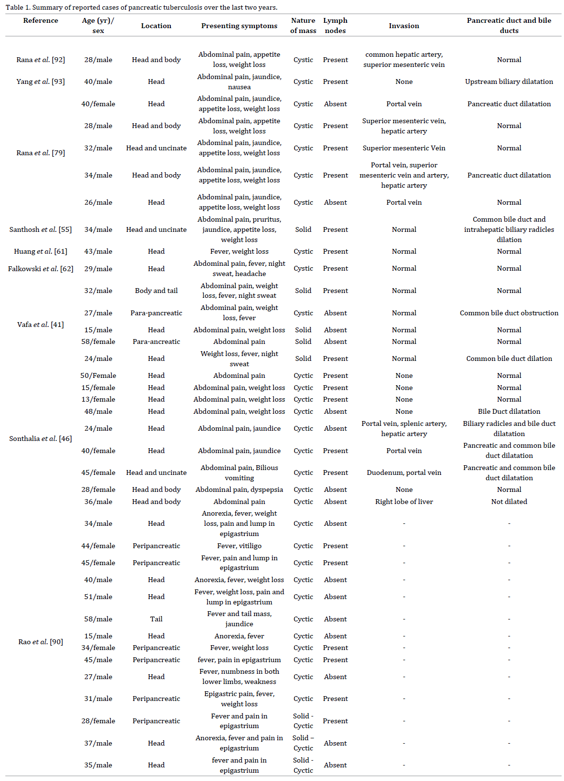
Three patients had the past history of pulmonary tuberculosis and lymphadenopathy was present in 22 patients. Pancreatic tuberculosis leaded to local vascular invasion in 10 patients also causing diagnostic confusion with locally advanced or metastatic pancreatic malignancy. Portal vein, superior mesenteric vein and hepatic artery involvement were reported in 6 (15.4%), 4 (10.3%), and 4 (10.3%) patients, respectively.
Diagnosis
Diagnosing pancreatic TB is challenging due to its nonspecific clinical, laboratory or radiological features. Since pancreatic TB may present as cystic or solid pancreatic masses, abscesses, lymphomas, pseudocysts or acute or chronic pancreatitis, cytological or histopathological as well as bacteriological confirmation is necessary for the diagnosis of pancreatic tuberculosis.
Pancreatic tuberculosis is a potential mimic of pancreatic malignancy. Although local vascular invasion is often considered to be an imaging feature of malignant lesions, the presence of vascular invasion cannot exclude possibility of pancreatic tuberculosis [60]. Also, the dilation of the pancreatic main or branch duct does not distinguish one condition from the other. However, there was other evidence against pancreatic malignancy such as fever (unless pyogenic cholangitis develops due to biliary duct compression) and the normal values of tumor markers [61]. In patients without typically tuberculosis manifestations (such as fever, night sweats, weight loss or cough), a range of cystic pancreatic lesions, including common lesions (pseudocysts, serous or mucinous cystadenomas, intraductal papillary mucinous neoplasm) and rare lesions such as solid pseudopapillary tumors pancreatic epithelial cysts, may need to be considered [62].
Pancreatic TB can be classified radiologically into 3 groups: mass-forming (with or without diffuse pancreatic enlargement), a diffuse form and a small, nodular form. The mass-forming is the most common form that accounts for 94.4% of cases [63].
Several imaging methods like CT scan, MRI, transcutaneous ultra-sound and endoscopic ultrasound are used to assess pancreatic pathology. Ultra sound (US) scan is noninvasive, simple, readily available and cost-effective; thus, it is usually used as an initial diagnostic tool. CT scan is used to rule out associated pathologies and to plan for disease management. It is the investigation of choice for pathologies of pancreas because of its high sensitivity. Although imaging findings may suggest the possibility of tuberculosis, none of them are neither specific nor pathognomonic for pancreatic TB [64]. They can reveal diffuse enlargement of the pancreas [64-66], focal hypoechoic mass on ultrasound or cystic lesions [67-73] (mostly in the head and uncinate process), heterogeneously hypo-isoechoic or isodense lesions on ultrasound and CT Scan, respectively, (primarily in the head) [39, 64, 65, 67, 69, 74] and enlarged peripancreatic [67, 70, 75] and other abdominal LNs [45, 64, 69, 70, 76, 77].
In pancreatic TB common bile duct and the pancreatic duct appear normal in images, even if the mass is localized centrally in the head of the pancreas. Contrary to pancreatic TB, in pancreatic adenocarcinoma the pancreatic duct is dilated in the tumors that are positioned centrally in the head region. Pancreatic enlargement with narrowing of the main pancreatic duct and heterogeneous enhancement is the characteristic of the diffuse form of pancreatic tuberculosis [78]. Bile cytology on endoscopic retrograde cholangiopancreatography (ERCP) may help in establishing the diagnosis [10, 56].
However, EUS-FNA sampling is essential for establishing the diagnosis of pancreatic tuberculosis. The samples are evaluated by staining, cytology, bacteriology, culture and polymerase chain reaction assay [79-86]. A definitive decision is usually made based on a histopathological or microbiological examination of a specimen that is obtained from the pancreas or based on peripancreatic LNs. In a study, Song et al. [87] were able to diagnose pancreatic/ peri-pancreatic tuberculosis in 76.2% of patients using EUS-FNA. The microscopic features of tuberculosis are granuloma, caseation necrosis (seen in 75%-100% of cases) and presence of acid fast bacilli (identified in 20%-40% of cases) [84]. EUS is a reliable technique for differentiating pancreatic lesion from peripheral structures [87]. It is also preferred for tissue biopsy because of less chances of needle tract dissemination especially when the mass seems malignant. Rana et al. found that the majority of patients with pancreatic tuberculosis (83.3%) had granulomas with culture for Mycobacterium tuberculosis being positive in 1 of 2 patients (50.0%) tested and acid fast bacilli being seen in only 1 of 6 (16.7%) [79]. In addition, acid fast bacilli smear using auramine or Ziehl-Neelsen staining can also be employed for this purpose.
As a result, and given the simplicity of the test and the rapid results that it produces, EUS-FNA with an acid-fast smear should be required for pancreatic TB diagnosis. A PCR assay yields highly specific same-day results, when used to detect mycobacterial DNA. Although its sensitivity to TB in FNA specimens has not yet been established, the PCR assay is increasingly used adjunctive to special staining techniques and mycobacterial cultures. It may show positive results even when specimen cultures are negative [77].
Therefore, diagnosis is a challenge, calling for a team approach with the goal of making the diagnosis noninvasively.
Treatment
Most cases of pancreatic TB respond well to ATD [32, 71, 88-90]. Therefore invasive treatments such as surgery and the drainage of fluid are not necessary most of the times [32]. Directly observed therapy with a standard multiple ATD regimen including isoniazid, rifampicin, pyrazinamide, and ethambutol or streptomycin for 6-12 months, is usually effective [74]. However for those patients whose tuberculous pancreatic mass is enlarged and causes symptoms even after ATDs therapy for a reasonable period of time, we should consider minimally invasive procedures [32] such as endoscopic internal drainage, percutaneous catheter drainage or biliary stenting. Percutaneous catheter drainage is preferably used in patients with no pancreatic duct strictures and pancreatic duct-pseudocyst communications and in those with immature or infected pseudocysts, or when the patient is at high surgical risk, or exhibits malnourishment [91, 92]. Also ATDs have hepatotoxic effects, and malnourished patients with jaundice are at higher risk. Therefore, patients with underlying liver disease might require modification of ATDs [79].
Conflicting Interest
The authors had no conflicts of interest
References
- Cherian JV, Somasundaram A, Ponnusamy RP,Venkataraman J .Peripancreatictuberculous lymphadenopathy. An impostor posing diagnostic difficulty. JOP 2007;8:326-329. [PMID: 17495362]
- Ahlawat SK, Charabaty-Pishvaian A, LewisJH, et al. Pancreatic tuberculosis diagnosed with endoscopic ultrasound guided fine needle aspiration . JOP 2005;6:598-602. [PMID:16286712]
- Floyd K1, Pantoja A.Financial resources required for tuberculosis control to achieve global targets set for 2015.Bull World Health Organ. 2008;86:568-76. [PMID: 18670669]
- Kaushik N, Schoedel K, Mc Grath K.Isolated pancreatic tuberculosis diagnosed by endoscopic ultrasound-guided fine needle aspiration: a case report. JOP 2006; 7:205-10. [PMID: 16525205]
- 1993 revised classification system for HIV infectionand expanded surveillance case definition for AIDS among adolescents and adults. MMWR Recomm Rep 1992; 41(RR-17):1-19.[https://goo.gl/aVcOKw]
- D'Cruz S, Sachdev A, Kaur L, Handa U, Bhalla A, Lehl SS. Fine needle aspiration diagnosis of isolated pancreatic tuberculosis: a case report and review of literature. JOP 2003; 4:158-62. [PMID: 12853684]
- Schneider A, Birgelen CV, Dührsen U,Gerken G, Rünzi M. Two cases of pancreatic tuberculosis in non-immunocompromised patients: a diagnostic challenge and a rare cause of portal hypertension. Pancreatology 2002; 2:69-73. [PMID: 12120010]
- Mellick SA. Sir Kenelm Digby (1603–1665):diplomat, entrepreneur, privateer, duellist, scientist and philosopher. ANZ J Surg 2011; 81: 911–4. [PMID: 22507419]
- Le Claire L. Kenelm Digby – ‘The Mirandola of his age’. The Wilkinson Lecture 2005. Worcester College Record 2005:51–69. [https://goo.gl/hT0q5Q]
- Sachdev A, D’Cruz S, Chauhan S, Thakur R,Kapoor V, Handa U. Pancreaticobiliary tuberculosis diagnosed by endoscopic brushings. JOP 2006; 7: 665-669. [PMID: 17095849]
- Echenique Elizondo M, Amondarain ArratíbelJ, Compton CC, Warshaw AL. Tuberculosis of the pancreas. Surgery 2001; 129:114-6. [PMID: 11150042]
- Pramesh CS, Heroor AA, Gupta SG, Krishnamurthy S, Shukla PJ, Jagannath P, et al. Pancreatic tuberculosis:an elusive diagnosis. HPB (Oxford) 2003; 5:43-5. PMID: 18332958]
- Khan R, Abid S, Jafri W, et al.Diagnostic dilemma of abdominal tuberculosis in non-HIV patients: anongoing challenge for physicians. World J Gastroenterol 2006;12:6371-5. [PMID: 17072964]
- Bhansali SK. Abdominal tuberculosis.Experiences with 300 cases. Am J Gastroenterol 1977;67:324-37. [PMID:879148]
- Yokoyama T, Miyagawa S, Noike T, et al.Isolated pancreatic tuberculosis. Hepatogastroenterology 1999;46:2011-4. [PMID: 10430386]
- Puri R, Thandassery RB, Eloubeidi MA, Sud R. Diagnosis of isolated pancreatic tuberculosis: the role of EUS-guided FNA cytology .Gastrointest Endosc. 2012;75:900-4.[ PMID: 22440205]
- Chen CH, Yang CC, Yeh YH, Yang JC, Chou DA. Pancreatic tuberculosis with obstructive jaundice--a case report. Am J Gastroenterol 1999; 94:2534-2536. [PMID: 10484020]
- Tosun S, Tosun A. Imaging of malignancy-suspected pancreatic involvement of extrapulmonary tuberculosis. Turk J Gastroenterol 2010;21:54-9.[ PMID: 20533115]
- Sharma SK, Mohan A. Extra pulmonary tuberculosis. Indian J Med Res 2004; 120: 316-353. [PMID: 15520485]
- Sharma MP, Bhatia V. Abdominal tuberculosis.Indian J Med Res 2004; 120: 305-315. [PMID: 15520484]
- Turan M, Sen M, Koyuncu A, et al. Pancreatic pseudotumor due to peripancreatictuberculous lymphadenitis.Pancreatology 2002;2:561-4. [PMID: 12435870]
- Evans JD, Hamanaka Y, Olliff SP,Neoptolemos JP. Tuberculosis of the pancreas presenting as metastatic pancreatic carcinoma. A case report and review of the literature. Dig Surg 2000;17:183-7. [PMID: 10781988]
- Turan M, Sen M, Koyuncu A, Aydin C,Elaldi N, Arici S. Pancreatic pseudotumor due to peripancreatic tuberculous lymphadenitis. Pancreatology 2002;2:561-4. [PMID: 12435870]
- Khaniya S, Koirala R, Shakya VC, Adhikary S, Regmi R, Pandey SR, et al. Isolated pancreatic tuberculosis mimickingas carcinoma: a case report and review of the literature. Cases J 2010;3:18. [PMID: 20205859]
- Tan KK, Chen K, Liau KH, Ho CK. Pancreatic tuberculosis mimicking pancreatic carcinoma: Series of three cases. Eur J Gastroenterol Hepatol 2009;21:1317-9. [PMID: 19474749]
- Pereira JM, Madureira AJ, Vieira A, RamosI. Abdominal tuberculosis: imaging features. Eur J Radiol 2005;55:173–80. [PMID: 2653722]
- Meinke AK. Pancreatic tuberculousabscess. Conn Med 1989; 53: 139-141. [PMID: 2653722]
- Auerbach O. Acute Generalized Miliary Tuberculosis. Am J Pathol 1944; 20:121-136. [PMID: 19970738]
- Fee MJ, Oo MM, Gabayan AE, Radin DR,Barnes PF. Abdominal tuberculosis in patients infected with the human immunodeficiency virus. Clin Infect Dis 1995; 20: 938-944. [PMID: 7795098]
- Sunderam G, McDonald RJ, Maniatis T,Oleske J, Kapila R, Reichman LB. Tuberculosis as a manifestation of the acquired immunodeficiency syndrome (AIDS). JAMA 1986; 256:362–6. [PMID:3723722]
- Xia F, Poon RT, Wang SG, Bie P, Huang XQ,Dong JH. Tuberculosis of pancreas and peripancreatic lymph nodes inimmuno competent patients: experience from China. World J Gastroenterol 2003;9:1361-4. [PMID: 12800257]
- Meesiri S. Pancreatic tuberculosis withacquired immuno deficiency syndrome: a case report and systematic review.World J Gastroenterol 2012; 18:720-726. [PMID: 22363146]
- Maniar JK, Kamath RR, Mandalia S, Shah K,Maniar A. HIV and tuberculosis: partners in crime. Indian J Dermatol Venereol Leprol 2006; 72: 276-282. [PMID: 16880573]
- Woodfield JC, Windsor JA, Godfrey CC, etal. Diagnosis and management of isolated pancreatic tuberculosis: recentexperience and literature review. ANZ J Surg 2004;74:368–371. [PMID:15144259]
- Paraf A, Ménager C, Texier J.Tuberculosis of the pancreas and tuberculosis of the lymph nodes of theupper region of the abdomen. Rev Med Chir Mal Foie 1966;41:101–126. [PMID:5944700]
- Mansoor J, Umair B. Primary pancreatic tuberculosis: a rare and elusive diagnosis . J Coll Physicians Surg Pak. 2013;23:226-8. [PMID: 23458052]
- Brugge WR, Mueller PR, Misdraji J Case8-2004 N Engl J Med 2004 350: 1131-1138. [PMID: 15014187]
- Knowles KF, Saltman D, Robson HG, Lalonde R. Tuberculous pancreatitis. Tubercle 1990; 71:65-68. [PMID: 2371763]
- Nagar AM, Raut AA, Morani AC, Sanghvi DA, Desai CS, Thapar VB. Pancreatic tuberculosis: a clinical and imaging review of 32 cases. J Comput Assist Tomogr 2009; 33: 136-141. [PMID:19188801]
- Echenique Elizondo M, Amondarain Arratibel JA, IribarrenLoyarte J. Pancreatic tuberculosis: the rebirth of a time worn problem. Cir Esp 2000; 67:103-105. https://goo.gl/dDQRcm
- Vafa H, Arvanitakis M, Matos C, et al. Pancreatic tuberculosis diagnosed by EUS: one disease, many faces. JOP 2013;14:256-260. [PMID: 23669474]
- D’Cruz S, Sachdev A, Kaur L, Handa U, Bhalla A, Lehl SS. Fine needle aspiration diagnosis of isolated pancreatic tuberculosis. A case report and review of literature. JOP 2003; 4:158-162. [PMID: 12853684]
- Weiss ES, Klein WM, Yeo CJ. Peripancreatic tuberculosis mimicking pancreatic neoplasia. J Gastrointest Surg 2005;9:254-62. [PMID: 15694822]
- Chaudhary A, Negi SS, Sachdev AK, Gondal R. Pancreatic tuberculosis: still a histopathological diagnosis. Dig Surg 2002; 19:389-392. [PMID: 12435910]
- Ezratty A, Gumaste V, Rose E, Sachar DB,Tiscornia-Wasserman P. Pancreatic tuberculosis: a frequently fatal butpotentially curable disease. J Clin Gastroenterol 1990; 12:74-77. [PMID:2105992]
- Sonthalia N, Ray S, Pal P, Saha A,Talukdar A . Fine needle aspirationdiagnosis of isolated pancreatic tuberculosis: A case report . World J Clin Cases 2013 16;1:181-6. [PMID: 24303497]
- Crowson MC, Perry M, Burden E.Tuberculosis of the pancreas: a rare cause of obstructive jaundice. Br J Surg 1984;71:239. [PMID: 6697132]
- Teo LL, Venkatesh SK, Ho KY. Clinics indiagnostic imaging. Singapore Med J 2007; 48:687-692. [PMID: 17609835]
- Abid M, Guirat A, Ayadi L, Mzali R, BenAmar M, Beyrouti MI. Pancreatic tuberculosis: a rare cause of pseudoneoplasic obstructive jaundice. Presse Med 2009;38:e7–10. [PMID:19167862]
- Jenney AW, Pickles RW, Hellard ME, Spelman DW, Fuller AJ, Spicer WJ. Tuberculous pancreatic abscess in an HIV antibody-negative patient: case report and review. Scand J Infect Dis 1998; 30:99-104.[ PMID: 9730291]
- Porter AE The Bacteriolytic Action of Gland Extracts on Tubercle Bacilli. J Hyg (Lond) 1917; 16:55-65. [PMID:20474643]
- Day AA, Gibbs WM The action of pancreaticjuice on bacteria .The Journal of Infectious Diseases 1930:26-30. [https://goo.gl/6ZfPoi]
- Franco-Paredes C, Leonard M, Jurado R,Blumberg HM, Smith RM. Tuberculosis of the pancreas: report of two casesand review of the literature. Am J Med Sci 2002; 323:54-8. [PMID: 11814144]
- Cho SB. Pancreatic tuberculosis presenting with pancreatic cystic tumor: a case report and review of the literature. Korean J Gastroenterol 2009; 53:324-8. [PMID: 19458471]
- Santhosh S, Bhattacharya A, Rana SS, Bhasin DK, Srinivasan R, Mittal BR. Pancreatic tuberculosis: Evaluation oftherapeutic response using F-18 fluoro-2-deoxy-D-glucose positron emission tomography/computed tomography. Indian J Nucl Med 2014;29:257-9. [PMID: 25400368]
- Saluja SS, Ray S, Pal S, Kukeraja M, Srivastava DN, Sahni P, Chattopadhyay TK. Hepatobiliary and pancreatic tuberculosis: a two decade experience . BMC Surg 2007;7:10 . [PMID: 17588265]
- Sinan T, Sheikh M, Ramadan S, Sahwney S, Behbehani A: CT features in abdominal tuberculosis: 20 years experience. BMC Med Imaging 2002, 2:3-10. [PMID: 12427257]
- Stock KP, Riemann JF, Stadler W, Rösch W. Tuberculosis of the pancreas. Endoscopy 1981; 13:178-180.[ PMID: 7250088]
- Rana SS, Bhasin DK, Rao C, et al. Isolated pancreatic tuberculosis mimicking focal pancreatitis and causing segmental portal hypertension. JOP 2010 11:393-5. [PMID: 20601818]
- Rana SS, Sharma V, Sampath S, Sharma R, Mittal BR, Bhasin DK. Vascular invasion does not discriminate between pancreatic tuberculosis and pancreatic malignancy: a case series . Ann Gastroenterol 2014;27:395-398. [PMID: 25331582]
- Huang CT, Lo CY, Lee TH. Isolated peripancreatic tuberculous lymphadenopathy: a rare manifestation of abdominal tuberculosis mimicking pancreatic cystic neoplasm . J Dig Dis 2013;14:105-8. [PMID: 23121697]
- Falkowski AL, Graber J, Haack HG, TarrPE, Rasch H . Isolated pancreatic tuberculosis: a case report and radiological comparison with cystic pancreatic lesions . J Radiol Case Rep 2013;7:1-11. [PMID: 23372869]
- Pombo F, DíazCandamio MJ, Rodriguez E, Pombo S. Pancreatic tuberculosis: CT findings. Abdom Imaging 1998;23:394-397. [PMID: 9663275]
- Takhtani D, Gupta S, Suman K, Kakkar N,Challa S, Wig JD, Suri S. Radiology of pancreatic tuberculosis: a report of three cases. Am J Gastroenterol 1996; 91:1832-1834. [PMID: 8792708]
- Pateron D, Naveau S, Brivet F, Bedossa P,Poynard T, Chaput JC. Acute tuberculous pancreatitis in a patient with acquired immuno deficiency syndrome. Gastroenterol Clin Biol 1990;14:80-83. [PMID: 2179013]
- Caminal Montero L, Rubiales AS, MarroquínAG, Trapiella Martínez L, Telenti Asensio M. Tuberculous pancreatitis in HIV infection. An Med Interna 1998; 15:338-339. [PMID: 9656519]
- Cappell MS, Javeed M. Pancreatic abscessdue to mycobacterial infection associated with the acquire dimmuno deficiency syndrome. J Clin Gastroenterol 1990; 12:423-429. [PMID: 2204652]
- Cho KC, Lucak SL, Delany HM, Morehouse HT, Jennings TA. CT appearance in tuberculous pancreatic abscess. J Comput Assist Tomogr 1990; 14: 152-154. [PMID: 2298985]
- Desmond NM, Kingdon E, Beale TJ, Coker RJ, Tanner AG, Harris JW. Tuberculous pancreatic abscess: an unusual manifestation of HIV infection. J R Soc Med 1995; 88: 109-110. [PMID:7769586]
- Molina M, Ortega G, Redondo C, Galera C,Pérez Luján YR. Tuberculous abscess of the pancreas as initial manifestation of AIDS. Gastroenterol Hepatol 1997; 20:165-166. [PMID:9162543]
- Cheng J, Tadi K, Halpern M, Feurdean M, Mc Nelis J, Brensilver J. Pancreatic tuberculosis in a human immunodeficiency virus positive patient: a case report. World J Gastroenterol 2008; 14:939-940. [PMID: 18240354]
- Eyal AS, Karusseit VO. Tuberculosis ofthe pancreas mimicking carcinoma. Int J Infect Dis 2008; 12:108-110. [PMID:17587622]
- Fenkel JM, Spodik M, Singu BS, Infantolino A, Deshmukh SP, Loren DE. Tubercular pancreatic abscess presenting as Fever and cystic pancreatic lesion with endoscopic management. Dig Dis Sci 2010; 55:2118-2120. [PMID: 19693668]
- Jaber B, Gleckman R. Tuberculous pancreatic abscess as an initial AIDS-defining disorder in a patientinfected with the human immuno deficiency virus: case report and review.Clin Infect Dis 1995; 20:890-894. [PMID: 7795090]
- Jena GP, Manoharan GR, Mbete DL, PillaySS. Tuberculous pancreatic abscess in HIV-positive patients. A report of 3cases and a review of the literature. S Afr J Surg 1999; 37:69-71. [PMID:10540573]
- Bascuñana A, Torres Tortosa M, PérezPérez M, López Cano A, Rendón P, Pérez Millán E. Abdominal tuberculous abscesses in patients with acquired immuno deficiency syndrome: descriptionof 5 cases. Med Clin (Barc) 1990; 95:259-261. [PMID: 15717764]
- Loya AC, Prayaga AK, Sundaram C,Shantharam V, Kumar A. Cytologic diagnosis of pancreatic tuberculosis inimmuno competent and immuno compromised patients: a report of 2 cases.Acta Cytol 2005; 49:97-100. [PMID: 15717764]
- De Backer AI, Mortelé KJ, Bomans P, De Keulenaer BL, Vanschoubroeck IJ, Kockx MM. Tuberculosis of the pancreas: MRI features. AJR Am J Roentgenol 2005; 184:50-54. [PMID: 15615950]
- Rana SS, Bhasin DK, Srinivasan R, SampathS, Mittal BR, Singh K. Distinctive endoscopic ultrasound features ofisolated pancreatic tuberculosis and requirements for biliary stenting.Clin Gastroenterol Hepatol 2012;10:323-325. [PMID: 22037426]
- Sharma V, Rana SS, Bhasin DK. Endoscopic ultrasound guided fi ne needle aspiration for diagnosis of pancreatic tuberculosis. JOP 2013;14:521. [PMID: 24018601]
- Rao C, Rana SS, Bhasin DK. Endoscopic ultrasound and pancreatic tuberculosis. J Gastrointestin Liver Dis 2012;21:439-440. [PMID: 23256131]
- MartínezAQ, DeCruz P, Murugananthan A,Desmond P, Chen R. Endoscopic ultrasound-guided fine needle aspirate: auseful method in the diagnosis of pancreatic tuberculosis. ANZ J Surg2012; 82:282-283. [PMID: 22510191]
- Chatterjee S, Schmid ML, Anderson K,Oppong KW. Tuberculosis and the pancreas: a diagnostic challenge solved byendoscopic ultrasound. A case series. J Gastrointestin Liver Dis 2012;21:105-107. [PMID: 22457868]
- Levine R, Tenner S, Steinberg W, GinsbergA, Borum M, Huntington D. Tuberculous abscess of the pancreas. Case reportand review of the literature. Dig Dis Sci 1992; 37:1141-1144. [PMID:1618064]
- Ahlawat SK. EUS-guided FNA diagnosis of pancreatic tuberculosis. Gastrointest Endosc 2007; 65:557-558. [PMID:17321273]
- Coriat R, Latournerie M, Galtier JB, Michiels C, Hillon P, Manfredi S. The role of echoendoscopic biopsy indiagnosing tuberculosis of pancreas. Gastroenterol Clin Biol 2008;32:98-101. [PMID:18341981]
- Song TJ, Lee SS, Park do H, Lee TY, Lee SO, Seo DW, Lee SK, Kim MH. Yield of EUS-guided FNA on the diagnosis ofpancreatic/peripancreatic tuberculosis. Gastrointest Endosc 2009; 69:484-491. [PMID:19231490]
- Raghavan P, Rajan D. Isolated pancreatic tuberculosis mimicking malignancy in an immuno competent host. Case Rep Med2012; 2012:501246. [PMID: 22761624]
- Yavuz A, Buluş H, Aydin A, Coşkun A.Pancreatic tuberculosis mimicking inoperable pancreatic cancer. Turk J Gastroenterol 2012; 23:95-97. [PMID:22505394]
- Rao RN, Pandey R, Rana MK, Rai P, GuptaA. Pancreatic and peripancreatic tuberculosis presenting as hypoechoicmass and malignancy diagnosed by ultrasound-guided fine-needle aspirationcytology . J Cytol 2013;30:130-5. [PMID:23833404]
- Beger HG, Warshaw AL, Büchler MW, Kozarek RA, Lerch MM, Neoptolemos JP, Shiratori K, Whitcomb DC, Rau BM, editors.The Pancreas: An Integrated Textbook of Basic Science, Medicine, and Surgery. 2nd ed. Oxford: Blackwell Publishing, 2008: 321-330. https://goo.gl/g6PaOV.
- Rana SS, Chaudhary V, Gupta N, Sampath S,Mittal BR, Bhasin DK . Pancreatic tuberculosis presenting as an unusual head mass . Endoscopy. 2013;45 Suppl 2UCTN:E317-8. [PMID: 24008485]
- Yang YJ, Li YX, Liu XQ, Yang M, Liu K.Pancreatic tuberculosis mimicking pancreatic carcinoma duringanti-tuberculosis therapy: A case report . World J Clin Cases 2014;2:167-9. [PMID: 24868519]

