Keywords
Pancreas
INTRODUCTION
Multiple terms have been used to describe fat accumulation
in the pancreas, such as pancreatic steatosis, fatty pancreas,
pancreatic lipomatosis, fatty infiltration, lipomatous
pseudohypertrophy of pancreas, and non-alcoholic fatty
pancreas (NAFP). Gastroenterologists should be informed
of this condition and its clinical correlations. Here, we
review the literature regarding this pancreatic condition in
terms of epidemiology, characteristics in imaging studies,
etiology, clinical significance, and clinical correlation
including treatment and prevention. This condition was
first reported in 1993 by Ogilvie [1] who revealed that a
higher incidence of fatty pancreas was observed in obese
compared to lean cadavers (17% vs. 9%, respectively). Up
to the present, the true incidence of fatty pancreas was still
unclarified, however, there were only two studies from
Choi CW et al. [2] and Seppe PS et al. [3] demonstrate
as high as 27.8-46% of the patients who underwent EUS
evaluation for some other reasons who had the evidence
of hyperechogenic pancreas. While Wong VW et al.
reported that 16.1% of Hongkong-Chinese population
had pancreatic steatosis(cut-off level at higher than 10%
of pancreatic fat infiltration which diagnosed by Fat-water
MRI technique) [4]. There have subsequently been many
clinical and basic science studies published regarded this
condition. A PubMed-library based search , restricted
to only human studies available in English articles, was
carried out for the period between 2005 and December
2014 . The following individual and combined keywords
were used: fatty pancreas, pancreatic steatosis, pancreatic
fat infiltration, pancreatic lipomatosis, non-alcoholic-fatty
pancreas, NAFP. The referenced obtained from the articles’
citations were also reviewed for other potential sources of
information. From a total of 958 articles , only 94 related
articles were obtained. All the case report and case series,
retrospective cohort studies, cross section and prospective
studies which all the abstracts and a total of 72 full text
manuscripts were reviewed. Figure 1 shows the diagram
for demonstration of the review process [5].
Figure 1. The diagram for demonstration of the review process
CLINICAL PRESENTATION, DIAGNOSIS AND
IMAGING STUDIES
Most of the patients who had pancreatic steatosis or
fatty pancreas were asymptomatic. They were diagnosed
by abnormal imaging studies of the pancreas. Only
a handful of case reports presented with pancreatic
bulging or mass-like lesion or abnormal pancreatic
imaging studies after surgery such as pancreatic
transplantation or chemotherapy. There is no gold
standard for histopathological diagnosis of fatty pancreas
due to limited tissue acquisition and few studies in living
patients. Therefore, the diagnosis of pancreatic steatosis
mainly depends on noninvasive imaging studies using
transabdominal ultrasound (US), computed tomography
(CT), magnetic resonance imaging (MRI), and recently,
EUS. The typical fatty tissue characteristic revealed by
US is diffuse hyperechoic pancreatic parenchyma when compared to the kidneys. However, if the ultrasonographer
would conclude that hyperechogenic pancreas found on
transabdominal ultrasonography were all fatty pancreas,
they might be wrong. Gullo et al. [6] reported , a small
study of 9 patients, that none of the patients who had
hyperechogenic pancreas from ultrasound was found to
have more fat infiltration in the pancreas when using a
regular MRI technique( which in the authors opinion it was
also not very sensitive). While hypo-attenuation signal
intensity of the pancreas when compared to the spleen on
a CT scan can also indicate pancreatic fat accumulation. To
our knowledge, MRI is considered the most accurate , noninvasive,
method to identify fat accumulation in visceral
organs, but there are also other techniques which are
more sensitive for detection of fat component in the tissue
including T2-weighted imaging which shows prominent
reduction in signal intensity of the fat replacement of
pancreas in opposed - phase MR imaged [7], chemical
shift imaging [8], fat-water MRI, and study of iterative
decomposition with echo asymmetry and least squares
estimation(IDEAL technique)[4-6], a spectral-spatial
excitation technique, which combines chemical shift
selectivity with simultaneous slice-selective excitation in
combination with another technique based on double-echo
chemical shift gradient-echo MR provides in- and opposedphase
images simultaneously [9]. All these MRI techniques
were all developed for early and accurate diagnosis of
fat within internal organs. The definitive diagnosis of
pancreatic steatosis is histopathology which usually
acquired from post mortem or surgical specimens. The
histopathology of pancreatic steatosis showed localized
or diffused replacement of pancreatic parenchyma by
mature adipose tissue[10]. Altinel and Yasuda et al. [11, 12] reported a rare condition of mass-like fat infiltration,
which could be missed diagnosis as pancreatic cancer,
called Lipomatous pseudohypertrophy of pancreas.
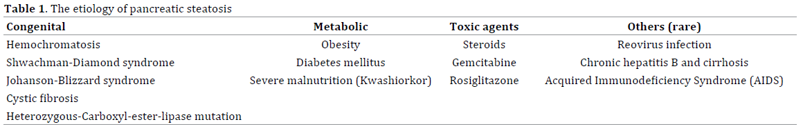
Etiology
More than 90% of population would have less than 5% of
fat infiltration in pancreas [13]. The etiology of pancreatic
steatosis varies from congenital related to acquired
conditions. However, it can be classified into 4 groups:
1) obesity and metabolic syndrome; there are some
clinical studies [2-5, 14] regarded the patients who were
diagnosed as fatty pancreas from endoscopic ultrasound,
MRI or CT scan which demonstrated that high body mass
index(BMI) and metabolic syndrome were associated
with fatty pancreas (Odd Ratio(OR) 1.05-3.13 while non
alcoholic fatty liver showed a 14-fold correlation with
pancreatic steatosis [15]. 2) congenital syndromes such
as cystic fibrosis, Shwachman–Diamond syndrome(which
was a rare autosomal recessive disorders characterized
by association of pancreatic exocrine insufficiency ,due to
fat infiltration and atrophy, bone marrow dysfunction and
skeleton abnormalities) [16-20], and Johanson–Blizzard
syndrome(a rare genetic disorder characterized by short
stature, mental retardation, pancreatic insufficiency,
sensorineural hearing lost, hypoplatic nasal alae, scalp
defect and dental abnormalies) [21, 22]. 3) toxic agents
and medications such as steroid therapy and gemcitabine
chemotherapy which all of these medication related cases
were reported case only [23-25] , and 4) other rare causes
such as reoviral infection [26], human immunodeficiency
virus infection that could cause pancreatic steatosis
through a combination of malnutrition-related and viralrelated
effects, and chronic hepatitis B infection [27]. A
summary of etiologies of pancreatic steatosis are provided
in Table 1.
Clinical Impact of Fatty Pancreas
The prevalence of NAFP was reported to be around 16% in
Hong Kong Chinese population [4]. There was a statistically
significant correlation between NAFP and non-alcoholic
fatty liver disease (NAFLD) (odds ratio [OR]=2.22; 95%
confidence interval [CI], 1.88–2.57; P<0.001), central
obesity (OR = 2.16; 95% CI, 1.85–2.52; P<0.001), age (OR
= 1.05; 95% CI, 1.04–1.05; P<0.001), hypertriglyceridemia
(OR = 1.32; 95% CI, 1.13–1.55; P=0.01), aspartate
aminotransferase and alanine transaminase level elevation
(OR = 1.29; 95% CI, 1.13–1.70; P=0.02), and diabetes
mellitus (DM) (OR = 1.59; 95% CI, 1.30–1.95; P<0.001).
Data suggest that fat accumulation in the pancreas may
lead to similar processes as in non-alcoholic steatohepatitis
(NASH). Patel et al. demonstrated in 2013 [28] that higher
pancreatic fat content correlated with a higher grade of
hepatic steatosis in patients with biopsy-proven NAFLD,
but did not correlate with body mass index (BMI) or
DM. This study also demonstrated no difference in the
distribution of fatty content among the pancreatic portions
(head, body, and tail). Although pancreatic steatosis was
reported as a clinical manifestation of metabolic syndrome
(Figure 1), other research indicates that this condition
might lead to beta-cell dysfunction, causing DM. There was
a significant difference between ethnicities (Hispanic >
African American > Caucasian) in the correlation between pancreatic steatosis, which detected by triglyceride
droplets in the cytosol of non-adipose cells in the pancreas
via positron emission scan, and beta-cell dysfunction and
compensatory insulin secretion. This correlations could
be used as a predictor for development of type 2 DM (prediabetic
state) [29].
Mirarrakhimov [30] proposed that obstructive sleep
apnea might predispose individuals to develop fatty
pancreas, which correlated with the etiology of metabolic
syndrome and DM, and was related to a greater risk of
cardiometabolic disease. Apart from metabolic-related
changes associated with pancreatic steatosis, patients with
NAFP had an increased risk of developing severe, acute
pancreatitis when pancreatitis occurred from any cause.
Van Greenen et al. [31] demonstrated a significant
correlation between pancreatic steatosis and the CT
severity index of pancreatitis.
Mathur et al. published in 2009 [32] a case controlled
study of 20 lymph node-negative and 20 lymph nodepositive
pancreatic cancer patients whose other factors
such as age, BMI, gender, tumor size, resection status,
and co-morbidity were matched. The study showed that
significantly more patients in the node-positive group
than the node-negative group had fatty pancreas (46.4±8.7 vs. 21.4±4.8; P<0.02). Patients in the node positive group also had less fibrosis than node-negative patients (1.7±0.3 vs. 2.7±0.3; P<0.02). The mean survival was also reduced
in the node-positive compared to that in the node-negative
group (18.9±2.7 months vs. 30.8±4.8 months; P<0.04). The
hypothesis was that pancreatic steatosis altered the tumor
microenvironment, enhanced tumor spread, and contributed
to early death of the pancreatic cancer patients.
Mathur et al. reported in 2007 [33] that the presence of
pancreatic fat significantly increased the risk of developing
a pancreatic fistula. A subsequent study [34] showed
similar results, as did the 2010 study by Gaujoux et al. [35],
which concluded that pancreatic fat was a more reliable
risk factor for developing pancreatic fistula than soft
pancreas.
However, the data regarded natural history of pancreatic
steatosis is still unknown due to lack of supportive
evidences.
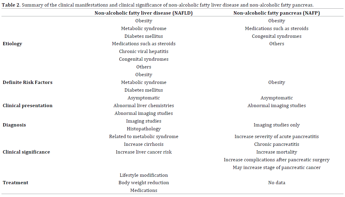
Correlation Between Non-Alcoholic Fatty Liver and
Non-Alcoholic Fatty Pancreas
As mentioned above, Erwin-Jan et al. demonstrated a
correlation between NAFLD and NAFP (both interlobular
fat infiltration and total pancreatic fat) from material
collected from 80 patients postmortem. There were
some evidence demonstrated that chronically increase in
level of plasma nonesterified fatty acid and triglyceride-riched lipoprotein impaired beta cell function and lead
to apoptosis(lipotoxicity process) in the animal model, it
is still inconclusive in human studies [36]. Since NAFLD
is well known and presents clinical concerns [37-41],
we summarize the clinical manifestations and clinical
significance of these two conditions in Table 2.
TREATMENT AND PREVENTION
Treatment of fatty pancreas depends upon the etiology.
If the etiology is identified and found to be correctable,
it may help reduce pancreatic fat infiltration. General
lifestyle modifications such as weight reduction, exercise,
or dietary restrictions can improve patients with metabolic
syndrome. However, there is no specific treatment for fatty
pancreas. In some particular patients such as the patients
who undergo pancreatic surgery, the higher risk of fistula
formation should be aware by the surgeon.
CONCLUSION
Pancreatic steatosis is a common, benign pancreatic
condition observed in clinical practice. Clinical knowledge of
this condition is essential for gastroenterologists to be able to
care for their patients. Identification of the exact etiology and
correction would help reduce pancreatic fat infiltration.
Future Research
A large cohort study should be conducted to definitively
determine the clinical significance of pancreatic steatosis,
its correlation with metabolic syndrome and DM, and
its association with pancreatic cancer risk. Potential
treatments such as lifestyle modification, weight reduction,
and medications used in NAFLD should be investigated.
Conflicting Interest
The authors had no conflicts of interest
References
- Ogilvie RF. The islands of Langerhans in 19 cased of obesity. J Pathol
1933; 37: 473-81.
- Choi CW, Kim GH, Kang DH, Kim HW, Kim DU, Heo J, Song GA, Park
do Y, Kim S. World J Associated factors for a hyperechogenic pancreas on
endoscopic ultrasound. Gastroenterol 2010; 16: 4329-34. [PMID: 20818817]
- Sepe PS, Ohri A, Sanaka S, Berzin TM, Sekhon S, Bennett G, Mehta G, et
al. A prospective evaluation of fatty pancreas by using EUS. Gastrointest
Endosc 2011; 73: 987-93.
- Wong VW, Wong GL, Yeung DK, Abrigo JM, Kong AP, Chan RS, et al.
Fatty pancreas, insulin resistance, and β-cell function: a population study
using fat-water magnetic resonance imaging. Am J Gastroenterol 2014;
109: 589-97. [PMID: 24492753]
- Hu HH, Kim HW, Nayak KS, Goran MI. Comparison of fat-water MRI
and single-voxel MRS in the assessment of hepatic and pancreatic fat
fractions in humans. Obesity (Silver Spring) 2010; 18: 841-7.
- Gullo L, Salizzoni E, Serra C, Calculli L, Bastagli L, Migliori M. Can
pancreatic steatosis explain the finding of pancreatic hyperenzymemia in
subjects with dyslipidemia? Pancreas 2006; 33: 351-3.
- Kim HJ, Byun JH, Park SH, Shin YM, Kim PN, Ha HK, Lee MG. Focal
fatty replacement of the pancreas: usefulness of chemical shift MRI. AJR
Am J Roentgenol. 2007; 188: 429-32. [PMID: 17242252]
- Li J, Xie Y, Yuan F, Song B and Tang C. Noninvasive Quantification of
Pancreatic Fat in Healthy Male Population Using Chemical Shift Magnetic
Resonance Imaging: Effect of Aging on Pancreatic Fat Content. Pancreas
2011; 40: 295-9. [PMID: 21178651].
- Schwenzer NF, Machann J, Martirosian P, Stefan N, Schraml C, Fritsche
A, Claussen CD, Schick F. Quantification of pancreatic lipomatosis and
liver steatosis by MRI: comparison of in/opposed-phase and spectralspatial
excitation techniques. Invest Radiol 2008; 43: 330-7.
- Smits MM, van Geenen EJ. The clinical significance of pancreatic
steatosis. Nat Rev Gastroenterol Hepatol 2011; 8: 169-77. [PMID:
21304475].
- Altinel D, Basturk O, Sarmiento JM, Martin D, Jacobs MJ, Kooby
DA, Adsay NV. Lipomatous pseudohypertrophy of the pancreas: a
clinicopathologically distinct entity. Pancreas 2010; 39: 392-7.
- Yasuda M, Niina Y, Uchida M, Fujimori N, Nakamura T, Oono T,
Igarashi H, et al. A case of lipomatous pseudohypertrophy of the pancreas
diagnosed by typical imaging. JOP. 2010; 11: 385-8.
- Lingvay I, Esser V, Legendre JL, Price AL, Wertz KM, Adams-Huet
B, Zhang S, Unger RH, Szczepaniak LS. Noninvasive quantification of
pancreatic fat in humans. J Clin Endocrinol Metab 2009; 94: 4070-6.
- Lee JS, Kim SH, Jun DW, Han JH, Jang EC, Park JY, et al. Clinical
implications of fatty pancreas: correlations between fatty pancreas and
metabolic syndrome. World J Gastroenterol 2009; 15: 1869-75.
- Al-Haddad M, Khashab M, Zyromski N, Pungpapong S, Wallace MB,
Scolapio J, et al. Risk factors for hyperechogenic pancreas on endoscopic
ultrasound: a case-control study. Pancreas 2009; 38: 672-5.
- Feigelson J, Pécau Y, Poquet M, Terdjman P, Carrère J, Chazalette
JP, Ferec C. Imaging changes in the pancreas in cystic fibrosis: a
retrospective evaluation of 55 cases seen over a period of 9 years. J
Pediatr Gastroenterol Nutr 2000; 30: 145-51. [PMID: 10697132].
- Ruggiero A, Molinari F, Coccia P, Attinà G, Maurizi P, Riccardi R,
Bonomo L. MRI findings in Shwachman diamond syndrome. Pediatr
Blood Cancer 2008; 50: 352-4. [PMID:17183583]
- Gana S, Sainati L, Frau MR, Monciotti C, Poli F, Cannioto Z, Comelli
M, Danesino C, Minelli A. Shwachman-Diamond syndrome and type 1
diabetes mellitus: more than a chance association? Exp Clin Endocrinol
Diabetes 2011; 119: 610-2.
- Sanklecha M, Balani K . Chronic pancreatic insufficiency-think of
Shwachmann Diamond Syndrome. Indian Pediatr. 2012; 49: 417-8.
[PMID:22700671]
- Nakaya T, Kurata A, Hashimoto H, Nishimata S, Kashiwagi Y, Fujita K,
Kawashima H, Kuroda M. Young-age-onset pancreatoduodenal carcinoma
in Shwachman-Diamond syndrome. Pathol Int 2014; 64: 75-80.
- Hoffman WH, Lee JR, Kovacs K, Chen H, Yaghmai F. Johanson-Blizzard
syndrome: autopsy findings with special emphasis on hypopituitarism
and review of the literature. Pediatr Dev Pathol 2007; 10: 55-60.
[PMID: 17378628]
- Godbole K, Maja S, Leena H, Martin Z. Johanson-blizzard syndrome.
Indian Pediatr. 2013; 50: 510-2. [PMID:23778732]
- Makay O, Kazimi M, Aydin U, Nart D, Yilmaz F, Zeytunlu M, Goker
E, Coker A. Fat replacement of the malignant pancreatic tissue after
neoadjuvant therapy. Int J Clin Oncol 2010; 15: 88-92.
- Lin WC, Chen JH, Lin CH, Shen WC. Rapidly progressive pancreatic
lipomatosis in a young adult patient with transfusion-dependent
myelodysplastic syndrome. J Formos Med Assoc 2007; 106: 676-9.
[PMID: 17711803]
- Oliveira NM, Ferreira FA, Yonamine RY, Chehter EZ. Antiretroviral
drugs and acute pancreatitis in HIV/AIDS patients: is there any
association? A literature review. Einstein (Sao Paulo). 2014; 12: 112-9.
[PMID: 24728257]
- Walters MN, Leak PJ, Joske RA, Stanley NF, Perret DH. Murine
infection with reovirus 3. pathology of infection with types 1 and 2. Br J
Exp Pathol 1965; 46: 200-12. [PMID: 14286949]
- Sasaki M, Nakanuma Y, Ando H. Lipomatous pseudohypertrophy of
the pancreas in a patient with cirrhosis due to chronic hepatitis B. Pathol
Int 1998; 48: 566-8. [PMID: 9701022]
- Patel NS, Peterson MR, Brenner DA, Heba E, Sirlin C, Loomba R.
Association between novel MRI-estimated pancreatic fat and liver
histology-determined steatosis and fibrosis in non-alcoholic fatty liver
disease. Aliment Pharmacol Ther 2013; 37: 630-9. [PMID: 23383649]
- Szczepaniak LS, Victor RG, Mathur R, Nelson MD, Szczepaniak EW,
Tyer N, et al. Pancreatic steatosis and its relationship to β-cell dysfunction
in humans: racial and ethnic variations. Diabetes Care 2012; 35: 2377-83.
[PMID: 22968187]
- Mirrakhimov AE. Nonalcoholic fatty pancreatic disease and cardiometabolic
risk: is there is a place for obstructive sleep apnea? Cardiovasc
Diabetol 2014; 13: 29. [PMID: 24475948]
- van Geenen EJ, Smits MM, Schreuder TC, van der Peet DL, Bloemena
E, Mulder CJ. Nonalcoholic fatty liver disease is related to nonalcoholic
fatty pancreas disease. Pancreas 2010; 39: 1185-90. [PMID: 20871475]
- Mathur A, Zyromski NJ, Pitt HA, Al-Azzawi H, Walker JJ, Saxena R,
Lillemoe KD. Pancreatic steatosis promotes dissemination and lethality of
pancreatic cancer. J Am Coll Surg 2009; 208: 989-94. [PMID: 19476877]
- Mathur A, Marine M, Lu D, Swartz-Basile DA, Saxena R, Zyromski NJ,
Pitt HA. .Nonalcoholic fatty pancreas disease. HPB (Oxford) 2007; 9: 312-
8. [PMID: 18345311]
- Tranchart H, Gaujoux S, Rebours V, Vullierme MP, Dokmak S, Levy
P, et al. Preoperative CT scan helps to predict the occurrence of severe
pancreatic fistula after pancreaticoduodenectomy. Ann Surg 2012; 256:
139-45. [PMID: 22609844]
- Gaujoux S, Torres J, Olson S, Winston C, Gonen M, Brennan MF,
et al. Impact of obesity and body fat distribution on survival after
pancreaticoduodenectomy for pancreatic adenocarcinoma. Ann Surg
Oncol 2012; 19: 2908-16. [PMID: 22411205]
- van Raalte DH, van der Zijl NJ, Diamant M. Pancreatic steatosis in
humans: cause or marker of lipotoxicity? Curr Opin Clin Nutr Metab Care
2010; 13: 478-85.
- Roberts KJ, Storey R, Hodson J, Smith AM, Morris-Stiff G. Pre-operative
prediction of pancreatic fistula: is it possible? Pancreatology 2013; 13:
423-8. [PMID: 23890142]
- Cusi K. Role of obesity and lipotoxicity in the development of
nonalcoholic steatohepatitis: pathophysiology and clinical implications.
Gastroenterology 2012; 142: 711-25.e6. [PMID: 22326434]
- Fisher CP, Kierzek AM, Plant NJ, Moore JB. Systems biology approaches
for studying the pathogenesis of non-alcoholic fatty liver disease. World J
Gastroenterol 2014; 20: 15070-78. [PMID: 25386055]
- Lonardo A, Ballestri S, Targher G, Loria P. Diagnosis and management
of cardiovascular risk in nonalcoholic fatty liver disease. Expert Rev
Gastroenterol Hepatol 2014; 20: 1-22. [PMID: 25327387]
- Machado MV, Cortez-Pinto H. Non-alcoholic fatty liver disease: What
the clinician needs to know. World J Gastroenterol 2014; 20: 12956-80.
[PMID: 25278691]


