Beyza Ozcinar1, Ilgin Ozden1, Orhan Bilge1, Ali Emre1, Arzu Poyanli2, Atilla Okten3
Departments of General Surgery1, Radiology2 and Gastroenterohepatology3,
Istanbul Faculty of Medicine, Istanbul University. Istanbul, Turkey
- *Corresponding Author:
- Beyza Ozcinar
Ordek Kasap Mah. Bezmi Alem Cad. Ziya sitesi
Cihan Apt. No: 4 D:3
Findikzade, 34093
Fatih-Istanbul
Turkey
Phone: +90-212.635.3082
Fax: +90-212.635.3082
E-mail: beyzazcinar@yahoo.com
Received November 18th, 2004 - Accepted November 30th, 2004
Keywords
Pancreas; Portal Vein; Thrombosis
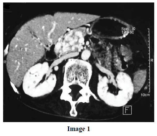
A 65 year-old-woman was examined for upper abdominal pain and weight loss. She was referred for distal common bile duct carcinoma diagnosed by magnetic resonance imaging and percutaneous transhepatic cholangiography and drainage. She had no jaundice and presented the following biochemical profile: SGOT 20 IU/L (reference range: 5-42 IU/L), SGPT 12 IU/L (reference range: 5-45 IU/L), alkaline phosphatase 75 IU/L (reference range: 0-135 IU/L), GGT 14 IU/L (reference range: 5-85 IU/L), total bilirubin 0.6 mg/dL (reference range: 0.2-1.0 mg/dL), and direct bilirubin 0.1 mg/dL (reference range: 0-0.3 mg/dL). Computed tomography revealed portal vein occlusion and extensive collateral formation completely filling the pancreatic head and extending to the hepatic hilum (Image 1: portal collaterals in the pancreas; Image 2: portal collaterals around the pancreas). Follow-up cholangiography showed a ‘seesaw’ appearance due to the impressions of the collaterals and the obstruction of the distal common bile duct (Image 3). Tests for Leiden genotype, anticardiolipin antibodies, protein C, protein S, antithrombin III, homocysteine, prothrombin gene mutations, and biochemical analysis yielded normal results. No esophagogastric varices were detected by endoscopy.


Portal cavernoma is a collateral network which develops due to the occlusion of the extrahepatic portal system. It consists of multiple, usually millimetric veins which partially maintain the hepatopedal portal blood [1]. It occurs predominantly in the suprapancreatic part of the common bile duct and causes stenosis and angulation [2]. Clinically significant cholestasis may develop due to biliary tract compression [3, 4, 5]. Ischemic biliary stricturing due to venous thrombosis has also been suggested [6]. To the best of our information, only one case of portal cavernoma producing a pancreatic mass has previously been reported [7].
References
- Vibert E, Azoulay D, Castaing D, Bismuth H. Portal cavernoma: diagnosis, aetiologies and consequences. Ann Chir 2002; 127:745-50. [PMID 12538094]
- Condat B, Vilgrain V, Asselah T, O'Toole D, Rufat P, Zappa M, et al. Portal cavernoma- associated cholangiopathy: A clinical and MR cholangiography coupled with MR portography imaging study. Hepatology 2003; 37:1302-8. [PMID 12774008]
- Bayraktar Y, Ozturk MA, Egesel T, Cekirge S, Balkanci F. Disappearance of ?pseudocholangiocarcinoma sign? in a patient with portal hypertension due to complete thrombosis of left portal vein and main portal vein web after web dilatation and transjugular intrahepatic portosystemic shunt. J Clin Gastroenterol 2000; 31:328-32. [PMID 11129276]
- Colle I, Van Vlierberghe H, Pattyn P, Troisi R, Vogelaers D, de Hemptinne B, De Vos M. Cholestasis as presenting symptom of portal cavernoma. Hepatol Res 2003; 25:32-7. [PMID 12644036]
- Chandra R, Kapoor D, Tharakan A, Chaudhary A, Sarin SK. Portal biliopathy. J Gastroenterol Hepatol 2001; 16:1086-92. [PMID 11686833]
- Dhiman RK, Puri P, Chawla Y, Minz M, Bapuraj JR, Gupta S, et al. Biliary changes in extrahepatic portal venous obstruction: compression by collaterals or ischemic? GastrointestEndosc 1999; 50:646-52. [PMID 10536320]
- Ragozzino A, De Ritis R, Gurdascione MA, Amitrano L. A portal cavernoma mimicking a pancreatic mass. J Hepatol 2003; 38:372. [PMID 12586306]

