Keywords
Carcinoma, Pancreatic Ductal; Chemotherapy, Adjuvant; Disease Management; Early Diagnosis; Incidence; Follow-Up Studies; Neoplasms, Cystic, Mucinous, and Serous; Pancreas
Abbreviations
BD: branch duct; MD: main duct; MUC5AC: mucin-5AC
What Did We Know Before the 2013 ASCO Gastrointestinal Cancers Symposium?
Intraductal papillary mucinous neoplasms (IPMNs) of the pancreas represent 20-50% of all pancreatic cystic neoplasms [1], but only about 1% of all pancreatic cancers [2]. Since this type of neoplasm can be small and asymptomatic, the true incidence of IPMN is not known, but it is believed to be increasing [1, 3]. This might result from the increasing frequency with which these neoplasms are being diagnosed worldwide [4]. More specifically, Klibansky et al. performed a retrospective cohort study to calculate a trend in reported incidence of IPMN. The authors used data collected from the Olmsted database (Rochester Epidemiology Project: https://www.rochesterproject.org), from 1985 to 2005 and concluded that the increased incidence of IPMN during this period results from an increase in diagnostic scanning rather than greater number of patients with clinically relevant disease [3].
IPMNs represent a heterogeneous group of neoplasms that can be classified anatomically as main duct (MD)- type, branch duct (BD)-type or mixed-type [5, 6]. As far as histology is concerned, four histological subtypes of IPMN have been described: intestinal type, pancreatobiliary, oncocytic type and gastric type [1]. IPMNs are potentially malignant neoplasms, graded to low-grade dysplasia (adenoma), moderate dysplasia (borderline), high-grade dysplasia (carcinoma in situ) and invasive carcinoma [1]. BD-IPMN is characterized by much lower malignancy rates than MD-IPMN, which justifies the worse prognosis for MD-IPMN, as reported in most clinical series [1,7].
Since patients with IPMN of the pancreas are at risk for both pancreatic and extrapancreatic malignancies, early recognition, treatment and systemic surveillance are of great importance [8, 9, 10].
What Did We Learn at 2013 ASCO Gastrointestinal Cancers Symposium?
Evaluating Malignant Intraductal Papillary Mucinous Neoplasm: A Population-Based Study (Abstract #324 [11])
Luu et al. performed a retrospective study in which they analyzed incidence, prognostic factors, treatment and survival of 2,987 patients diagnosed with invasive IPMN from 1988 to 2009. During this period there was a decrease in age-adjusted incidence. The overall resection rate was 20.6% with an increase in annual resection rates. Age greater than 65 years, tumor location, poorly differentiated tumor grade, higher T stage and positive lymph nodes predicted worse survival at the univariate analysis, while more recent diagnosis, higher number of lymph nodes examined and surgery indicated improved survival. Curative surgery proved to be predictive of survival at the multivariate analysis, as well. The median survival rates for patients with stage I, II and III disease who underwent surgery were 87, 18 and 14 months respectively, compared to 6, 7 and 5 months in the no surgery group (Table 1).

This study supports the necessity for early identification of patients with invasive IPMN, so that they may have survival benefit from surgical resection. Moreover, although recent reports show increasing incidence of IPMN, this study demonstrates decreasing incidence of malignant IPMN, which, according to the authors, may result from increased detection of nonmalignant disease.
Clinical Implications of Luminal Membrane Expression of Mesothelin in Intraductal Papillary Mucinous Neoplasms (Abstract #179 [12])
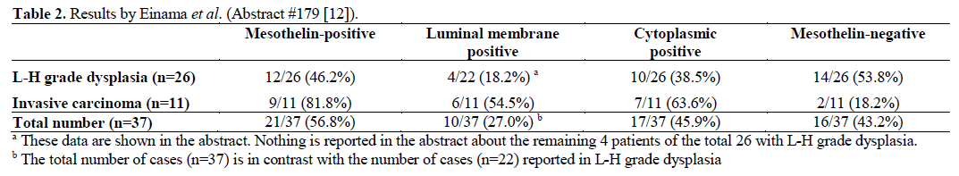
Einama et al. investigated the immunohistochemical analysis of mesothelin expression in IPMNs, as well as the localization of mesothelin. More specifically, the authors examined tissue samples from 37 IPMNs, which were divided into two groups: low, intermediate and high-grade dysplasia (L-H grade dysplasia group) and IPMN with an associated invasive carcinoma (invasive carcinoma group). The proportion and intensity of constituent tumor cells with mesothelin expression were analyzed and classified as ‘positive expression’ or ‘negative expression’. Moreover, the staining localization of mesothelin was evaluated as ‘luminal membrane positive’ and/ or ‘cytoplasmic positive’ in the ‘positive expression group’.
Mesothelin expression was observed in 21 of 37 cases (56.8%), 46.2% (12 of 26) of L-H grade dysplasia and 81.8% (9 of 11) of invasive carcinoma (P=0.071). Luminal membrane positive was observed in 10 cases (27.0%), 18.2% (4 of 22; nothing is reported in the abstract about the remaining 4 patients of the total 26 with L-H grade dysplasia) of L-H grade dysplasia and 54.5% (6 of 11) of invasive carcinoma (P<0.001). Cytoplasmic positive was observed in 17 cases (45.9%), 38.5% (10 of 26) of L-H grade dysplasia and 63.6% (7 of 11) of invasive carcinoma (P=0.28). Six of 37 cases (16.2%) showed recurrence after surgery. Five cases out of 6 cases that showed recurrence after surgery (83.3%) exhibited mesothelin expression, and all of them were luminal membrane positive (Table 2). This study supports the clinicopathological significance of the luminal membrane expression of mesothelin in IPMNs. According to the authors, the immunohistochemical examination of mesothelin expression has clinical significance in prognosis and decision making about further treatment options in patients with IPMN who have undergone curative surgical resection.
Association of Dilated Main Pancreatic Duct with Biological High Proliferative Activity in Intraductal Papillary Mucinous Neoplasm (Abstract #187 [13])
Eto et al. studied the biological significance of clinical malignant predictive markers, such as diameter of main pancreatic duct, cystic size and a mural nodule, in cell proliferative activity. More specifically, surgically resected pancreatic specimens of IPMN were obtained from 49 patients, during the period 2002-2011. Adenoma was present in 27 of 49 pancreatic specimens (55.1%), while carcinoma was present in 22 of 49 specimens (44.9%). Cell proliferative activity was evaluated by MIB-1 index with the use of Ki67 immunostaining.
The MIB-1 index was found significantly higher in the “carcinoma” group (20.1±13.2% vs. 9.0±6.8% for the “adenoma” group; P=0.004). When the groups with high MIB-1 index (greater than 15%) and low index (less than 15%) were compared, the diameter of main pancreatic duct in the high MIB-1 index group was found significantly larger than that in the low MIB-1 index group (9 mm with range 1-27 mm vs. 4.5 mm with range 2-15 mm, respectively; P<0.05). As far as the cystic size and a mural nodule are concerned, no significant differences were found between the high MIB-1 index group and the low MIB-1 index group. With regard to the type of IPMNs (main- or branchduct type), 60.9% of main-duct type and 19.2% of branch-duct type IPMN belong in the high MIB-1 index group.
This study supports the positive correlation between an increased diameter of main pancreatic duct and high cell proliferative activity in IPMNs. So the authors concluded that dilatation of main pancreatic duct, due to mucin production, may predict the biologically high tumor proliferative activity.
Discussion
The studies by Luu et al., Einama et al. and Eto et al. presented above, demonstrate the necessity for clinical and molecular markers for prognosis and decision making as far as treatment of patients with IPMN of the pancreas is concerned.
Since a great proportion of IPMN patients have only nonspecific clinical signs, imaging techniques, cytology and laboratory markers are useful tools in the diagnosis and surgical decision making of patients with IPMN [1]. US, CT, MRI, ERCP, MRCP, EUS with possibility of performing FNA and FDG-PET can be used in order to diagnose and characterize IPMN [14, 15, 16, 17, 18].
As far as serum tumor markers are concerned, serum levels of carbohydrate antigen (CA) 19-9 and carcinoembryonic antigen (CEA) may aid in differentiating between invasive and benign IPMN. In 142 patients undergoing surgical resection for IPMN, raised CEA and CA 19-9 serum levels were significantly associated with invasive IPMN [19]. Moreover, mucin-5AC (MUC5AC) serum levels may help in differentiating between high-risk and low-risk IPMN patients. A study by Marker et al. found increased serum levels of MUC5AC in patients with high-grade dysplasia or carcinoma compared to those with low-grade or moderate dysplasia [20].
Molecular pathogenesis of IPMNs is another aspect which might interfere with early diagnosis [21]. K-ras mutations are found in approximately 50% of IPMN cases, although further analysis is required in order to determine the K-ras mutation rates in different subtypes of IPMNs [1]. Mutations in the tumor suppressor gene CDKN2a have also been seen in association with IPMNs, while only a minority of these tumors displays aberrant expression of p53 and SMAD4 [22]. IPMNs have been identified in a subset of patients with Peutz-Jeghers syndrome who have mutations of STK11, indicating that STK11 mutations might contribute to sporadic IPMNs [1].
Furthermore, several genes are known to undergo aberrant methylation in IPMNs, including cyclin D2, SOCS-1 and TFPI-2 [1, 21]. The detection of these aberrantly methylated genes might help early recognition and diagnostic evaluation of pancreatic neoplasms [21]. Aberrant microRNA (miRNA) expression in IPMNs was evaluated in a study by Habbe et al [23]. The authors demonstrated that miR- 155 and miR-21 are significantly upregulated in the majority of non-invasive IPMNs and also concluded that the role of miR-155 as a biomarker for IPMNs in clinical samples should be further evaluated [23]. Lastly, MUC2 mucin and MUC5 mucin miRNA are also highly expressed in IPMNs [24, 25].
Mainly due to their malignant potential, IPMNs, once recognized, should be treated with pancreatic resection according to the Sendai Consensus Guidelines [4]. The role of adjuvant chemoradiotherapy for patients with invasive carcinoma associated with IPMN who underwent surgical resection, has been addressed in a retrospective study by Swartz et al [26]. The authors found that adjuvant chemoradiotherapy was associated with 57% decrease in the relative risk of mortality after pancreaticoduodenectomy [26].
Patients with IPMN who do not have indications for surgery are still at risk for developing cancer. Furthermore, patients who were surgically treated are at risk of recurrence of IPMN [27, 28]. Patients with IPMN may also be at increased risk for extrapancreatic malignancies [8, 9]. These data suggest that long-term surveillance is critical to the patients with IPMN [27]. As far as prognosis is concerned, invasive IPMN has a favorable prognosis compared with pancreatic ductal adenocarcinoma, which is likely due to the less aggressive nature of the disease [29].
Since their first description in 1982, a great progress has been done in the management of IPMNs of the pancreas. There are many things still to learn, though, about the biology of IPMN tumorigenesis. Further studies, especially in this field, are necessary in order safe conclusions to be drawn. Additionally, a multidiscipline team of oncologists, pathologists, surgeons and gastroenterologists would ensure the optimal therapeutic results for IPMN patients.
Conflict of interest
The authors have no potential conflicts of interest
References
- Grützmann R, Niedergethmann M, Pilarsky C, et al. Intraductal Papillary Mucinous Tumors of the Pancreas: Biology, Diagnosis and Treatment. Oncologist.2010;15(12):1294
- Freeman HJ. Intraductal papillary mucinous neoplasms and other pancreatic cystic lesions. World J Gastroenterol.2008;14(19):2977
- Klibansky DA, Reid-Lombardo KM, Gordon SR, Gardner TB. The clinical relevance of the increasing incidence of intraductal papillary mucinous neoplasm. ClinGastroenterol Hepatol.2012;10(5):555
- Tanaka M, Chari S, Adsay V, et al. International consensus guidelines for management of intraductal papillary mucinous neoplasms and mucinous cystic neoplasms of the pancreas. Pancreatology.2006;6(1-2):17
- Kobari M, Egawa S, Shibuya K, et al. Intraductal papillary mucinous tumors of the pancreas comprise 2 clinical subtypes: differences in clinical characteristics and surgical management. Arch Surg.1999;134(10):1131
- Terris B, Ponsot P, Paye F, et al. Intraductal papillary mucinous tumors of the pancreas confined to secondary ducts show less aggressive pathologic features as compared with those involving the main pancreatic duct. Am J Surg Pathol.2000;24(10):1372
- Nagai K, Doi R, Kida A, et al. Intraductal papillary mucinous neoplasms of the pancreas: clinicopathologic characteristics and long-term follow-up after resection. World J Surg.2008;32(2):271
- Sugiyama M, Atomi Y. Extrapancreatic neoplasms occur with unusual frequency in patients with intraductal papillary mucinous tumors of the pancreas. Am J Gastroenterol.1999;94(2):470
- Choi MG, Kim SW, Han SS, et al. High incidence of extrapancreatic neoplasms in patients with intraductal papillary mucinous neoplasms. Arch Surg.2006;141(1):51
- Kamisawa T, Tu Y, Egawa N, et al. Malignancies associated with intraductal papillary mucinous neoplasm of the pancreas. World J Gastroenterol.2005;11(36):5688
- Luu C, Nelson RA, Lee B, Singh G, Kim J. Evaluating malignant intraductal papillary mucinous neoplasm: A population-based study. J Clin Oncol.2012;30: Abstract 324
- Einama T, Kamachi H, Nishihara H, Homma S, Kawamata F, Tahara M, Taniguchi M, Furukawa H, Kamiyama T, Taketomi A, Matsuno Y, Todo S. Clinical implications of luminal membrane expression of mesothelin in intraductal papillary mucinous neoplasms. J Clin Oncol.2012; 30 (Suppl. 34): Abstarct #179.
- Eto T, Hayashi H, Kuroki H, Nakagawa S, Hashimoto D, Ikuta Y, Chikamoto A, Beppu T, Baba H. Association of dilated main pancreatic duct with biological high proliferative activity in intraductal papillary mucious neoplasm. J Clin Oncol.2012;30: Abstarct 187
- Grützmann R, Bunk A, Kersting S, et al. Prospective evaluation of ultrasound and colour duplex imaging for the assessment of surgical resectability of pancreatic tumors. Langenbecks Arch Surg.2003;388:392
- Brugge WR. The use of EUS to diagnose cystic neoplasms of the pancreas. Gastrointest Endosc.2009;69(2 suppl):S203
- Sahani DV, Kadavigere R, Blake M, et al. Intraductal papillary mucinous neoplasm of the pancreas: Multi-detector row CT with 2D curved reformations-correlation with MRCP. Radiology. 2006;238:560
- Yamada Y, Mori H, Matsumoto S. Intraductal papillary mucinous neoplasms of the pancreas: Correlation of helical CT and dynamic MR imaging features with pathologic findings. Abdom Imaging.2008;33:474
- Baiocchi GL, Portolani N, Bertagna F, et al. Possible additional value of 18FDG-PET in managing pancreas intraductal papillary mucinous neoplasms: Preliminary results. J ExpClin Cancer Res.2008;27:10
- Fritz S, Hackert T, Hinz U, et al. Role of serum carbohydrate antigen 19-9 and carcinoembryonic antigen in distinguishing between benign and invasive intraductal papillary mucinous neoplasm of the pancreas. Br J Surg.2011;98(1):104
- Maker AV, Katabi N, Gonen M, et al. Pancreatic cyst fluid and serum mucin levels predict dysplasia in intraductal papillary mucinous neoplasms of the pancreas. Ann Surg Oncol.2011;18(1):199
- Hong SM, Kelly D, Griffith M, Omura N, Li A, et al. Multiple genes are hypermethylated in intraductal papillary mucinous neoplasms of the pancreas. Modern Pathology.2008;21:1499
- Furukawa T, Fujisaki R, Yoshida Y, et al. Distinct progression pathways involving the dysfunction of DUSP6/MKP-3 in pancreatic intraepithelial neoplasia and intraductal papillary mucinous neoplasms of the pancreas. Modern Pathology.2005;18:1034
- Habbe N, Koorstra JB, Mendell JT, et al. MicroRNA miR-155 is a biomarker of early pancreatic neoplasia. Cancer Biol Ther.2009;8(4):340
- Yonezawa S, Sato E. Expression of mucin antigens in human cancers and its relationship with malignancy potential. Pathol Int.1997;47(12):813
- Yonezawa S, Horinouchi M, Osako M, et al. Gene expression of gastric type mucin (MUC5AC) in pancreatic tumors: its relationship with the biological behavior of the tumor. Pathol Int.1999;49(1):45
- Swartz MJ, Hsu CC, Pawlik TM, et al. Adjuvant chemoradiotherapy after pancreatic resection for invasive carcinoma associated with intraductal papillary mucinous neoplasm of the pancreas. Int J RadiatOncolBiol Phys.2010;76(3):839
- Sohn TA, Yeo CJ, Cameron JL, et al. Intraductal papillary mucinous neoplasms of the pancreas: an updated experience. Ann Surg.2004;239(6):788
- Chari ST, Yadav D, Smyrk TC, et al. Study of recurrence after surgical resection of intraductal papillary mucinous neoplasm of the pancreas. Gastroenterology.2002;123(5):1500
- Murakami Y, Uemura K, Sudo T, et al. Invasive intraductal papillary mucinous neoplasm of the pancreas: comparison with pancreatic ductal adenocarcinoma. J Surg Oncol.2009;100(1):13

