Keywords
Gene Expression; Immunotherapy; Pancreatic Neoplasms
Abbreviations
EGF epidermal growth factor; FGF fibroblast growth factor; NPC nasopharyngeal carcinoma; VEGFR vascular endothelial growth factor receptors
INTRODUCTION
With the increase of morbidity and mortality, cancer has become a major problem with public health in humans [1, 2, 3]. Pancreatic cancer is the most fatal malignancy with a 5-year survival rate of less than 5%, which is the eighth leading cause of cancer-related deaths worldwide [4] and the sixth in China [5]. Of all forms of pancreatic malignancies, 85–95% were pancreatic ductal adenocarcinoma [6, 7]. Surgical resection is the only curative treatment of pancreatic cancer but 80% of pancreatic adenocarcinoma is not resectable. There is an urgent need to develop powerful treatment of such a malignant disease.
Angiogenesis is a crucial event for tumor growth and is regulated predominantly by several different growth factors such as vascular endothelial growth factor (VEGF), epidermal growth factor (EGF) and fibroblast growth factor (FGF). Monoclonal antibodies against VEGF and against EGF receptors have been used clinically for systemic anticancer treatment [8, 9, 10] although detrimental side-effects have been observed [11, 12]. VEGF is highly expressed in most tumors and its expression has been found to be associated with tumor progression [13]. VEGF is thus likely to play an important role in stimulating the growth of new blood vessels within tumor tissues and promoting tumor progression by interacting with their corresponding receptors on the surfaces of cancer cells [14]. Accordingly, blockage of the VEGF signaling pathway might represent an effective anti-angiogenic therapy for most types of tumors [15]. Bevacizumab, a humanized anti-VEGF monoclonal antibody that has been approved by the USA Food and Drug Administration (FDA), was the first monoclonal antibody for treatment of solid tumors. It has also been studied for possible use as an anti-angiogenic agent in patients with pancreatic cancer although clinical outcomes did not appear to be satisfactory [15, 16, 17, 18]. Possibly, direct inhibition of VEGF receptor function would be more powerful in treatment of such a malignant disease.
Natural antibodies are part of the humoral immune system and there is evidence that natural antibodies are able to kill distinct human tumor cells both in vivo and in vitro [19, 20]. In a recent study, we found that plasma natural IgG antibodies against VEGF receptor 1 (VEGFR1) significantly inhibited the proliferation of cancer cell lines derived from nasopharyngeal carcinoma [21]. Accordingly, the present work was designed to examine if plasma anti- VEGFR1 IgG could also inhibit the proliferation of cancer cells derived from pancreatic cancer.
METHODS
Detection of Plasma Anti-VEGFR1 IgG
A total of 150 plasma samples collected from healthy blood donors were kindly provided by Dongguan Blood Center, Guangdong Province, China. Pooled plasma from 20 randomly selected healthy individuals was used as a reference sample (RS) to detect plasma rich in anti-VEGFR1 IgG. This work was approved by the Institutional Review Board of Guangdong Medical University, Dongguan, China.
An enzyme-linked immunosorbent assay (ELISA) was used to detect plasma that abundantly contained natural IgG antibodies against the extracellular domain of human VEGFR1 protein (NCBI accessionNP_002010). The ELISA antibody test kit provided by Hailanshen Biotechnology Ltd, Qingdao, China, mainly comprised the following components: (1) Assay Buffer that was 0.1 M phosphate buffer containing 0.15 M NaCl and 10 mM EDTA, pH 7.2, (2) Wash Buffer that was 0.1 M phosphate buffer containing 0.15 M NaCl and 0.05% Tween-20, pH 7.2, and (3) foil film-sealed 96-well plates coated with specific antigens. In brief, the antigen-coated plate was washed twice with 200 μL Wash Buffer before use; 50 μL plasma sample diluted 1:200 in Assay Buffer was added to each sample well, and 50 μL Assay Buffer was added to each negative control (NC) well. After incubation at room temperature for 2 hours, the plate was washed three times with 200 μL Wash Buffer, and 100 μL peroxidase-conjugated goat anti-human IgG antibody (A8667, Sigma-Aldrich, Shanghai, China), which was diluted 1:30000 in Assay Buffer, was then added to each well. Following incubation at room temperature for 2 hours, 50 μL Stabilized Chromogen (SB02, Life Technologies, Guangzhou, China) was added to develop color; 25 minutes (min) later, 50 μL Stop Solution (SS04, Life Technologies) was added to terminate the color development. The optical density (OD) was measured by a microplate reader within 10 min at 450 nm with a reference wavelength of 620 nm. All the samples were tested in duplicate and the positive sample ratio (PSR) was used to represent antibody levels. Calculation of PSR is as follows:
PSR= (ODSample – ODNC) / (ODRS – ODNC)
Cell Proliferation Assay
Two cell lines, PANC-1 and SW1990 derived from human pancreatic cancer, were used for this in vitro study. These two cell lines were purchased from Shanghai Cell Bank of Chinese Academy of Sciences. PANC-1 cells were initially seeded in a 96-well plate, 100 μL/well with a density of 2.5×10⁴ cells/ml Dulbecco’s Modified Eagle Medium (DMEM, GIBCO, Guangzhou, China) containing 10% fetal calf serum (FCS), and cultured in humidified atmosphere with 5% CO2 at 37°C for 24 hours. SW1990 cells were initially cultured in RPMI 1640 medium (1640, GIBCO, Guangzhou, China) supplemented with 10% FCS in the same condition as mentioned above. After incubation for 24 hours, the cells were cultured in DMEM or RPMI 1640 medium containing 15% human plasma either positive or negative for anti-VEGFR1 IgG and cells for 48 hours in the same conditions as mentioned above; cell viability was then detected with cell counting kit-8 (CCK-8, Sigma-Aldrich). According to the manufacturer’s instruction, 10 μL CCK-8 solution was added to each well; after incubation at 37oC for 1.5 hours, OD of each well was measured on a microplate reader at a wavelength of 450 nm. The complete medium was used as blank. Cell viability was used to present data and calculated as follows:
Cell viability = (OD positive – OD blank) / (OD negative – OD blank),
where OD positive is defined as OD measured in cancer cells cultured with 15% human plasma positive for anti- VEGFR1 IgG, and OD negative is defined as OD measured in cancer cells cultured with 15% plasma negative for anti- VEGFR1 IgG.
Analysis of Apoptosis
The cells were seeded in 12-well plates, 1 mL/well with a density of 1×10⁵ cells/ml DMEM or RPMI1640 medium containing 10% FCS. After incubation in humidified atmosphere with 5% CO2 at 37°C for 24 hours, the cells continued to were cultured with DMEM or RPMI 1640 medium containing 15% human plasma positive or negative for anti-VEGFR1 IgG. Cultured cells were collected at 24 hours and 48 hours, respectively, for analysis of apoptosis.
Annexin V-FITC Apoptosis Detection Kit I (BD Biosciences, USA) was used to analyze apoptosis of pancreatic cancer cells. According to the manufacturer’s instruction, cells were harvested and washed twice with cold PBS and then resuspended in 1X Binding Buffer at a density of 1×10⁶ cells/ml, and 100 μL cells (1×10⁵ cells) were transferred to a 5 mL tube; 400 μL 1X Binding Buffer was then added to each tube, and a 5 μL volume of each FITC Annexin V and PI was added. The cells were vortexed gently and incubated for 15 min at room temperature (25°C) in the dark and then analyzed by FACSCalibur flow cytometer (Becton Dickinson, UK).
Analysis of VEGFR1 Gene Expression
Total RNA was extracted from PANC-1 and SW1990 cells using TRIzol reagent (TaKaRa Bio-technology, Dalian, China). To eliminate DNA contamination, total RNA samples were treated with a DNA-free kit (Fermentas, Hanover, USA) and then reversely transcribed into cDNA using PrimeScript™ RT Master Mix (TaKaRa Bio-technology, Dalian, China). Quantitative real-time PCR assay with SYBR Premix Ex Taq kit (TaKaRa Bio-technology, Dalian, China) was performed to detect expression of the VEGFR1 gene on the ABI 7500 Real-Time PCR system. Primers’ sequences used for PCR amplification are as follows: 5’-TTAGGACCAGGAAGCAGCAC-3’ (forward) and 5’-CCGAGGTTCCTTGAACAGTGA-3’ (reverse). Glyceraldehydes-3-phosphate dehydrogenase (GAPDH), purchased from QIAGEN (Shanghai, China), was used as a housekeeping gene for normalization. Relative quantity of gene expression was calculated using the comparative Ct method; fold change (FC) was used to present data and worked out based on the formula: FC=2-ΔΔCt.
STATISTICS
All experimental data were expressed as mean±standard deviation (SD). GraphPadInStat™ (version 5.02) statistical software was used to perform Student’s t-test (two-tailed) to examine the differences in cell viability and proportions of apoptotic cells between cells treated with anticancer IgG positive plasma and those treated with anticancer IgG negative plasma as well as in mRNA expression between two cell lines.
RESULTS
These 150 plasma samples showed a mean PSR of 1.01±0.33 ranging from 0.36 to 1.63. Of these plasma samples, 2 with the highest PSR values of 1.63 and 1.62 and 2 with the lowest PSR values of 0.36 and 0.37 were selected for cell culture work. The 2 plasma samples with the highest PSR values were used as anti-VEGFR1 IgG positive plasma (namely A for 1.63 and B for 1.62); the 2 plasma samples with the lowest PSR values were mixed and then used as anti-VEGFR1 IgG negative plasma.
Inhibitory Effects of Plasma Anti-VEGFR IgG on the Proliferation of Pancreatic Cancer Cells
As shown in Figure 1, when compared with anti- VEGFR1 IgG negative plasma, anti-VEGFR1 IgG positive plasma significantly inhibited the proliferation of PANC- 1 cells (0.52±0.09, t=-8.98, df=16, P<0.0001 for plasma A; 0.47±0.13, t=-8.39, df=16, P<0.0001 for plasma B; and 0.50±0.09, t=9.44, df=16, P<0.0001 for positive A+B). The results also showed, that anti-VEGFR1 IgG positive plasma A inhibited the proliferation of SW1990 cells (0.88±0.03, t=7.71, df=16, P<0.0001) but plasma B appeared to stimulate cell proliferation although positive A+B plasma failed to influence the proliferation of SW1990 cells (1.03±0.03, t=-1.56, df=16, P=0.1376).
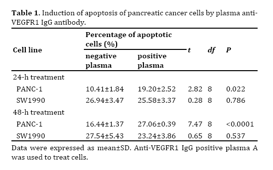
Effect of Plasma Anti-VEGFR1 IgG on Apoptosis of Pancreatic Cancer Cells
Anti-VEGFR1 IgG positive plasma A was used to treat both PANC-1 and SW1900 cell lines. As shown in Table 1, the percentage of apoptotic cells was significantly higher in PANC-1 cells treated with anticancer IgG positive plasma than those treated with negative plasma (t=-2.82, df=8, P=0.022 for 24-h treatment and t=-7.82, df=8, P<0.0001 for 48-h treatment). However, the percentage of apoptotic cells of SW1990 cells showed no significant differences between anti-VEGFR1 IgG positive plasma and negative plasma treated cells (t=-0.28, df=8, P=0.787 for 24-h treatment and t=-0.65, df=8, P=0.534 for 48-h treatment).
Expression of VEGFR1 mRNA in Pancreatic Cancer Cells
As shown Figure 2, PANC-1 and SW1990 cell lines were used to investigate the expression of VEGFRI mRNA was detected in both PANC-1 and SW1990 cell lines; the results showed that expression levels of VEGFR1 mRNA were significantly higher in PANC-1 cells than SW1990 cells (t=7.01, df=10, P<0.0001).
DISCUSSION
Natural antibodies secreted primarily by B1 lymphocytes in the absence of external antigen stimulation or immunization are defined as a subset of immunoglobulins [22, 23], which are composed of different classes known as isotypes IgA, IgM and IgG. Most natural antibodies are IgM, but IgA and IgG isotypes have also been reported [24]. It has been indicated that natural antibodies play an important role not only in elimination of invading pathogens but also in maintaining homeostasis of the immune system and destruction of cancer cells formed in our body [20, 25]. In a previous study, we found that plasma anti-VEGFR1 IgG antibodies significantly inhibited the proliferation of cancer cell lines derived from nasopharyngeal carcinoma [21]. Similarly, the present work also showed an inhibitory effect of plasma anti- VEGFR1 IgG on the proliferation of pancreatic cancerderived cell lines (Figure 1). The mechanism behind anticancer activity of plasma anti-VEGFR1 IgG is due to its induction of apoptosis of pancreatic cancer cells (Table 1).
Both PANC-1 and SW1990 cells were tested in this study but they had different responses to plasma anti-VEGFR1 IgG. Both plasma A and B decreased PANC-1 cell viability by 50% but only plasma A (not plasma B) decreased cell viability by 12% in SW1990 cells (Figure 1). Analysis of apoptosis suggests that plasma A could induce apoptosis of PANC-1 cells but not SW1990 cells (Table 1). Because SW1990 cells express a significantly lower level of VEGRF1 mRNA than PANC-1 cells (Figure 2), this may be the major reason why the response of SW1990 cells to plasma anti- VEGFR1 IgG was much weaker than PANC-1 cells.
Figure 1. Effects of anti-VEGFR1 IgG positive plasma on the proliferation of pancreatic cancer cell lines.
The data of cell viability were expressed as mean±SD. Anti-VEGFR1 IgG positive plasma A was used to treat cells.
Figure 2. Expression of VEGFR1 mRNA in PANC-1 and SW1990 cells.
The data of VEGFR1 gene expression were expressed as mean±SD. Student’s t-test showed a significant difference in VEGFR1 mRNA expression between PANC-1 and SW1990 cells (t=7.01, df=10, P<0.0001)
CONCLUSION
PANC-1 cell line was derived from epithelioid carcinoma while SW1990 cell line from adenocarcinoma. Because more than 85% patients with pancreatic malignancies may suffer from adenocarcinoma, further screening of target molecules expressed by pancreatic adenocarcinoma cells and their corresponding natural antibodies in human plasma is needed. In addition, there may be some growth factors exiting in the circulation; these growth factors may stimulate the growth of malignant tumors and partially counteract the inhibitory effects of plasma anticancer antibodies on cancer cells. It is possible that gamma-globulins purified from plasma rich in anticancer antibodies may become a promising agent for immunotherapy of pancreatic cancer and other types of cancer in the future.
Acknowledgements
We thank Li Zhang for the detection of plasma rich in anti-VEGFR1 IgG antibodies. This work was supported by Hailanshen Biomedical Technology Ltd, Shenzhen, China.
Conflict of interest
All authors declared no financial conflict of interest.
References
- Peery AF, Crockett SD, Barritt AS, Dellon ES, Eluri S, Gangarosa LM, Jensen ET, et al. Burden of gastrointestinal, liver, and pancreatic diseases in the United States. Gastroenterology 2015; 149:1731-41. [PMID: 26327134]
- Quaresma M, Coleman MP, Rachet B. 40-year trends in an index of survival for all cancers combined and survival adjusted for age and sex for each cancer in England and Wales, 1971–2011: a population-based study. Lancet 2015; 385:1206-18. [PMID: 25479696]
- Zheng R, Zeng H, Zhang S, Chen T, Chen W. National estimates of cancer prevalence in China, 2011. Cancer Lett 2016; 370:33-8. [PMID: 26458996]
- Jemal A, Siegel R, Ward E, Hao Y, Xu J, Thun MJ. Cancer statistics, 2009. CA Cancer J Clin 2009; 59:225-49. [PMID: 19474385]
- He Y, Zheng R, Li D, Zeng H, Zhang S, Chen W. Pancreatic cancer incidence and mortality patterns in China, 2011. Chin J Cancer Res 2015; 27:29-37. [PMID: 25717223]
- Torre LA, Bray F, Siegel RL, Ferlay J, Lortet-Tieulent J, Jemal A. Global cancer statistics, 2012. CA Cancer J Clin 2015; 65:87-108. [PMID: 25651787]
- Siegel RL, Miller KD, Jemal A. Cancer statistics, 2015. CA Cancer J Clin 2015; 65:5-29. [PMID: 25559415]
- Larsen JE, Cascone T, Gerber DE, Heymach JV,Minna JD. Targeted therapies for lung cancer:clinical experience and novel agents. Cancer J 2011; 17:512. [PMID: 22157296]
- Jawa Z, Perez RM, Garlie L, Singh M, Qamar R, Khandheria BK, Jahangir A, et al. Risk factors of trastuzumab-induced cardiotoxicity in breast cancer: A meta-analysis. Medicine (Baltimore) 2016; 95:e5195. [PMID: 27858859]
- Jomrich G, Schoppmann SF. Targeted therapy in gastric cancer. Eur Surg 2016; 48:278-84. [PMID: 27795701]
- Economopoulou P, Kotsakis A, Kapiris I, Kentepozidis N. Cancer therapy and cardiovascular risk: focus on bevacizumab. Cancer Manag Res 2015; 7:133-43. [PMID: 26082660]
- Lambertini M, Pondé NF, Solinas C, de Azambuja E. Adjuvant trastuzumab: a 10-year overview of its benefit. Expert Rev Anticancer Ther 2017; 17:61-74. [PMID: 27883296]
- Fukumura D, Xavier R, Sugiura T, Chen Y, Park EC, Lu N, Seed B. Tumor induction of VEGF promoter activity in stromal cells. Cell 1998; 94:715-25. [PMID: 9753319]
- Hicklin D J, Ellis L M. Role of the vascular endothelial growth factor pathway in tumor growth and angiogenesis. J Clin Oncol 2005; 23:1011-27. [PMID: 15585754]
- Vasudev NS, Reynolds AR. Anti-angiogenic therapy for cancer: current progress, unresolved questions and future directions. Angiogenesis 2014, 17:471-94. [PMID: 24482243]
- Ferrara N, Gerber HP, Le Couter J. The biology of VEGF and its receptors. Nat Med 2003; 9:669-76. [PMID: 12778165]
- Presta LG, Chen H, O'connor SJ, Chisholm V, Meng YG, Krummen L, Winkler M, et al. Humanization of an anti-vascular endothelial growth factor monoclonal antibody for the therapy of solid tumors and other disorders. Cancer Res 1997; 57:4593-9. [PMID: 9377574]
- Crane CH, Ellis LM, Abbruzzese JL, Amos C, Xiong HQ, Ho L, Charnsangavej C, et al. Phase I trial evaluating the safety of bevacizumab with concurrent radiotherapy and capecitabine in locally advanced pancreatic cancer. J Clin Oncol 2006, 24:1145-51. [PMID: 16505434]
- Sun Y, Parmentier HK, Frankena K, Van der Poel JJ. Natural antibody isotypes as predictors of survival in laying hens. Poultry Sci 2011; 90:2263-74. [PMID: 21934009]
- Schwartz-Albiez R, Laban S, Eichmüller S, Kirschfink M. Cytotoxic natural antibodies against human tumours: an option for anti-cancer immunotherapy?. Autoimmun Rev 2008; 7:491-5. [PMID: 18558368]
- Cai W, He Z, Wu L, Wang Y, Meng Q, Mustard CJ, Wei J. Inhibitory effects of natural IgG antibodies against VEGFR1 and HER2 on the proliferation of nasopharyngeal carcinoma cells. Oncol Lett 2016 (in press).
- Panda S, Ding JL. Natural antibodies bridge innate and adaptive immunity. J Immunol 2015; 194:13-20. [PMID: 25527792]
- Ochsenbein AF, Fehr T, Lutz C, Suter M, Brombacher F, Hengartner H, Zinkernagel RM. Control of early viral and bacterial distribution and disease by natural antibodies. Science 1999; 286:2156-9. [PMID: 10591647]
- Avrameas S. Natural autoantibodies: from ‘horror autotoxicus’ to ‘gnothi seauton’. Immunol Today 1991; 12:154-9. [PMID: 1715166]
- Vollmers HP, Brändlein S. Natural antibodies and cancer. N Biotechnol 2009; 25:294-8. [PMID: 19442595]


