Keywords
Adenocarcinoma; Hepatic Artery; Pancreaticoduodenectomy
Abbreviations
PA pancreatic adenocarcinoma; PD pancreaticoduodenectomy; SMA superior mesenteric artery
INTRODUCTION
Because a replaced right hepatic artery (r-RHA) or accessory RHA, which has been reported to be present in 10-18% [1, 2, 3, 4] of the population, is not rare, it is important to know precisely the anatomic variation of the artery preoperatively and plan a proper therapeutic strategy for pancreatic adenocarcinoma (PA) to obtain negative surgical margins.
The r-RHA arises from the superior mesenteric artery (SMA), and it has an intimate relationship with the head of the pancreas, since it frequently runs directly adjacent to and occasionally through the pancreatic parenchyma or extrapancreatic nerve plexus [4]. When the distance between the tumor and the r-RHA is small, the risk of positive surgical margins might increase. In some cases, even when the tumor is not abutting or adjacent to the rRHA, the surgical margins might be positive by dissecting the r-RHA from the extrapancreatic nerve plexus due to perineural invasion of the PA. Therefore, it is occasionally thought that sacrifice of the r-RHA is necessary during pancreaticoduodenectomy (PD) [5, 6]. However, ligation of the r-RHA is reported to cause hepatic ischemia and biliary anastomotic complications, such as biliary fistulas and biliary stenosis, because the RHA becomes the predominant vascular source for the distal common bile duct during PD [7, 8]. On the other hand, a recent study reported that preservation of the r-RHA in patients who underwent PD for PA did not show increased positive margins [2, 9]. Thus, the indication for preservation or en bloc resection of the r-RHA remains unclear.
Invasion by carcinoma of the head of the pancreas via the extrapancreatic nerve plexus is reported to be divided into 2 patterns based on the embryological structure of the pancreas and the location of the tumor [10]. Namely, patients with carcinoma in the uncinate process frequently have pancreatic head plexus and SMA plexus invasion, while patients with carcinoma in the dorsal pancreas have invasion into the common hepatic artery plexus and the plexus within the hepatoduodenal ligament. It is assumed that the risk of positive surgical margins would increase with dissection of the r-RHA from the pancreatic head plexus in patients who have tumor in the uncinate process. Therefore, tumor location may strongly affect margin status in patients with an r-RHA who undergo PD for PA. The aim of this study was to elucidate the impact of tumor location on surgical margins and to consider the optimal resection strategy in patients with an r-RHA who undergo PD for PA.
PATIENTS AND METHODS
A total of 117 patients with T3 PA who underwent curative PD in our institution from January 2001 to December 2015, including 91 with a normal RHA and 26 with RHA variations (24 with an r-RHA and two with a replaced CHA from the SMA), were retrospectively analyzed. All patient data were entered retrospectively into clinical databases approved by our institutional review boards. The study protocol was approved by the Clinical Research Ethics Committee of our hospital (protocol number: 28-187). Written, informed consent was obtained from all patients in the study. Patients with PA of the body or tail of the pancreas, intraductal papillary mucinous adenocarcinoma, common bile duct carcinoma, and ampulla of Vater adenocarcinoma were excluded from this study. All patients were examined preoperatively by computed tomography (CT), and arterial variations were evaluated. All patients underwent PD with reconstruction using a 70-cm roux-en-Y loop of jejunum for pancreatic and biliary anastomosis. An extended lymphadenectomy was not routinely performed in our institution. The r-RHA was generally dissected along its entire course up to its origin from the SMA and preserved. When tumor encasement of the r-RHA or remarkable extrapancreatic nerve plexus invasion was suspected on preoperative CT, preoperative coil embolization of the r-RHA was performed to promote collateral pathways and prevent ischemia-related complications, as well as en bloc resection of the r-RHA during PD.
Patients were defined as potentially resectable, borderline resectable in accordance with the National Comprehensive Cancer Network (NCCN) criteria. During the period between January 2001 to December 2004, no patients with PA received neoadjuvant therapy. During the period between January 2005 to December 2011, the patients with borderline resectable PA received gemcitabine standard-dose chemotherapy for 8 weeks concurrent with a total dose of 50 Gy of radiation. Since January 2012, patients with borderline resectable PA were planned to receive S-1 standard-dose/gemcitabine 800 m/m2 chemotherapy for 8 weeks as neoadjuvant therapy. When a histopathological examination revealed R1 resections, we administered systemic chemotherapy postoperatively.
Variables that included age, sex, maximal tumor size, location of the tumor, operative duration, blood loss during operation, morbidity, perineural invasion, lymph node status, surgical margin status including the pancreatic resection margin, biliary margin, posterior margin, and retroperitoneal margin were evaluated. Median overall survival was also determined. Margin status and survival were compared between the patients with the r-RHA (r-RHA(+) group, n=26) and the patients without the r-RHA (r-RHA(-) group, n=91) according to the tumor location, which involved the uncinate process and the dorsal pancreas. The uncinate process and dorsal pancreas were distinguished using the duct of Santorini, the duct of Wirsung, the portal vein (PV)/superior mesenteric vein (SMV), and the bile duct as landmarks on the CT images. The head of the pancreas was divided into the uncinate process and dorsal pancreas by a line linking the PV/SMV and the anterior edge of the intrapancreatic bile duct (Figure 1) [11]. In addition, the margin status was assessed according to the distance between tumor and the r-RHA in the r-RHA (+) group. A tumor located within 10 mm of the r-RHA was considered adjacent tumor, and tumor located more than 10 mm from the r-RHA was considered distant tumor in this study [12]. In this study, we defined R1 as the presence of tumor cells within 1mm of resection margin (1 mm rule) [13]. Considering a long study period, we reassessed all pancreatic resection specimens according to 1mm rule.
Figure 1: Distinguishing between the uncinate process and dorsal pancreatic. The head of the pancreas is divided into the dorsal pancreas and uncinate process by a line linking the PV/SMV and the anterior edge of the intrapancreatic bile duct.
Continuous data are expressed as means ± SD and were compared using the Mann-Whitney U test. Categorical data were assessed using the chi-squared test. Patient survival and recurrence rates were estimated by the Kaplan-Meier method, and differences between survival curves were tested by the log-rank test. Statistical analysis was carried out using JMP software (version 9.0; SAS Institute, Inc., Cary, NC, USA).
RESULTS
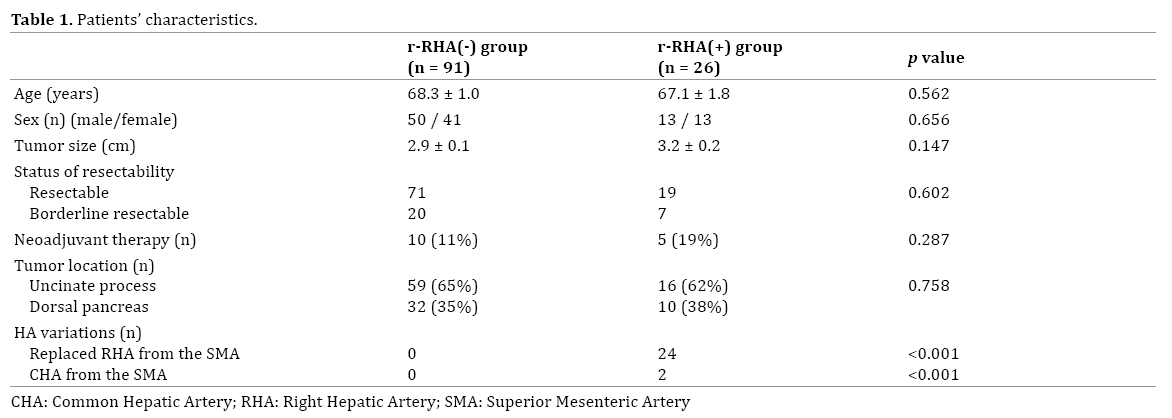
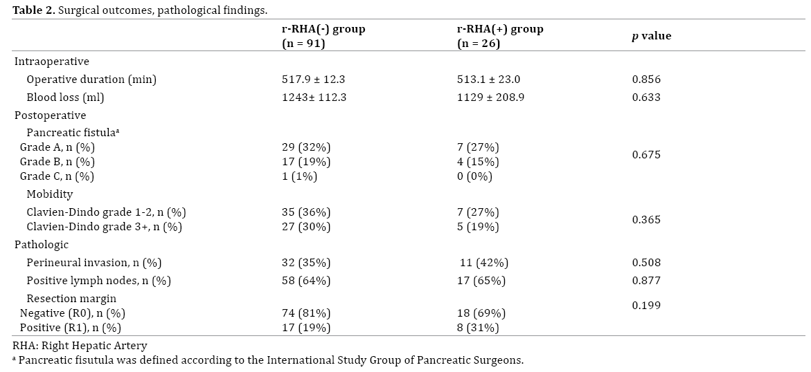
RHA variation was identified in 26 patients (22.2%) on preoperative CT. The variations of the RHA included 24 (20.5%) r-RHAs from the SMA and two (1.7%) replaced CHAs from the SMA. The r-RHA was preserved in 22 patients. There were no significant differences in age, sex, status of resectability, number of patients who received neoadjuvant therapy, tumor size, and proportion of tumor locations between the r-RHA(-) group and the r-RHA(+) group (Table 1). There were also no significant differences in operative duration, blood loss, and the incident of postoperative pancreatic fistula and the grading of the overall postoperative complications evaluated by Clavien- Dindo’s classification revealed no significant differences between the two groups (Table 2). On pathological examination, there were no differences in the rates of perineural invasion and positive lymph nodes between the two groups. A positive resection margin was seen in 17 patients (18.7%) in the r-RHA(-) group and 8 patients (30.8%) in the r-RHA(+) group; the difference was not significant (P=0.199).
Subgroup analyses of resection margin status were performed by tumor location (the uncinate process and dorsal pancreas). In the r-RHA(-) group, 59 patients (64.8%) had tumor in the uncinate process. On the other hand, in the r-RHA(+) group, 15 patients (57.7%) had tumor in the uncinate process. When tumor was located in the uncinate process, positive microscopic surgical margins were seen significantly more frequently in the r-RHA(+) group (6 of 15, 40%) than in the r-RHA(-) group (9 of 59, 15%) (P=0.048). However, when tumor was located in the dorsal pancreas, there was no significant difference in surgical margin status between the r-RHA(+) group (2 of 11, 18%) and the r-RHA(-) group (8 of 32, 25%) (P=0.638) (Figure 2).
Figure 2: Resection margin status by tumor location. When tumor was located in the uncinate process, positive microscopic surgical margins were seen significantly more frequently in the r-RHA(+) group (6 of 15, 40%) than in the r-RHA(-) group (9 of 59, 15%) (P=0.048). However, when tumor was located in the dorsal pancreas, there was no significant difference in surgical margin status between the r-RHA(+) group (2 of 11, 18%) and the r-RHA(-) group (8
of 32, 25%) (P=0.638).
The median survival times were 26 months in the r-RHA(-) group and 32 months in the r-RHA(+) group; the difference was not significant (p=0.430) (Figure 3a). Similarly, the overall survival of patients with tumor located in the dorsal pancreas (median survival time: 28 months) was similar to that of patients with tumor located in the uncinated process (median survival time: 27 months) (P=0.752: Figure 3b). However the patients who underwent R0 resections had a significantly better overall survival than those who underwent R1 resections (median survival time: 31 months vs. 18 months, p=0.019) (Figure 3c).

Figure 3: Kaplan-Meier curves for survival analysis according to presence/absence of r-RHA, tumor location, R0/R1 margins. (a). Curves comparing the r-RHA(-) group (solid) versus the r-RHA(+) group (broken). The median survival times were 26 months in the r-RHA(-) group and 32 months in the r-RHA(+) group (P=0.430). (b). Curves comparing the patients with tumor located in the dorsal pancreas (solid) versus the patients with tumor located in the uncinate process (broken). The overall survival of patients with tumor located in the dorsal pancreas (median survival time: 28 months) was similar to that of patients with tumor located in the uncinated process (median survival time: 27 months) (P=0.752). (c). Curves comparing the patients who underwent R0 resection (solid) versus the patients who underwent R1 resection (broken). The patients who underwent R0 resections had a significantly
better overall survival than those who underwent R1 resections (median survival time: 31 months vs. 18 months, p=0.019).
The enrolment period was quite huge in this study; therefore, we performed sub-analysis of survival by period. All patients were divided into the early period (2001-2008) and the late period (2009-2015). The median survival time was 24 months in the early period, and 31 months in the late period, respectively. The patients in the late period showed a better survival than those in the early period (P=0.174) (Figure 4a). However, when survival in each period was analyzed by presence or absence of the r-RHA, there were no significant differences between the r-RHA(-) group and the r-RHA(+) group in survival (P=0.585 in the early period and P=0.768 in the late period (Figure 4b).
Figure 4: Kaplan-Meier curves for survival analysis according to the period. (a). Curves comparing the patients in the early period (solid) versus the late period (broken). The outcomes of the late period were improved versus those of the early period (P=0.174) (b). Curves comparing the r-RHA (-) group (solid) versus the r-RHA(+) group (broken) in each period. There were no significant differences between the r-RHA(-) group and the r-RHA(+) group in survival (P=0.585 in the early period and P=0.768 in the late period).
Of the 15 patients with tumor located in the uncinate process in the r-RHA group, one of three patients (33.3%) with adjacent tumor showed a positive resection margin, while 5 of 12 patients (41.7%) with distant tumor showed a positive resection margin (Figure 5).
Figure 5: A diagnostic and therapeutic flowchart of 117 patients with pancreatic ductal adenocarcinoma. Of the 15 patients with tumor located in the ventral pancreas domain in the r-RHA group, one of three patients with adjacent tumor shows a positive resection margin, while 5 of 12 patients with
distant tumor show a positive resection margin.
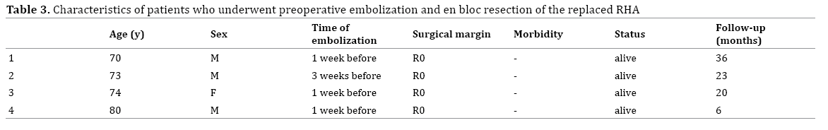
In the r-RHA(+) group, four patients underwent preoperative coil embolization and en bloc resection of the r-RHA during PD, because one patient had tumor adjacent to the r-RHA, and three patients had pancreatic head plexus invasion though the main tumor was not adjacent to the r-RHA on preoperative CT. Microscopically negative margins were identified in all four patients (Table 3). In two of three patients who had pancreatic head plexus invasion on preoperative CT, the histopathological examination showed that perineural invasion to the extrapancreatic nerve plexus was adjacent to the r-RHA that was resected by en bloc resection, even though the r-RHA was not adjacent to the main tumor. Hepatic abscesses and biliary anastomotic complications due to ischemia did not occur in these four patients.
DISCUSSION
Previous studies reported that R1 resection was associated with poor overall survival [14, 15]. Though the presence of an r-RHA could present a difficult surgical situation, whether preservation of the r-RHA affects surgical margin status during PD still remains unclear. This study demonstrated that positive microscopic surgical margins were seen significantly more frequently in the r-RHA(+) group than in the r-RHA(-) group when tumor was located in the uncinate process, though there was no difference in surgical margin status between the two groups when tumor was located in the dorsal pancreas.
One possible explanation could account for the fact that R1 resection rates increased in the r-RHA(+) group with the tumor located in the uncinate process. The head of the pancreas arises from two anlagen on an embryological basis [16]. The smaller ventral bud forms the caudal part of the head of the pancreas and uncinate process, whereas the cephalic part of the head of the pancreas, as well as the body and tail, are derived from the larger dorsal bud. The distribution of the ventral pancreas after fusion is the dorsal portion of the head containing the area surrounding the intrahepatic common bile duct and the uncinate process. There was a significant correlation between tumor location considering the two anlagen of the pancreas and the site of extra pancreatic nerve plexus invasion. Tumor located in the uncinate process tends to spread toward pancreatic head plexus 1 (PL ph1) and pancreatic head plexus 2 (PL ph2), while tumor located in the dorsal pancreas frequently spreads toward the common hepatic artery plexus and the plexus within the hepatoduodenal ligament. Therefore, in patients with tumor located in the uncinate process, radical dissection of the PL ph1 and PL ph2 should be performed. Considering that the r-RHA runs directly adjacent to and occasionally through these nerve plexuses, it is likely that the risk of positive surgical margins would increase by dissecting the r-RHA from these nerve plexuses into which carcinoma might infiltrate.
Some authors have insisted that the presence of the r-RHA itself does not affect R1 resection [4, 12, 17]. Jah et al. reported that the surgical and oncological outcomes of PD remained unaffected by the presence of the r-RHA provided that the anatomy was recognized and appropriately managed [17], which was similar to the present results for all patients, but they did not look at the pancreas domain. In our experience, though the presence of the r-RHA did not adversely affect surgical outcomes such as intraoperative blood loss, operative time, and morbidity, the rate of R1 resection was significantly higher with tumor located in the uncinate process than in the dorsal pancreas. On the other hand, Okada et al. insisted that the proximity of the pancreatic carcinoma to the r-RHA would be expected to yield a poor prognosis due to an increased R1 resection rate or invasion of the r-RHA [12]. Although the proximity of the tumor to the r-RHA certainly seems to be a risk factor for R1 resection, in the present study, histopathological examination showed that perineural invasion to the extrapancreatic nerve plexus was adjacent to the r-RHA in two of three patients who underwent en bloc resection of the r-RHA, even though the tumor seemed to be distant from the r-RHA. The fact that 5 of 12 patients with distant tumor showed positive resection margins also supports the hypothesis that the surgical margins can be positive by dissecting the r-RHA from the extrapancreatic nerve plexus due to perineural invasion of the PA even when the tumor is not abutting or adjacent to the r-RHA.
When extra pancreatic nerve plexus invasion is strongly suspected on preoperative CT in patients with tumor located in the uncinate process, preservation of the r-RHA might lead to R1 resection, which indicates that en bloc resection of the r-RHA should be considered. Although r-RHA ligation and reconstruction may be safe and feasible [18], several reports have suggested that preoperative embolization of the r-RHA to increase liver blood flow though the left hepatic artery can be useful [19, 20, 21]. The liver can tolerate considerable hepatic arterial embolization without serious complications because of the collateral pathways [22, 23, 24]. Therefore, some authors considered that preoperative embolization was unnecessary [2]. However, Mehdi et al. reported that hepatic ischemia was observed on CT performed 1 day after embolization, demonstrating the real existence of ischemia and it is easily presumed that morbidity such us cholangitis and biloma could occur under the conditions that liver ischemia exists after PD if the r-RHA would have been sacrificed without reconstruction [19]. Mimyamoto et al. also noted that a collateral pathway via the left and right gastric arteries was seen immediately after embolization, and, 10 days later, this collateral pathway was more clearly developed [21]. Preoperative embolization seems to be desirable considering that there are some risks of hepatic ischemia because the collateral pathway is usually narrow and it needs some time to develop. In the present study, embolization was performed 1-3 weeks before PD, and no ischemia-related complications occurred in the four patients who underwent en bloc resection of the r-RHA. However, there are still only a few cases of r-RHA embolization before PD reported in the literature. Further studies are needed to clarify the safety and usefulness of preoperative embolization.
In the present study, histological examination confirmed R0 resections in all four patients with tumor located in the uncinate process that underwent preoperative embolization and en bloc resection of the r-RHA. On the other hand, in patients with preservation of the r-RHA, positive microscopic surgical margins were seen significantly more frequently in the r-RHA(+) group than in the r-RHA(-) group when the tumor was located in the uncinate process. When extrapancreatic nerve plexus invasion is suspected, avoiding unnecessary dissection of the r-RHA from the extrapancreatic nerve plexus into which carcinoma may infiltrate might contribute to R0 resection.
Limitation of our study includes its retrospective design small sample size and its fairly long study period. Additional larger multi-institutional trials are needed to further validate outcomes in the patients with the r-RHA.
CONCLUSION
In conclusion, the results of the present study demonstrated that positive microscopic surgical margins were seen significantly more frequently in patients with the r-RHA who underwent r-RHA-preserving PD for PA when tumor was located in the uncinate process. Based on the spreading patterns of carcinoma via the extra pancreatic nerve plexus, it might be better to consider en bloc resection of the r-RHA to improve the R0 resection rate in the patients with tumor located in the uncinate process when extra pancreatic nerve plexus invasion is strongly suspected.
Conflict of Interest Statement
There are no conflicts of interest and no financial or material support.
References
- Hiatt JR, Gabbay J, Busuttil RW. Surgical anatomy of the hepatic arteries in 1000 cases. Ann Surg. 1994; 220:50-52. [PMID: 8024358].
- Turrini O, Wiebke EA, Delpero JR, Viret F, Lillemoe KD, Schmidt CM. Preservation of replaced or accessory right hepatic artery during pancreaticoduodenectomy for adenocarcinoma: impact on margin status and survival. J Gastrointest Surg 2010; 1:1813-1819. [PMID: 20697832].
- López-Andújar R, Moya A, Montalvá E, Berenguer M, De Juan M, San Juan F, et al. Lessons learned from anatomic variants of the hepatic artery in 1,081 transplanted livers. Liver Transpl 2007; 13:1401-1404. [PMID: 17902125].
- Yang SH, Yin YH, Jang JY, Lee SE, Chung JW, Suh KS, et al. Assessment of hepatic arterial anatomy in keeping with preservation of the vasculature while performing pancreatoduodenectomy: an opinion. World J Surg 2007; 31:2384-2391. [PMID: 17922256].
- El Amrani M, Leteurtre E, Sergent G, Ernst O, Maunoury V, Branche J, et al. Pancreatic head carcinoma and right hepatic artery: embolization management-A case report. J Gastrointest Oncol 2014; 5:E80-83. [PMID: 25083312].
- Cloyd JM, Chandra V, Louie JD, Rao S, Visser BC. Preoperative embolization of replaced right hepatic artery prior to pancreaticoduodenectomy. J Surg Oncol 2012; 106:509-512. [PMID: 22374866].
- Traverso LW, Freeny PC. Pancreaticoduodenectomy. The importance of preserving hepatic blood flow to prevent biliary fistula. Am Surg 1989; 55:421-426. [PMID: 2742226].
- Northover JM, Terblanche J. A new look at the arterial supply of the bile duct in man and its surgical implications. Br J Surg 1979; 66:379-384. [PMID: 466017].
- Lee JM, Lee YJ, Kim CW, Moon KM, Kim MW. Clinical implications of an aberrant right hepatic artery in patients undergoing pancreaticoduodenectomy. World J Surg 2009; 33:1727-1732. [PMID: 19459000].
- Makino I, Kitagawa H, Ohta T, Nakagawara H, Tajima H, Ohnishi I, et al. Nerve plexus invasion in pancreatic cancer: spread patterns on histopathologic and embryological analyses. Pancreas 2008; 37:358-65. [PMID: 18972625].
- Kitagawa H, Ohta T, Makino I, Tani T, Tajima H, Nakagawara H, et al. Carcinomas of the ventral and dorsal pancreas exhibit different patterns of lymphatic spread. Front Biosci 2008; 13:2728-2235. [PMID: 17981748].
- Okada K, Kawai M, Hirono S, Miyazawa M, Shimizu A, Kitahata Y, et al. A replaced right hepatic artery adjacent to pancreatic carcinoma should be divided to obtain R0 resection in pancreaticoduodenectomy. Langenbecks Arch Surg 2015; 400:57-65. [PMID: 25359559].
- The Royal College of Pathologists. Standards and Datasets for Reportiong Cancers. In: Dataset for Histopathological Reporting of Carcinomas of the Pancreas, Ampulla of Vater and Common Bile Duct, 2nd edn. London: The Royal College of Pathologists..
- Konstantinidis IT, Warshaw AL, Allen JN, Blaszkowsky LS, Castillo CF, Deshpande V, et al. Pancreatic ductal adenocarcinoma: is there a survival difference for R1 resections versus locally advanced unresectable tumors? What is a "true" R0 resection? Ann Surg 2013; 257:731-736. [PMID: 22968073].
- Howard TJ, Krug JE, Yu J, Zyromski NJ, Schmidt CM, Jacobson LE, et al. A margin-negative R0 resection accomplished with minimal postoperative complications is the surgeon's contribution to long-term survival in pancreatic cancer. J Gastrointest Surg 2006; 10:1338-1345. [PMID: 17175452].
- Suda K, Mizuguchi K, Hoshino A. Differences of the ventral and dorsal anlagen of pancreas after fusion. Acta Pathol Jpn 1981; 31:583-589. [PMID: 7025573].
- Jah A, Jamieson N, Huguet E, Praseedom R. The implications of the presence of an aberrant right hepatic artery in patients undergoing a pancreaticoduodenectomy. Surg Today 2009; 39:669-674. [PMID: 19639433].
- Sarmiento JM, Panneton JM, Nagorney DM. Reconstruction of the hepatic artery using the gastroduodenal artery. Am J Surg 2003; 185:386-387. [PMID: 12657395].
- El Amrani M, Leteurtre E, Sergent G, Ernst O, Maunoury V, Branche J, et al. Pancreatic head carcinoma and right hepatic artery: embolization management-A case report. J Gastrointest Oncol 2014; 5:E80-83.
- Cloyd JM, Chandra V, Louie JD, Rao S, Visser BC. Preoperative embolization of replaced right hepatic artery prior to pancreaticoduodenectomy. J Surg Oncol 2012; 106:509-512. [PMID: 22374866].
- Miyamoto N, Kodama Y, Endo H, Shimizu T, Miyasaka K, Tanaka E, et al. Embolization of the replaced common hepatic artery before surgery for pancreatic head cancer: report of a case. Surg Today 2004; 34:619-622. [PMID: 25083312].
- Allison DJ. Therapeutic embolization. Br J Hosp Med 1978; 20:707-715..
- Charnsangavej C, Chuang VP, Wallace S, Soo CS, Bowers T. Angiographic classification of hepatic arterial collaterals. Radiology 1982; 144:485-494. [PMID: 6285413].
- Takeuchi Y, Arai Y, Inaba Y, Ohno K, Maeda T, Itai Y. Extrahepatic arterial supply to the liver: observation with a unified CT and angiography system during temporary balloon occlusion of the proper hepatic artery. Radiology 1998; 209:121-128. [PMID: 9769822].


