Warda Makhloufi*, Chouaib Sayah, Hichem Choutri and Zineddine Soualili
Department of Pediatric Surgery, Setif, Algeria
*Corresponding Author:
Warda Makhloufi
University of Farhat Abbas-Setif
Women's and Children's Hospital
Department of Pediatric Surgery
El-Bez, Setif-19000, Algeria
Tel: +213773852781
E-mail: sayahchouaib@gmail.com
Received date: 26 July, 2016; Accepted date: 2 November, 2016; Published date: 4 November, 2016
Citation: Makhloufi W, Sayah C, Choutri H, et al. Giant Soft Fibroma Located in Labia Majora: Pediatric Case Report. Gynecol Obstet Case Rep.2016, 2:3. doi: 10.21767/2471-8165.1000034
Keywords
Soft fibroma; Fibrous tissue; Tumors of the vulva
Introduction
Vulvar fibroma is a rare and the most common benign tumor that is predominantly found in women of reproductive age group. They have been reported in infants, postmenopausal and pregnant women, Soft fibromas are connective-tissue benign tumors that have multiple synonyms (Naevus molluscum, acrochordon, templeton skin tag, and fibroepithelial polyp). This case report describes giant soft fibroma located in labia majora in a girl aged 03 years.
Case Report
History
A 03-year-old girl with no particular history that is presented to our pediatric surgery department with huge mass of the left labia majora whose appearance comes at birth with slow gradual growth.
Physical examination
The girl had a good health condition, well developed. The left labia majora was increased size without inflammatory and scratching lesions, painless, without palpable inguinal lymphadenopathy (Figure 1). The rest of the vulva was quite normal.
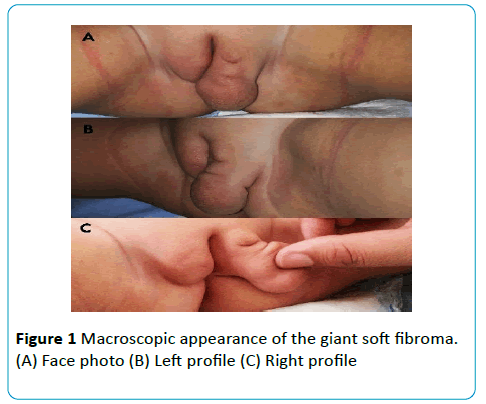
Figure 1: Macroscopic appearance of the giant soft fibroma. (A) Face photo (B) Left profile (C) Right profile.
Lab and imaging studies, histologic findings
An echo-Doppler coupled objective of soft tissue found a pseudo cystic fatty cellular thickening of the labia majora, avascular, with homogeneous content. The therapeutic decision was a surgical biopsy resection with reconstruction of the affected labia for diagnostic and therapeutic purposes (Figure 2).
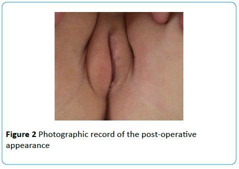
Figure 2: Photographic record of the post-operative appearance.
Histological study shows the presence of a fibrous connective tissue hyalinized, edematous well vascularized compatible with a soft fibroma without evidence of malignancy
The cosmetic result after three months is considered very well by the child's parents.
Discussion
Soft fibromas are described as pedunculated tumors due to an elongation in their conjunctive tissue, especially in superficial tumors [1]. When located in the vulva, they occur more often in the labia majora and less frequently in the labia minora, clitoris, vestibule, and posterior commissure. In tumors of long clinical duration, ulcerations with superficial bleeding, often due to repeated trauma, are often observed [2]. To start with, the tumor may be asymptomatic but has the potential to grow to huge sizes. Apart from causing physical signs due to its size and location, important differential diagnosis includes Lipoma, Inguinal hernia, vulvovaginal cysts, vulval elephantiasis, and fibro epitheloid tumours [3]. According to the histological variants, fibromas of the vulva can be classified as either pure or mixed. The pure form may be hard or soft. The pure-hard variant presents fibrous and hard conjunctive tissue, with a white or pink hue, and a lobulated and well-differentiated appearance [4].
The treatment of malignant tumors include: surgery, radiotherapy and chemotherapy. The recommended treatment for each case depends on several factors such as the stage of cancer, the exact subtype or "quality" of cancer, and overall health [5].
The total vulvectomy with bilateral inguinal lymphadenectomy in one piece is the intervention of choice in cases of malignancy, the treatment of benign tumors involves the surgical removal: cryo-surgery, electro-coagulation or laser surgery [6].
For our patient, we realize excision biopsy with a cosmetic purpose, knowing that our attitude will be final in case of benign tumor, will not delay treatment if it were a malignant tumor or will hinder the quality of supported cases of malignant tumor.
Conclusion
Giant soft fibromas of the vulva are benign tumors that in some cases, due to their macroscopic appearance, can lead to a mistaken diagnosis of malignancy. Biopsy should be considered as a possible first step of oncological care. However, respect for cosmetics is easy to achieve in an excisional biopsy, which can be as definitive for our case.
Conflict of interest: None
Ethical approval: Necessary approval was taken from the Institution and the patients for carrying out this work.
Acknowledgement
I would like to express my sincere gratitude to my Prof. Z. SOUALILI and all pediatric surgery team for the continuous support, for patience, motivation, and immense knowledge.
References
- Najam R, Chowdhury HH, Awasthi S (2013) A Large Fibroma Polyp of Labia Majora? A Case Report. J Clin Case Rep 3:297.
- Hernandes VMV (2007)Vulva fibroma. A case report. J Low Genit Tract Dis 1:23-6.
- De Lima A (1952) Fibroma of the vulva (Molluscum Pendulum). ObstetGynaecolsurv 7: 845-846
- Roberto Netto A, Focchi GRA, Ribalta JCL, Giannotti Filho O, Focchi J, et al. (2001) Fibroma de Vulva (Molluscum Pendulum): Relato de Caso. Rev Bras Ginecol Obstet. 23: 187-90
- Guidozzi F, Sadan O, Koller AB, Marks SR (1987) Combined chemotherapy and irradiation therapy after radical 98 K. A. Behranwala et al. surgery for leiomyosarcoma of the vulva. S Afr Med J71:327.
- Nucci MR, Fletcher CDM (2000) Vulvovaginal soft tissue tumors. update and review. Histopathology 36: 97-108.

