Keywords
Skeletal muscles; Sensory acquisition Blinking; Blood coagulation (DRG) ; Disgust; Dorsal respiratory group
Introduction
This study was conducted to explain the significance of five new reflexes, especially with respect to the unexpected relationship to blinking. It shows of how the brain’s reflex centers regulate internal hemostasis in response to environmental changes. The thalamus (Figure 1) receives most of the body’s sensory input. It generates the defensive startle reflex in response to sudden, intense environmental threats by causing arousal, stiffening of the limbs, and an instantaneous blink within 3-8 ms [1]. This blink protects the eye causing lid closure by contraction of the orbicularis muscle (Figure 1). It covers the eye; helps shift attention from the previous focus to the new, more threatening target [2]; stimulates five of the six extra ocular muscles responsible for moving the eye to cocontract resulting in globe retraction 0.7-1.0 mm into the orbit [3]; causes the eye to rotate upward under the upper lid thus protecting the cornea (Bell’s sign) and causes constriction of the pupil, which decreases photophobia [4]. Within 300 ms of the onset of this startle reflex, the amygdala is stimulated (Figure 1) and it is determined whether the conscious sympathetic emotion of fear, with the unconscious reflexes for freeze-fight-flight should be generated [5].
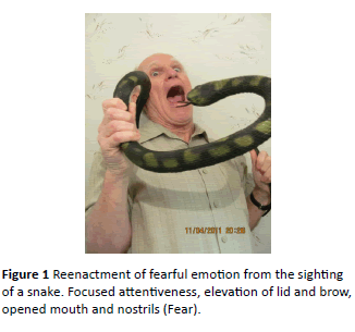
Figure 1: Reenactment of fearful emotion from the sighting of a snake. Focused attentiveness, elevation of lid and brow, opened mouth and nostrils (Fear).
If the amygdala determines a fear response is warranted, it causes the skeletal muscles to freeze, a deep in-breath, and cessation of eye movement with focus on the danger. The hypothalamus (Figure 1) activates the sympathetic nervous system causing the release of epinephrine and norepinephrine (Figure 2). These hormones prepare the body for fight-or-flight by increasing respiration, speeding up the heart, raising blood pressure, increasing blood coagulation, and constricting blood vessels in the skin to prevent bleeding.
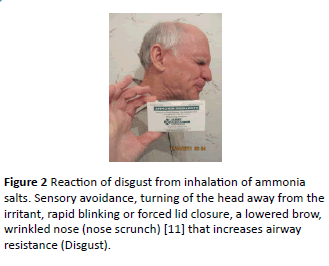
Figure 2: Reaction of disgust from inhalation of ammonia salts. Sensory avoidance, turning of the head away from the irritant, rapid blinking or forced lid closure, a lowered brow, wrinkled nose (nose scrunch) [11] that increases airway resistance (Disgust).
The diverted blood is sent instead to the brain, heart, and skeletal muscles. There is also an increase in mental alertness and dilation of the pupils [6,7]. Sensory acquisition is enhanced by a decreased rate of blinking and slowing of eye movement [8,9] (Figure 2). Susskind confirmed with MRI that humans exposed to fearful stimuli demonstrate an increase in inspiration volume and air velocity with enlarged nasal airways [4,5,10]. This study proposes for the first time that it is the initial deep inhalation that stops the blink and slows scanning eye movements so that the eyes can lock onto the new, potential threat. Descending fibers from the amygdala terminate in the solitary nucleus containing the dorsal respiratory group (DRG) (Figure 1). This may be the pathway causing the deep breath (Figure 1).
The length of time for one complete blink from beginning descent of the lid to its return to the original, elevated position is 0.30-0.90 s. During that time, it completely obscures vision for 0.15 s and partly reduces visual field during the entire blink [11]. During normal blinking, people are unaware of this loss of vision, since the brain creatively fills in the missing segments. This creativity could lead to a victim’s demise during a predatory attack. Fear decreases the blink rate in order to increase vision, focus, and concentration. This need for maximizing focus also causes slowing of the rapid scanning eye movements to slow pursuit.
The study presents evidence that the same neuro-pathway needed for the fear reflex is stimulated to a lesser degree for the concentration needed when reading, using computer [11-13] applying eye mascara or performing surgery. If true, the amygdala is closely tied to all types of mental concentration. Eye professionals routinely encourage computer users to voluntarily increase their rate of blinking to prevent drying of the eyes.
The levator palpebrae muscle is the primary lid elevator. Its effectiveness is closely related to the level of alertness being most stimulated during arousal, e.g. from fear or surprise. It is least active in fatigue and is totally inhibited during sleep [13,14]. The release of epinephrine by the fear response causes direct stimulation of Muller’s muscle, which also elevates the lid. The frontalis muscle also stimulated by fear or surprise primarily lifts the eyebrow, and, secondarily lifts the eyelid. The instantaneous need for three different muscles to elevate the lid, serve to increase the superior field of vision.
Threats from carnivores, often in the lower limbs of trees; and from raptors swooping down from the sky, at speeds up to 100 miles per h, mandated this evolutionary mechanism to instantaneously expand the superior field of vision [15]. It may also be lifesaving when awakened by nocturnal predators.
The aversive reflexes associated with the emotion of disgust, on the other hand, are parasympathetic reactions with most integrative reflex centers in the pons and medulla (Figure 1). They manifest an opposite behavior to fear, which is often the case with the reciprocally behaving sympathetic and parasympathetic systems [16] (Figure 2). Disgust typically causes an instantaneous reaction where one turns away from the unpleasant obnoxious danger, such as rotting food, excrement, irritating odors, airborne contaminants, or even a person or immoral act. Its rejection of the environment could result in sneezing, coughing, gagging, nausea, vomiting, bronchoconstriction, cessation of breathing, blinking, eye closure, tearing, and rejection of potentially dangerous food stuffs [17]. This rejection of incoming information could decrease the ability to learn in school [18].
Methods
The protocol for this observational study was directed at demonstrating five previously unreported reflexes associated with the emotions of fear and disgust.
Participants were randomly chosen from a population of adult male and female patients visiting this ophthalmologist’s office for a routine eye exam and seated comfortably in the examining chair. Every individual agreed to participate and received no monetary compensation. All observations and questioning were performed solely by the author of this paper. Subjects were told in advance that I was doing a study, but they were not given any clues as to its purpose. The study was terminated when evidence for each of the new reflexes was confidently confirmed. An intense literature search found no previous reports of these five reflexes or their clinical implications in humans.
All the results were statistically significant as determined by a one-directional chi-square test. Effect sizes were determined by calculating the phi coefficient.
Reflex I related to fear
The deep in-breath, already known to be associated with the onset of fear, was tested to see if it suppressed the spontaneous blink during the normal in-breath, and if the effect was even more pronounced during a deep in-breath.
Subjects were asked to look across the room and it was noted whether they blinked during a deep or normal inspiration. It was repeated at least five times. The first noticeable obstacle was the wide range of blink rates. They varied from one every two seconds up to one blink every sixty seconds, which his similar to previous reports [12]. This study tried to simulate spontaneous blinking, which occurs in the absence of external stimuli or internal effort, but some manipulation of the blink was required. Blinking is a function of many variables, the strongest of which is anxiety, which increases the rate; and concentrated focus on one target that decreases the rate [12,13]. It was difficult to test subjects that only blinked once every minute or anxious participant who blinked every 2 s, since the strong effect of concentration or anxiety masked the effect of inhalation. Therefore, rapid blinkers were asked to concentrate on an object across the room, which is known to slow the blink rate and remove contact lenses, since even minor eye irritations influence the spontaneous blink rate behavior [12]. Slow blinkers were asked to move their eyes while looking across the room, since this is known to increase the blink rate [12]. If the blink rate was still too slow, I touched the eyelashes, which always increases the rate (Table 1).
| Initial blink rate |
Study conditions |
Results |
| Less than every 3 seconds |
Anxious subjects wore glassesand were asked to concentrateon an object in order to slow their blink rate. |
43 of 45 subjects didnot blink during deep inspiration. |
| Every 3-10 seconds |
Subjects removed their glasses avoiding concentration on any fixed target. |
153 of 165 subjects didnot blink during deep inspiration. |
| Less than every 10 seconds |
Subjects were asked to move their eyes back-and-forth, since this increases the rate of blinking.If the subjects still didn’t blink often enough, I would touch, or tug on their lashes to elicit the blink. |
42 of 45 subjects didnot blink during deep inspiration.In ten subjects, when the lashes ere stimulated, the blink occurred 100% of the time during exhalation. |
Table 1: Results of suppression of blibking on inspiration.
This study shows a cause and effect relationship between deep inhalation and blink suppression. Two hundred and thirty-eight out of 255 participants did not blink during deep inhalation.
Χ2 (1, N=255) =192.29, p<0.001, Φ=0.75. In most of the subjects, a shallow breath was even enough to suppress the blink, although exact statistics were difficult to evaluate because of the subjective nature of determining what constitutes a shallow breath.
Discussion
Reflex I
This is the first report showing the blink can be suppressed by inhalation. It could prove useful in portrait photography, sports, use of firearms, and in ocular procedures, such as placement of contact lenses. Optical coherent tomography (OCT) is a new, increasingly common, test for imaging ocular structures. The test takes three seconds to perform and a blink distorts the image.
Reflex II – Related to fear
Eye movement is suppressed during a quick, deep inhalation. Seventy subjects were asked to rapidly, and repeatedly, look from left to right and then take a deep breath.
Results of Reflex II
Eye movement was suppressed during a quick, deep, inbreath in 64 out of 70 subjects (91%), as noted by the participant or an observer.
There was a significant relationship between deep inhalation and suppression of eye scanning. Χ2 (1, N=70) =48.06, p<0.001, Φ=0.69.
Discussion of Reflex II
The senses of sight, hearing, and smell continually monitor our cluttered environment with little emotional or behavioral response to most incoming information [19]. Vision accounts for most of this sensory input. The visual orienting reflex unconsciously scans the surroundings determining if a target warrants attention or avoidance [20]. These quick eye movements scan large areas using the less visually acute peripheral retina. Targets that are deemed significant are then focused onto the fovea, which is the most sensitive part of the retina [21]. These rapid scanning eye movements then concentrate the focus with a tunnel-like vision and slower, smooth pursuit movements. The stimuli that are most successful in causing arousal and drawing our eyes’ attention are those that contribute to survival and reproduction, such as predators, sexual partners, food sources, and cues to foster parental responsibilities [22]. Frightening stimuli, indicative of a predator, such as sudden, small movement in the peripheral visual field, as subtle as the asymmetric rustling of a few blades of grass, or unexpected sounds or lights, are most salient [11].
Fixation on sexual targets are less intense than dangerous ones, but powerful enough to be exploited by the advertising industry [23]. Less important, but also attention grabbing, are new, novel, pleasurable, moving, loud, or bright, colorful targets. This study presents evidence that the scanning eye movements are suppressed by a deep in-breath that accompanies the onset of the fear response.
Reflex III related to fear
The mouth opens when applying mascara. Sixty women were asked “do you open your mouth while applying eyelash mascara?” Those women who weren’t sure, or never thought about it, were eliminated from the study. Forty-five women offering a yes or no opinion were office patients. Fifteen were cosmetologists from local retail stores who assisted many hundreds of clients in applying eyelash mascara. Forty of 45 women in the office setting admitted to opening their mouths. All 15 cosmetologists agreed they opened their mouths and all of them additionally stated it was a trait common to most of their clients. Fifty-five out of 60 (91%) participants reported that they opened their mouth while applying mascara. There was a significant relationship between opening of the mouth and applying mascara, Χ2 (1, N=60-4.167, P<0.001, Φ=0.69.
Discussion of Reflex III
Vision is vital to survival. Threats to the vulnerable eye, therefore, are a cause for much arousal, as evidenced by the startle reflex. Wary, fearful, focused attention is required when a make-up brush approaches the eye. Opening of the mouth is common to fear and serves to decrease airway resistance, which could increase oxygen intake in the case of increased brain activity or if fight-or-flight is necessary. I consistently find myself open-mouthed intensely concentrating during eye surgery or examining fine details at the slit lamp. The brain represents only 2% of body weight, but accounts for 20% of the body’s oxygen consumption, and the need increases with metabolic demand [24,25]. This study’s findings indicate that the amygdala, which is involved in the fear response causing slowing of eye scanning, decreased blink rate, and more focused concentration, might be responsible for similar actions needed for reading and learning. Catecholamine-like chemicals, such as caffeine and amphetamines, similar in action to those released with fear, have been well documented in helping focus concentration and learning (www.hightimes.com), (www.emedicine.medscape), (www.luxury.rehabs.com), (www.medicineabuseproject.org).
In the book, “Primate Communication”, in the chapter on facial expressions, it states that Barbary macques monkeys - when threatened – raise their eyebrows, stare, and open their mouths [26]. Another, less important, possible reason for opening of the mouth is that predators prefer unsuspecting prey (Andrew). Exaggerated facial expressions, such as staring open-mouthed at the predator communicates the non-verbal message that the potential victim is aware of the predator’s presence and is prepared to ward off an attack [27].
Reflex IV related to disgust
Rapid blinking suppresses diaphragmatic in-breathing. Eighty subjects were asked to breathe normally with their hands on their abdomen so that they could feel their bellies rise with each abdominal breath.
Then, while blinking quickly, they were asked whether it was harder or easier to breathe, and whether they felt their belly expand. Seventy-six out of 80 (92%) participants reported that it was more difficult to breathe and detected less movement of their bellies. Rapid blinking significantly inhibited diaphragmatic breathing. Χ2 (1, N=80) =64, p<0.001, Φ 0.81.
Discussion of Reflex IV
The surface of the eye is sensitive to noxious irritants and responds with tearing, an increased rate of blinking, and bronchoconstriction mediated by the parasympathetic nervous system [28]. The blink reflex arc (trigeminal-solitariifacial reflex) consists of the afferent trigeminal nerve (CNV), the solitary tract as the central integrating center, and the facial nerve (CN VII) stimulation of the orbicularis oculi muscle causing lid closure. It has also already been shown that air pollution slows the rate of breathing and that the tidal volume of each breath decreases with increased nasal and eye irritation [28]. It is likely that the blink suppresses inspiration by stimulating the pontine respiratory group [29].
(Figure 1) Inhalation is caused mostly by downward movement of the diaphragm, but also by expansion of the rib cage by the external intercostal muscles. This is the first study to show that regardless of any noxious stimuli, the physical act of rapid blinking bypasses the sensory CN V of the blink reflex and suppresses the diaphragm muscle by stimulating the efferent loop of this reflex arc (Figure 1). Disgust is already known to cause rapid blinking, and bronchoconstriction [30,31].
Disgust’s role in suppression of diaphragmatic respiration is especially important to understand in light of the growing incidence of asthma. It affects 8% of children and 6.7% of adults in the U.S. However, prevalence rose to 10.3% in families with incomes below the federal poverty level, according to a 2004 study from the Center for Disease Control (CDC). There has been a higher incidence of emotion-induced asthma reported in poorer, crime ridden, vermin infested, neighborhoods, where chronic disgust from unpleasant surroundings prevail [32]. Visual stimuli of disgusting images, or even imagined aversive images, or the psychological effects of air pollution have been shown to cause the bronchial airways to constrict. A dirty toilet, blood, and viewing of injuries are most salient [31]. Functional magnetic resonance imaging of humans shown disgusting images, reveals cessation of breathing, initial closure of the glottis, and, then, sudden opening with a vigorous expiration, which clears air from the respiratory tract, as for example, in the case of sneezing [33]. Disgust - a parasympathetic response – decreases concentration and intrinsic motivation to learn. Contrast this with fear’s sympathomimetic response that increases focus. Caffeine, the most widely used psychoactive substance, and Allderall, the most common amphetamine, are both sympathomimetics used to enhance concentration and alertness. Non-medical use of prescription stimulants by college students in 2011 was 4.1-10.8% (www.hightimes.com), (www.emedicine.medscape), (www.luxury.rehabs.com), (www.medicineabuseproject.org).
Reflex V related to disgust
Thirty-eight adult subjects were requested to take some water in their mouths. Thirty-five of thirty-eight patients noticed difficulty swallowing while blinking rapidly. There was a significant relationship between rapid blinking and suppression of swallowing. Χ2 (1, N=38) =26.95, p<0.001, Φ =0.71.
Discussion of Reflex IV and V related to disgust
The solitary tract is the main sensory neuropath way receiving information needed for triggering the reflexes of the disgust response (Figure 1). It lies in the dorsal medulla (Figure 1) and receives visceral, gustatory, respiratory, and orotactile information in all mammals [34]. It provides the first central relay for most primary sensory axons of the trigeminal (CN V), facial (CN VII), glossopharyngeal (CN IX), and vagus (CN X) (Beckstead and Norgren), whose nuclei are closely approximated in the dorsal pons and medulla (Figure 1). These four nerves are stimulated by taste, irritant receptors in the larynx, nose, trachea, carina, large bronchi, and gastrointestinal tract and rapidly respond to smoke, ozone, sulphur dioxide, cold air, water, and other health threatening substances. The swallowing center and breathing center, called the dorsal respiratory group (DRG), are located in the dorsal medulla within the solitary nucleus [35] (Figure 1). The inhibitory breathing center is located in the pons. It is called the pontine respiratory group (PRG) and is controlled by higher centers and acts on the DRG to decrease the rate and depth of breathing in response to emotion and other needs. It is essential for the cough reflex [29]. Blinking’s suppression of respiration is likely due to its effect on the PRG, but further studies are needed to confirm this.
CN V (trigeminal nerve) monitors the sensory receptors from the nose, eyes, and parts of the mouth and it may trigger the blink reflex.
CN VII (facial nerve) includes the taste receptors from anterior 2/3 of the tongue and efferent parasympathetic fibers to the lacrimal gland which cause tearing and to the orbicularis oculi muscle that causes blinking and eye closure (Figure 1).
CN IX (glossopharyngeal nerve) adjacent to medullary breathing and swallowing centers (Figure 1) includes taste receptors from posterior 1/3 of the tongue and contributes to closing of the airway while swallowing and contains afferents from the carotid body, which monitors carbon dioxide levels in the blood helping to regulate the rate of breathing.
CN X (vagus nerve) includes afferents from the larynx, pharynx, gastrointestinal tract, the lung and aortic body that monitor blood levels of carbon dioxide. Seventy-five percent of all parasympathetic fibers in the body lie in this nerve (Beckstead and Norgren). Its efferent fibers can cause bronchoconstriction and controls the epiglottis, which may divert air from entering the trachea [30,31] (Figure 1). Stimulation of this nerve has also been shown to suppress the diaphragm muscle [35-37]. Reflex IV and V reveal a reflex link between swallowing, blinking, and breathing. Swallowing suppresses breathing so that food is not aspirated. The odor of rotten food causes blinking [35] and the rate of blinking increases when sour or bitter tasting solutions are placed on the tongue [38]. This gustatory-facial reflex receives most of its afferent sensory input from CN VII receptors in the oral cavity. The efferent part of this reflex causing lid closure and blinking [39] also originate from the CN VII nucleus. The integrating center appears to be the CN VII nucleus. Rapid, intentional blinking that suppresses swallowing, as shown in this study,appears to be simulating the efferent part of the gustatoryfacial reflex arc thus bypassing the afferent taste receptors. The new finding is that the resulting blinking, irregardless of any adverse gustatory or olfactory stimuli, prevents swallowing. The swallowing center and the dorsal respiratory group (DRG), responsible for inspiration, both lie within the solitary nucleus (Figure 1). The pontine respiratory group (PRG), which inhibits respiration, lies in the dorsal pons adjacent to the CN VII nucleus responsible for blinking and eye closure [29] (Figure 1).
The solitary tract begins at the lower end of the CN VII nucleus in the medulla and terminates at the phrenic nucleus in the spinal column at the level of C-3 to C-6 [35,39] (Figure 1). The solitary tract interconnects the network of nerves that mediate the parasympathetic disgust response to irritants from the respiratory and gastrointestinal systems and is involved in blinking and connects to the vomiting center which also lies in the dorsal medulla [17]. The solitary tract and nucleus links the neuropathways responsible for respiration, swallowing, and blinking (Figure 1). It provides fibers to the phrenic nerve, which exits the spinal cord at Level C-3, C-4, and C-5 (Figure 1) and is the sole motor innervator to the diaphragm muscle involved in inspiration (Figure 1). Fibers from the vagus nerve (CN X), reach the phrenic nerve nucleus through the solitary tract, as confirmed by the fact that stimulation of CN X suppresses the diaphragm.
Irritation of the ocular or nasal surface both cause blinking which suppresses breathing. The blink acts to suppress breathing regardless of irritation to the eye or nose [29] and is likely the efferent part of the trigeminal-solitarii-facial reflex arc. Similarly, a response to very bright light (dazzle reflex) causes a strong disgust response with blinking and an unexpected sneeze [40-45] which is an even more extreme disgust response than suppression of breathing.
Conclusion
The eye scans the environment (orienting reflex) seeking an important target. Danger is most significant. The brain determines if the target should be focused on in fear or avoided in disgust. Intense fear causes a freeze, a deep breath and opening of the mouth, increased attention with a decrease in blink rate and eye movement, and significant sympathetic stimulation. A moderate attention-getting stimulus for tasks, such as applying make-up, also causes an increase in concentration and an open mouth, but with less sympathetic response needed for fight-or-flight. An even less attention-getting stimulus for tasks, such as reading and computer, also increases concentration with a decreased blink rate, with less little sympathetic activity than applying makeup. Disgust causes decreased focusing, a closed mouth, an increased blink rate and a decreased ability to learn. It also decreases respiration, which could aggravate asthma.
Acknowledgement
Jeannine Creazzo and Elizabeth Herron, Librarians at St. Peter’s University Hospital for helping to gather the references.Statistical and content analysis by Helene Sisti, Ph.D, Founder of RISEN: Research in Sport, Education, and Neuroscience, LLC. She suggested Reflex III. Andrea Kase for her patience in typing and retyping changes and updates. Michael Nissenblatt MD for suggesting suppression of the blink might be related to fear.
Patient consent
This was a survey and observational non-invasive study with no risks to any participants. The only photographs used were those of the author. The study received exempt status from St. Peter’s University Hospital’s Committee for the Protection of Human Subjects in Research. CPHSP Study #13128. No consents were required and the data was anonymized without distorting the scientific meaning.
Conflict of Interest Disclosure
This was an independent study that entailed no monetary gain for the author, and no competing interests were disclosed.
References
- Yeoman JS, Franklin PW (1995) The acoustic startle reflex: neurons and connections. Brain Res Review 21: 304-314.
- Nakano T (2012) Blink-related momentary activation of the default made network while viewing videos. Proceedings of the National Academy of Sciences 110: 702-706.
- Evinger C, Shaw MD, Peck CK, Manning KA, Baker R (1984) Blinking and associated eye movements in humans, guinea pigs, and rabbits. J. Neurophysiol 52: 323-339.
- Klooster J, Vrednsen G (1995) Pathways subserving the pupillary and lid closure reflex, a tracing study. Vision Research 35:121.
- Susskind JM, Lee D, Cuss A, Feiman R, Goedski W, et al. (2008) Expression, fear enhanced sensory acquisition. Nature Neuroscience 11: 7.
- Boiton FA, Frijda NH, Wientjes JE (1994) Emotions and respiratory patterns: review and critical analysis. Intl J of Psychophysiol 17: 103-128.
- Filion DL, Dawson M, Schell AM (1998) The psychological significance of human startle eye blink Modification: a review. Biological Psych 1998: 47.
- Cheney DL, Seyfarth RM, Fischer J (2003) Predation is a major cause of mortality in non-human primates. Intl J of Psychophysiol 25: 401-428.
- Phelps E, Ling S, Carasco M (2006) Emotion facilitates perception and potentiates the perceptual benefits of attention. PsycholSc 17: 292.
- Pochedly S (2012) The facial expression of disgust includes wrinkling of the top part of the nose, raising the upper lid called “nose scrunch”. Its role in avoidance of contaminents. Emotion 12: 1315-1319.
- Balaban MT, Taussig HN (1994) Salience of fear/threat in the effective modulation of the human startle blink. Biological Psychol 38: 117-131.
- Monster WW, Chan HC, O’Connor D (1978) Long term trends in human blink rate. Biotem Patient Monit 5: 206-216.
- Evinger C (1995) A brain stem reflex in the blink of an eye. News in Physiologic Sciences. 147-148.
- Schmidte K, Buttne-Ennever JA (1992) Nervous control of eyelid function. Brain 115: 227-247.
- Gursky S, Nekaris K (2010) Primate anti-predator strategies. Developments in Primatology. Springer Science 11: 50-70.
- Bernston G, Gacioppo JT, Quigley KS (1991) Autonomic determinism: The modes of autonomic Control. Psychological Review 4: 459-487.
- Miller AD, Leslie RA (1994) The area postrama and vomiting. Frontiers in Neuroendocrimology 4: 301-320.
- Krusemark E (2011) Do all threats see the same way? Divergent effects of fear and disgust on sensory perception and attention. The Journal of Neuroscience 31: 3429-3434.
- Boyle DJ, Schoeydor HB, Henaff MA, Krolak-Salmon P (2011) Emotional facial expression detection in the peripheral visual field. PLoS ONE 6: e21584.
- Whalen PJ, Kleck RE (2008) The shape of faces (to come) Have facial expressions evolved randomly? New work offers evidence of a selection pressure that may have shaped fearful and disgusting expressions. Nature Neuroscience 11: 7.
- O’Regan JK, Levy-Schoen A (1976) Eye Movements from Physiology to Cognition. Amsterdam, Holland, North Holland Publisher 129.
- Sherman GD, Haidt J (2011) Cuteness and disgust: the humanizing and dehumanizing effects of emotion. Emotion Review 3: 3.
- VanHooff JC, Crawford H, Van Vogt M (2010) The wandering mind of men: ERP evidence of gender differences in attention bias towards attractive opposite sex faces SocCogn Affect Neurosci.
- Siegel GJ, Agramoff BW (1999) Basic Neurochemistry: Molecular, cellular, and medical aspects. (6th Edn), Lippencott, Williams and Wilkins, Philadelphia, USA.
- Mayhew J, Johnston D, Martindale J, Jones M (2013) Increased oxygen consumption following activation of brain. Neuroimage 6: 975:987.
- Koljaliebal B, Walker M, Burrows A, Slocombe KE (2014) Private communication: Chapter on facial expressions. Cambridge University Press, Cambridge, United Kingdom.
- Isbell L (1994) Predation on primates: ecologic patterns and evoluationary consequences, predation pressure has an overwhelming influence on the behavior on non-human primates. Evolutionary Anthropology 3: 61-71.
- Walker JC, Kendall-Reed M, Utell MJ, Cain WS (2001) Human breathing and eye blink rate response to airborne chemicals. Environmental Health Perspectives Supplements 109: 507.
- Shannon R, Baekey DM, Morris KF, Noding SC, Segers LS, et al. (2004) Pontine respiratory group neuron discharge is altered during fictive cough. RespirPhysiolNeurobiol 142: 43-54.
- Ritz T (2009) Studying non-invasive indices of vagal control: The need for respiratory control and the problem of target specifity. Biologic Psychology 80: 158-168.
- Ritz T, Thons M, Fuhrenkrug S, Dahme B (2005) Airways, respiration, and respiratory sinus arrhythmia during picture viewing. Psychophysiology 42: 568-578.
- Isenberg SA, Lehrer PM, Hochron S (1992) The effects of suggestion and emotional arousal on pulmonary function. A review and hypothesis regarding vagal mediation. Psychosomatic Medicine 54: 192-216.
- Naverrette CD, Fessler DMT (2006) Disease avoidance and ethnocentrism: the effects of disease, vulnerability and disgust sensitivity on intergroup attitudes. Evolution and Human Behavior 27: 270-282.
- Bradley RM, King M (2007) The Role of the Nucleus of the Solitary Tract in Gustatory Processing.
- Jean A (2001) Brainstem control of swallowing: neuronal network and cellular mechanisms. Physiologic Reviews 2: 929-969.
- Sears TA (1964) The slow potentials of thoracic motoneurons and their relationship to breathing. J Physiology 175: 404-424.
- Berger A (1979) Phrenic Motoneurons in the CAT: Subpopulations of nature of respiratory drive Potentials. J Neurophysiol 1979.
- Ischiro A (2013) Eye blinks responses to the four basic taste stimuli in healthy young humans. J of Behavioral and Brain Science 3: 379-384.
- Weiland R, Ellgring H, Macht M (2010) Gustofacial and olfactofacial responses in human adults Chem Senses 35: 841-853.
- Angel-James J, DeBurgh DM (1973) The interaction of reflexes elicited by stimulation of carotid body chemoreceptors in the nasal affecting respiration and pulse interval in a day. J Physiol 229: 133-149.
- Andrew RJ (2010) Evolution of facial expressions. Science 142: 1034-1041.
- Beckstead RM, King M (2007) The Role of the Nucleus of the Solitary Tract in Gustatory Processing.
- Falt I (1992) Physiology of the Eye. Stoneham, MA: Butterworth Heineman. 240-241.
- Randler C, Eberhard H, Wüst-Ackerman P (2012) The influence of perceived disgust on student’s motivation and achievement. Intl Journal of Science Education 2012: 2839-2856.
- Yonas MA, Lang NE, Celedon JC (2012) Psychological stress and asthma morbitity. CurrOpin Allergy ClinImmunol 12: 202-210.

