Benedetto Mangiavillano, Silvia Carrara, Maria Chiara Petrone, Paolo Giorgio Arcidiacono, Pier Alberto Testoni
Gastroenterology and Gastrointestinal Endoscopy Unit, Vita-Salute San Raffaele University, San Raffaele Scientific Institute. Milan, Italy
- *Corresponding Author:
- Benedetto Mangiavillano
Division of Gastroenterology and Gastrointestinal Endoscopy
Vita-Salute San Raffaele University
IRCCS San Raffaele Hospital Via Olgettina, 60, 20132 Milan, Italy
Phone +39-02.2643.2145/2744/6303
Fax +39 02 2643.2504
E-mail mangiavillano.benedetto@hsr.it
Received: 24 February 2009 Accepted: 29 June 2009
Context Ascaris lumbricoides is the second most common intestinal parasite world-wide and, although the infection can be asymptomatic, in some cases it can present with complications, such as acute pancreatitis. Case report We describe the case of a 37- year-old man, with a history of travelling in Eastern countries who presented with Ascaris lumbricoides-induced acute pancreatitis mimicking a small pancreatic cancer, diagnosed during an upper EUS. The endoscopy revealeda roundworm floating in the duodenum; its endoultrasonographic appearance showed a diffuse inhomogeneous pattern, with hypoechoic echotexture, such as in acute pancreatitis. Microbiological examination of the worm revealed a 20 cm long Ascaris lumbricoides. Conclusion In non endemic countries, acute pancreatitis induced by Ascaris lumbricoides is an unusual diagnosis, and should be suspected especially in patients with history of traveling in endemic areas.
Keywords
Ascaris lumbricoides; Endosonography; Pancreatitis, Acute Necrotizing
Introduction
Ascaris lumbricoides is the second most common intestinal parasite world-wide and, although the infection can be asymptomatic, in some cases, it can present with complications, such as acute pancreatitis [1]. The most endemic areas are Asiatic countries such as India, and other developing countries. Ascaris lumbricoides infection can also occur in people residing in developed countries, especially in people who frequently travel to less developed regions where the infestation is endemic [2, 3]. We describe a case of Ascaris lumbricoides-induced acute pancreatitis mimicking a small pancreatic cancer, diagnosed during upper endoscopic ultrasonography (EUS).
Case Report
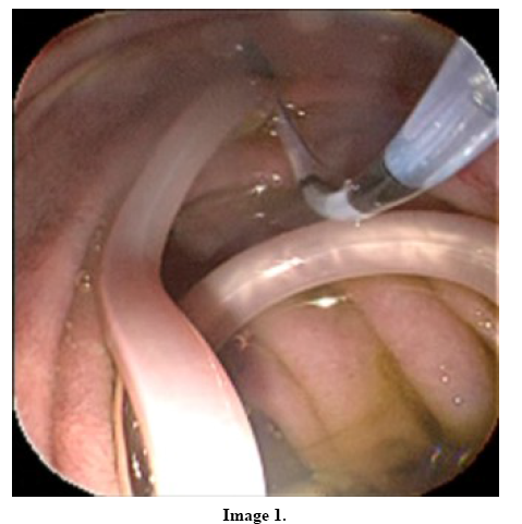
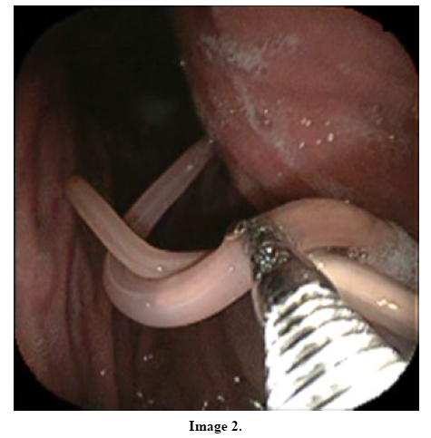
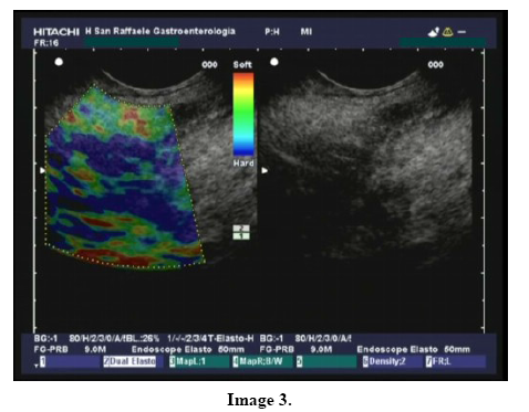
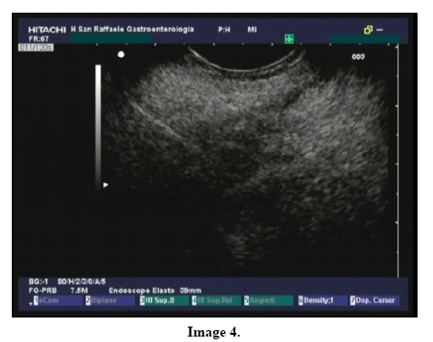
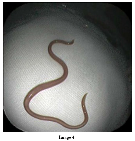
A 37-year-old man with a history of traveling in Eastern countries (5 years ago) and who usually ate fresh vegetables was referred to the emergency room for severe abdominal pain. There was no history of drugs or ethanol abuse. Family history was negative for pancreatic diseases. Physical examination disclosed pain in the upper abdomen while laboratory tests showed elevated amylasemia (508 U/L; reference range: 0-100 U/L) and leucocytosis (WBC 13.8 x10⁹/L; reference range: 6-10 x109/L) with eosinophilia (13.7%; reference range: 1-6%); reactive C protein was 29.6 mg/L (reference range: 2-6 mg/L). The patient underwent transabdominal ultrasonography (US) showing a dilated main pancreatic duct (6 mm) with a suspicious hypoechoic area (size 13 mm) at the pancreatic isthmus. A CT scan confirmed the previous US pattern with the suspicion of pancreatic malignancy. The patient then underwent EUS under deep sedation (propofol). The EUS was performed with a therapeutic video-echoendoscope (EG3830UT Pentax, Hamburg, Germany). The endoscopy revealed a roundworm floating in the duodenum (Image 1); a crocodile forceps was used for its removal (Image 2). The papilla of Vater was studied and presented a dilated orifice with “stagnant” dark bile flowing into the duodenal lumen. EUS showed a diffuse inhomogeneous pattern, with hypoechoic echotexture, such as in acute pancreatitis, and a suspected inhomogeneous focal area between the head and the isthmus. An elastosonography of the area confirmed a difference in elasticity between the suspicious mass (hard) and the surrounding tissue with signs of acute pancreatitis (soft) (Image 3). An EUS-guided fine needle aspiration biopsy (FNA) was performed with a 25 Gauge needle (Echo25, Cook Medical, Limerik,Ireland) (Image 4). Cytology showed ductal and acinar cells without atypia and a certain degree of acute inflammation. Microbiological examination of the worm revealed a20 cm long Ascaris lumbricoides (Image 5); antihelminthic therapy was administered. An EUS performed 6 months later showed normal dimensions of the pancreatic head with a slightly inhomogeneous echo structure without any focal lesions, or biliary or pancreatic duct dilation.
Discussion
This is the first case of acute pancreatitis related to Ascaris lumbricoides infection described in Italy whereas it is frequent in Asiatic countries. Treatment with antihelminthic drugs and endoscopic retrograde cholangiopancreatography (ERCP) with sphincterotomy (ES) often represents successful management. Moreover, US is an efficient, reliable and non-invasive diagnostic tool for hepatobiliary, enteric and pancreatic ascarisis [4]. The pathogenesis of Ascaris lumbricoides-induced acute pancreatitis is the same as the obstructive one; the worms can migrate from the gut to the main pancreatic duct or common bile duct, with a successive mechanical obstruction, causing severe pancreatitis or cholangitis.
The introduction of EUS in routine clinical practice has changed the management of various diseases, especially in the study of the pancreatic parenchyma [5]. In this particular case, EUS allowed us to diagnose the benign nature of the pancreatic lesion, considered to be malignant at US and CT scan.
In conclusion, in non-endemic countries, acute pancreatitis induced by Ascaris lumbricoides is an unusual diagnosis, and should be suspected especially in patients with a history of traveling in endemic areas. In our case, the diagnosis was missed at US and CT which diagnosed a pancreatic malignancy; instead, endoscopy yielded a correct diagnosis. FNA was performed to confirm the not-malignant nature of the disease, because previous imaging results had indicated a malignancy.
Conflict of interest The authors have no potential conflicts of interest
References
- Al-Qurashi A, Maklad KM, Al Abdulwahed O. Pancreatitis due to ascarislumbricoides, a case report. J Egypt SocParasitol 2003;33:657-62.
- Madsen RB, Djurhuus H. Acute pancreatitis caused by Ascarislumbricoides. UgeskrLaeger 2000;162;3730-1.
- Sandouk F, Haffar S, Zada MM, Graham DY, Anand BS. Pancreatic-biliary ascarisis: experience of 300 cases. Am J Gastroenterol 1997;92:2264-7.
- Ferreyra NP, Cerri GG. Ascariasis of the alimentary tract, liver, pancreas and biliary system; its diagnosis by ultrasonography. Hepatogastroenterology 1998;45:932-7.
- Sreenarasimhaiah J. The emerging role of endoscopic ultrasonography in cancer staging. Am J Med Sci 2005;329:247-58.

