Keywords
Early Diagnosis; Early Detection of Cancer; Metabolomics; Pancreatic Neoplasms
INTRODUCTION
Metabolomics is an emerging branch of studies focusing on phenotypic characteristics rather than genetic profiles of biological systems [1, 2]. Metabolomes are defined as all compounds generated by living organisms during their life cycles and reflect the function of the organisms as a whole [1]. This concept can be applied to single cells, organisms or larger biologic systems. Because of the dynamic profile of living entities, the intricate interactions of biochemical cycles and the uniqueness of each system, metabolomics is one of the most challenging disciplines of the family of “omics” [2].
Contrary to genomes that remain relatively stable over time, metabolomes are subject to environmental influences that can alter their profile in a very short period of time and the mere sampling, preparation and analysis of biological samples may alter their outlines [2]. This has proven to be one of the hardest tasks to overcome for the advancement of this field. Another challenge is the variability of the molecular size and the large number of metabolites within a given sample that requires very sophisticated methods for the extraction and for the data analysis [2]. Despite these challenges, the field of metabolomics has made significant advances, and in recent years, has become one of the most promising areas of research for some malignancies where pure genetic tests might fall short especially when exogenous predisposing factors play a role [3].
METABOLOMIC ANALYSIS METHODS
Metabolomic analysis can be applied to both in vitro and in vivo specimens by using body fluids, cells or tissues. Various methods have been utilized to detect and generate metabolic profiles. The two most common are nuclear magnetic resonance spectropscopy (NMR) and mass spectrometry (MS) [2-4]. These can be coupled with gas chromatography, liquid chromatography or capillary electrophoresis [2-4]. NMR has the benefit of requiring minimal sample preparation; it can analyze a wide range of metabolites and applies to both solids and liquids. On the other hand, MS has the benefit of being more sensitive than NMR but does require more extensive sample preparation that might lead to interactions between analysates and instruments [3, 4].
Metabolomics in Oncology
The use of metabolomics seems to have the greatest potential in oncological research, as the pathogenesis of cancer often is due to both genetic and environmental factors. Comparing variations of metabolic profiles occurring in neoplastic cells versus normal cells, can lead to the identification of various metabolites or combinations of metabolites that could potentially be used for the diagnosis of specific cancers or to monitor their response to treatments.
In addition, metabolomics can be used to analyze a large number of compounds and biological samples that can be used for for screening and early detection. This includes the use of a wide variety of methods such as obtaining tissues or using less invasive methods such as collecting biofluids like serum, saliva or urine [2-4]. Another benefit of metabolomics is the lower sample volume of no more than 0.5 ml for sufficient analysis.
In recent years, the study of metabolomics has expanded to other fields of research such as pharmacometabolomics where metabolomes are analyzed before, during and after treatment allowing researchers to better understand how patients respond to specific drugs and how neoplastic processes react to established or experimental therapies [5].
Metabolomics and Pancreatic Cancer
Pancreatic adenocarcinoma (PC) is the twelfth most common cancer in North America and Europe, and the fourth cause of cancer related deaths with an overall 5-year survival rate of 5 to 10% [6-9]. The high mortality rate of PC is multifactorial and when PC is diagnosed in early stage and patients undergo resection, 5-year survival can be as high as 20-35% [9]. However, the majority of patients become symptomatic late and are diagnosed with locally advanced or with metastatic disease.
Presently, no screening tests have shown any promising impact for the survival of PC patients [10]. Cross sectional imaging tests such as abdominal ultrasound (US), computerized tomography (CT-scan), magnetic resonance imaging (MRI), endoscopic ultrasound (EUS) and endoscopic retrograde cholangiopancreatography (ERCP) have failed to detect early stage tumors when used for screening purposes even in high risk populations [10-12]. Serologic markers such as carbohydrate antigen 19-9 (CA 19-9) lack enough specificity to differentiate between PC and other gastrointestinal malignancies or benign pancreaticobiliary diseases such as pancreatitis and cholangitis [10]. Newer serologic biomarkers (CA242, M2-PK, PBF-4, PNA-binding glycoprotein, hTert, MMP-2, synuclein-γ, ICAM-1, OPG, CEA, TIMP-1, MUC1, CEACAM1, MIC1) have been identified as potential screening tools but none has been proven to be better than CA 19-9 [10, 13-23].
Applications of Metabolomics in Pancreatic Cancer
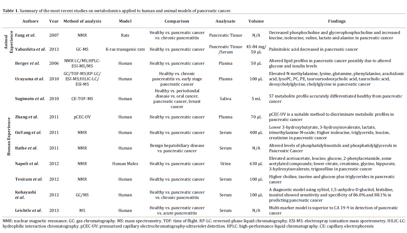
One of the main barriers to early detection of PC is differentiating it from benign pancreatic pathology most notably chronic pancreatitis. Numerous research groups have sought to differentiate between the two conditions using NMR and MS. NMR provides a broader approach for the identification of various metabolites whereas MS provides the ability to identify individual metabolite markers [3]. Each technique has individually been applied in the field of early detection of PC (Table 1).
Animal Experience
Animal studies have applied metabolomics mainly to rats for early diagnosis of PC. Yabushita et al. used K-ras 301 transgenic rats to compare metaobolic profiles of PC vs. controls. This rat model had previously shown to be a valuable model to study the development of PC once rats are treated with an adenoviral vector [24]. In their study, rats were confirmed to have PC without evidence of local invasion or metastases. The authors found that rats with PC had decreased serum levels of palmitoleic acid when compared to healthy controls on gas chromatographymass spectroscopic analysis [25]. Fang et al. also used rat models that were divided in three different groups: rats with PC, rats with chronic pancreatitis and healthy controls. PC rats developed moderately differentiated PCs and demonstrated differences in their metabolic profiles in comparison to rats with chronic pancreatitis and healthy controls as phosphocholine and glycerophosphocholine were decreased while leucine, isoleucine, valine, lactate and alanine were increased. The opposite trend was seen in chronic rats with pancreatitis [26].
Human Experience
Several research groups have applied metabolomics techniques to patients with PC and compared their profiles to healthy controls (Table 1). NMR technologies allowed Tesiram et al. to identify statistically significant different levels of choline, taurine glucose and triglycerides between the two groups. Studies were performed on patients using 100 μL of serum and tumor clinical stages varied from IIB to IV [27]. NMR was also used by Ou Yang et al. and reported altered levels of isoleucine, triglycerides, leucine, creatinine, lactate, 3-hydroxybutyrate, 3-hydroxyisovalerate, and trimethylamine-N-oxide. In this study, only 400 μL of serum was necessary [28]. Urayama et al. used multiple methods including gas chromatography/ time-of-flight mass spectrometry (GC/TOF-MS), reversedphase liquid chromatography/electrospray ionization mass spectrometry (RP-LC/ESI-MS), and hydrophilic interaction chromatography/electrospray ionization mass spectrometry (HILIC-LC/ESI-MS) to identify 24 metabolites that were abnormal in patients with PC. These patients had early stage PC (IB-IIB) and only 100 μL of serum was used [29]. Zhang et al. were able to demonstrate a novel method of pressurized capillary electrochromatography/ultraviolet detection (pCEC-UV) as a superior method than established chromatographic techniques for PC [30]. Berger et al. utilized 1D proton NMR techniques as well as liquid chromatography (LC/ MS), and high performance liquid chromatography (HPLCESI- MS/MS) to demonstrate the alterations of various lipid molecules in PC patients with tumor stages between I to IV where only 50 μL of plasma was needed [31]. These studies have shown that over time, techniques and volumes used for their analysis have changed, but no single metabolome has consistently shown to predict the presence of PC with enough accuracy. Therefore, further studies are necessary to figure out what analytical methods perform better in these settings.
Because no single molecule has shown to be promising enough for the discrimination of patients with early PC, Kobayashi et al. have used multiple logistic regression analysis to compare PC patients with healthy controls. PC patients varied from stage 0 to IV and 100 μL of plasma was used. Using GC/MS, 18 metabolites were identified as potential biomarkers and using 4 of these metabolites (xylitol, 1,5-anhydro-D-glucitol, histidine, and inositol), a statistical diagnostic model was generated. This model performed extremely well with an area under the receiveroperating characteristic curve (AUC) of 0.92. When compared to conventional tumor markers CA19-9 and CEA in the validation study, this model did not perform as well as the AUC went down to 0.76 but maintained acceptable sensitivity (71.4%) and specificity (78.1%). The sensitivity of this diagnostic model remained overall stable even when applied to early stage diseases (0-IIB) (77.8%) in comparison to CA 19-9 (55.6%) and CEA (44.4%) [32].
Differentiating Between Benign and Malignant Disease
Bathe et al. [33] used NMR spectroscopy to identify serum metabolites that could be used to differentiate various benign versus malignant pancreatic diseases. In their study, glutamate, acetone, and 3-hydroxybutyrate were strongly associated with malignancy and their multivariate model demonstrated good discrimination with an AUC of 0.83 [33]. The robustness of this model was further demonstrated via a feasibility test of 14 pairs matched for the presence of jaundice, pancreatic mass and diabetes with comparable AUC values. Similarly, Leichtle et al. [34] performed a three-class analysis using serum from individuals affected by PC (stages I-IV), pancreatitis and healthy controls. A significant difference among the three groups was found for 22 amino acids [34]. Using mass spectrometry both individually and in conjunction with CA19-9 levels, a multivariate model based on both specific amino acids in conjunction with CA19-9 provided a 3-Dimensional analogue of AUC (VUS) with good discrimination (VUS value=0.89) [34]. These studies have shown that metabolomics, both alone and in conjunction with other established serologic testing, can be used to improve our current ability to detect and discriminate PC from other benign conditions.
Early Detection of Pancreatic Cancer
Beyond serum, other biofluids have been used to study metabolic profiles for the diagnosis of early PC [35, 36]. Previous investigations of bile and pancreatic fluids had identified a range of proteins that could be used as markers [10]. Unfortunately these samples require invasive techniques that are costly and often not available in many centers. To overcome these limitations, several studies have focused on the use of biofluids that can be collected by noninvasive techniques. For example, 5 mL of unstimulated saliva obtained from patients with non-metastatic PC was analyzed using capillary electrophoresis time-of-flight mass spectrometry (CE/TOF-MS) and compared with samples from healthy controls, patients with oral or breast cancers and periodontal diseases. Using multiple linear regression analysis of a total of 48 potential metabolites, 5 were selected to construct a model that showed excellent accuracy for the detection of PC with AUC equal to 0.94, the highest value ever seen [35] for any of the diagnostic tests. Furthermore, Napoli et al. [36] performed a case controlled study using NMR spectrography on urine samples (630 μL) from PC patients (stages II-IV) and healthy individuals. Not only they observed a statistically significant different profile between the two groups, but they were also able to identify differences between different cancer stages [36]. This was the first study that was able to discriminate diverse metabolomic profiles for patients with different stages of PC. Although extremely promising, further studies are necessary to confirm if these results are reproducible in other populations.
Future Directions and Applications of Metabolomics in Pancreatic Cancer
The studies reviewed in this paper have all sought to identify useful bio-markers that could, one day, be used for the diagnosis of early PC either by improving the sensitivity and specificity of current tests or substitute them. So far, metabolomics have shown promising results but not sufficient to change current clinical practice and more studies are needed [33, 34]. Given the fact that metabolomic profiles incorporate both genetic and environmental factors that play a role in PC, it is important that future studies identify and control for some of these variables (e.g. age, sex, co-morbidities, and disease stage).
Another promising area where metabolomics can have a significant impact is the analysis of samples from groups with pre-malignant or predisposing factors for PC such as intraductal papillary mucynous neoplasms, familial PC, BRCA2 and other conditions.
The concept of pharmacometabolomics was introduced earlier in this review. Although pharmacometabolomics have not been widely used in clinical practice, expanding its approach to include changes following resection and/or chemoradiation therapy in PC would allow metabolomics not only to be applied to diagnosis and screening but also to surveillance.
Although metabolomics applied to PC is still in its infancy, it has the all the potentials of becoming an important field that could lead to the identification of better biomarkers to be used alone or in combination with other already available modalities for the screening, early detection, staging and treatment of this challenging tumor. Hopefully, this would translate in better prognosis for patients with PC whose survival continues to be unsatisfactory.
Conflicting Interest
The authors had no conflicts of interest
References
- Fiehn O. Metabolomics--the link between genotypes and phenotypes. Plant Mol Biol 2002; 48:155-171. [PMID: 11860207]
- Spratlin JL, Serkova NJ, Eckhardt SG. Clinical applications of metabolomics in oncology: A review. Clin Cancer Res 2009; 15:431-440. [PMID: 19147747]
- Armitage EG, Barbas C. Metabolomics in cancer biomarker discovery: Current trends and future perspectives. J Pharm Biomed Anal 2014; 87:1-11. [PMID: 24091079]
- Bu Q, Huang Y, Yan G, Cen X, Zhao YL. Metabolomics: A revolution for novel cancer marker identification. Comb Chem High Throughput Screen 2012; 15:266-275. [PMID: 22221059]
- Kaddurah-Daouk R, Weinshilboum RM. Pharmacometabolomics Research Network. Pharmacometabolomics: Implications for clinical pharmacology and systems pharmacology. Clin Pharmacol Ther 2014; 95:154-167. [PMID: 24193171]
6. Canadian Cancer Society's Advisory Committee on Cancer Statistics. Canadian cancer statistics 2013. 2013.
- Ferlay J, Parkin DM, Steliarova-Foucher E. Estimates of cancer incidence and mortality in europe in 2008. Eur J Cancer 2010; 46:765-781. [PMID: 20116997]
- Maisonneuve P, Lowenfels AB. Epidemiology of pancreatic cancer: An update. Dig Dis 2010;28:645-656. [PMID: 21088417]
- Siegel R, Ma J, Zou Z, Jemal A. Cancer statistics, 2014. CA Cancer J Clin 2014; 64:9-29. [PMID: 24399786]
- Gonda TA, Saif MW. Early detection and screening of pancreatic cancer. highlights from the "2011 ASCO gastrointestinal cancers symposium". san francisco, CA, USA. JOP 2011; 12:83-85. [PMID: 21386626]
- Ross WA, Wasan SM, Evans DB, et al. Combined EUS with FNA and ERCP for the evaluation of patients with obstructive jaundice from presumed pancreatic malignancy. Gastrointest Endosc 2008; 68:461-466. [PMID: 18384788]
- Savides TJ, Donohue M, Hunt G, et al. EUS-guided FNA diagnostic yield of malignancy in solid pancreatic masses: A benchmark for quality performance measurement. Gastrointest Endosc 2007; 66:277-282. [PMID: 17643700]
- Gold DV, Modrak DE, Ying Z, Cardillo TM, Sharkey RM, Goldenberg DM. New MUC1 serum immunoassay differentiates pancreatic cancer from pancreatitis. J Clin Oncol 2006; 24:252-258. [PMID: 16344318]
- Ni XG, Bai XF, Mao YL, et al. The clinical value of serum CEA, CA19-9, and CA242 in the diagnosis and prognosis of pancreatic cancer. Eur J Surg Oncol 2005; 31:164-169. [PMID: 15698733]
- Eguia V, Gonda TA, Saif MW. Early detection of pancreatic cancer. JOP 2012; 13:131-134. [PMID: 22406583]
- Fry LC, Monkemuller K, Malfertheiner P. Molecular markers of pancreatic cancer: Development and clinical relevance. Langenbecks Arch Surg 2008; 393:883-890. [PMID: 18266003]
- Ventrucci M, Cipolla A, Racchini C, Casadei R, Simoni P, Gullo L. Tumor M2-pyruvate kinase, a new metabolic marker for pancreatic cancer. Dig Dis Sci 2004; 49:1149-1155. [PMID: 15387337]
- Fiedler GM, Leichtle AB, Kase J, et al. Serum peptidome profiling revealed platelet factor 4 as a potential discriminating peptide associated with pancreatic cancer. Clin Cancer Res 2009;15:3812-3819. [PMID: 19470732]
- Koopmann J, Rosenzweig CN, Zhang Z, et al. Serum markers in patients with resectable pancreatic adenocarcinoma: Macrophage inhibitory cytokine 1 versus CA19-9. Clin Cancer Res 2006; 12:442-446. [PMID: 16428484]
- Ching CK, Rhodes JM. Enzyme-linked PNA lectin binding assay compared with CA19-9 and CEA radioimmunoassay as a diagnostic blood test for pancreatic cancer. Br J Cancer 1989; 59:949-953. [PMID: 2736232]
- Uehara H, Nakaizumi A, Tatsuta M, et al. Diagnosis of pancreatic cancer by detecting telomerase activity in pancreatic juice: Comparison with K-ras mutations. Am J Gastroenterol 1999; 94:2513-2518. [PMID: 10484017]
- Yokoyama M, Ochi K, Ichimura M, et al. Matrix metalloproteinase-2 in pancreatic juice for diagnosis of pancreatic cancer. Pancreas 2002; 24:344-347. [PMID: 11961486]
- Hibi T, Mori T, Fukuma M, et al. Synuclein-gamma is closely involved in perineural invasion and distant metastasis in mouse models and is a novel prognostic factor in pancreatic cancer. Clin Cancer Res 2009; 15:2864-2871. [PMID: 19351749]
- Ueda S, Fukamachi K, Matsuoka Y, et al. Ductal origin of pancreatic adenocarcinomas induced by conditional activation of a human ha-ras oncogene in rat pancreas. Carcinogenesis 2006; 27:2497-2510. [PMID: 16774944]
- Yabushita S, Fukamachi K, Tanaka H, et al. Metabolomic and transcriptomic profiling of human K-ras oncogene transgenic rats with pancreatic ductal adenocarcinomas. Carcinogenesis 2013; 34:1251-1259. [PMID: 23393225]
- Fang F, He X, Deng H, et al. Discrimination of metabolic profiles of pancreatic cancer from chronic pancreatitis by high-resolution magic angle spinning 1H nuclear magnetic resonance and principal components analysis. Cancer Sci 2007; 98:1678-1682. [PMID: 17727683]
- Tesiram YA, Lerner M, Stewart C, Njoku C, Brackett DJ. Utility of nuclear magnetic resonance spectroscopy for pancreatic cancer studies. Pancreas 2012; 41:474-480. [PMID: 22422139]
- OuYang D, Xu J, Huang H, Chen Z. Metabolomic profiling of serum from human pancreatic cancer patients using 1H NMR spectroscopy and principal component analysis. Appl Biochem Biotechnol 2011; 165:148-154. [PMID: 21505807]
- Urayama S, Zou W, Brooks K, Tolstikov V. Comprehensive mass spectrometry based metabolic profiling of blood plasma reveals potent discriminatory classifiers of pancreatic cancer. Rapid Commun Mass Spectrom 2010; 24:613-620. [PMID: 20143319]
- Zhang H, Wang Y, Gu X, Zhou J, Yan C. Metabolomic profiling of human plasma in pancreatic cancer using pressurized capillary electrochromatography. Electrophoresis 2011; 32:340-347. [PMID: 21298661]
- Beger RD, Schnackenberg LK, Holland RD, Li D, Dragan Y. Metabonomic models of human pancreatic cancer using 1D proton NMR spectra of lipids in plasma. Metabolomics. 2006; 2:125.
- Kobayashi T, Nishiumi S, Ikeda A, et al. A novel serum metabolomics-based diagnostic approach to pancreatic cancer. Cancer Epidemiol Biomarkers Prev 2013; 22:571-579. [PMID: 23542803]
- Bathe OF, Shaykhutdinov R, Kopciuk K, et al. Feasibility of identifying pancreatic cancer based on serum metabolomics. Cancer Epidemiol Biomarkers Prev. 2011; 20:140-147. [PMID: 21098649]
- Leichtle AB, Ceglarek U, Weinert P, et al. Pancreatic carcinoma, pancreatitis, and healthy controls: Metabolite models in a three-class diagnostic dilemma. Metabolomics 2013; 9:677-687. [PMID: 23678345]
- Sugimoto M, Wong DT, Hirayama A, Soga T, Tomita M. Capillary electrophoresis mass spectrometry-based saliva metabolomics identified oral, breast and pancreatic cancer-specific profiles. Metabolomics 2010; 6:78-95. [PMID: 20300169]
- Napoli C, Sperandio N, Lawlor RT, Scarpa A, Molinari H, Assfalg M. Urine metabolic signature of pancreatic ductal adenocarcinoma by (1)h nuclear magnetic resonance: Identification, mapping, and evolution. J Proteome Res 2012; 11:1274-1283. [PMID: 22066465]

