Keywords
Diabetic Nephropathies; Hyperglycemia; Oxidative Stress; Quercetin; Resveratrol
Abbreviations
LDH lactate dehydrogenase
Introduction
Diabetes mellitus is a common chronic disease that has been increasing in prevalence. It afflicts 7.7% of the population aged 20-79 years. In 2010, it was estimated that about 285 million people worldwide were diabetic; this is expected to increase to 439 million by 2030 [1].
Diabetic nephropathy is a serious complication of diabetes and the most common cause of end-stage renal disease in most developed countries [2]. Hyperglycemia is a crucial factor in the development of microvascular diabetic complications [3] and related mechanisms induce disorders in kidney cells that result in diabetic nephropathy [2].
Evidence suggests that oxidative stress plays a vital role in the pathogenesis of diabetic nephropathy [3]. NADPH oxidase (NOX) is a major source of reactive oxygen species (ROS) generation and is strongly expressed in the kidney, especially in renal vessels, mesangial, and podocyte cells and is implicated in diabetic nephropathy [4, 5]. Advanced glycation end-products (AGEs) are products of nonenzymatic glycation and oxidation of proteins and lipids, the generation of which increases significantly in various tissues under diabetic conditions [2].
AGEs interact with their cell surface receptors (RAGE) and induce oxidative stress that contributes to progression of diabetic renal disease [6]. NOX and RAGE contribute in the induction of oxidative stress and the resulting kidney damage associated with diabetes. One of the proteins that are affected by hyperglycemia and has a role in oxidative stress is senescence marker protein-30 (SMP30). SMP30 is an aging marker molecule for which expression decreases with age in a sex-independent fashion [7]. SMP30 binds with Ca2+ and regulation of its intracellular concentration plays a key role in cell protection against high intracellular Ca2+ injury, especially in renal tubule cells [8]. Recent reports demonstrate that SMP30 controls antioxidant enzyme activity and its age-dependent attenuation is closely associated with oxidative stress [9].
Antioxidants decrease oxidative stress and are a potential source of therapy for improving kidney function in diabetic patients [10]. Flavonoids are phenolic compounds with antioxidant properties and are widelydistributed secondary metabolites in plants. Quercetin (3,5,7,3 ÃÆÃââââ‰â¢ÃÆââ¬Å¡Ã¢ââ¬Ã ,4 ÃÆÃââââ‰â¢ÃÆââ¬Å¡Ã¢ââ¬Ã -pentahydroxyflavone; Q) is a flavonoid present in red wine and several types of fruit, vegetables, and nuts [11]. Recent studies have shown that Q is a potent scavenger of free radicals that affect antioxidant pathways both in vivo and in vitro [12]. Resveratrol (trans-3,5,4′- trihydroxystilbene; RSV) is a polyphenolicphytoalexin found in red grapes and numerous plant species having beneficial health properties. RSV is a potent antioxidant [13] that can directly scavenge ROS and has anti-inflammatory properties [14]. The present study evaluated and compared the protective effects of RSV, Q, and a mixture of the two (RSV/Q) against hyperglycemiamediated oxidative stress in a human embryonic kidney cell line (HEK-293) to elucidate the molecular mechanism of RSV and Q under high glucose conditions.
Chemicals
Dulbecco Modified Eagle Medium (DMEM), fetal bovine serum (FBS), phosphate buffered saline (PBS) and 0.05% trypsin/EDTA (1X) solution were purchased from Gibco (USA). MTT powder (3-(4,5-dimetylthiazol-2-Yl)–2,5– diphenyltetrazolium bromide), antibiotics (penicillin and streptomycin), RSV, and Q powder were purchased from Sigma (USA), dimethylsulfoxid (DMSO) were purchased from MP Biomedical (USA), and primer sequences for quantitative real-time PCR were purchased from Bioneer (Korea).
In vitro experiments
HEK-293 cells were acquired from Pasteur Institute of Iran (Tehran) and were cultured in DMEM supplemented with 10% FBS and 1% streptomycin and penicillin. The cells were incubated in a humidified atmosphere with 5% CO2 at 37°C. The culture medium was refreshed every 48 h.
Cell viability assay
HEK-293 cells with 80% confluence were trypsinized and seeded in 96-well cell culture microplates at a density of 7×10³ cell/well in DMEM supplemented with 2% FBS and 100 mM glucose for the high glucose group. DMEM with 25 mM glucose served as the control. After 6-8 h, the cells in the high glucose condition were treated with different concentrations of RSV, Q, and RSV/Q for 24 and 48 h. The treatments used in this study are listed in Table 1. Cell viability under normal and high glucose conditions before and after treatment with RSV and Q were evaluated by MTT assay. MTT solution (5 mg/ml in PBS) was added to the wells. After 4h of incubation in the dark at 37°C, the media was aspirated and insoluble crystal formazan was dissolved in DMSO (100 μL/well). Absorbance was measured at 570 nm by a plate reader (BioTek; Epoch; USA).

Cytotoxicity assay
The ability of RSV, Q and RSV/Q to support the cells from hyperglycemia-induced cell injury was assessed by lactate dehydrogenase (LDH) release. A Pierce LDH cytotoxicity assay kit (No. 88953) was used to measure LDH release. Cells (2×10⁵ cells/well) were plated in 24-well plates the day before the experiments. After treatment for 48 h, the medium from each well was collected to measure the amount of LDH released. The cells then were exposed to lysis buffer (9% Triton X 100) for 30 min at 37°C and the media was collected to measure the total amount of cellular LDH. Optical density was measured at a wavelength of 490 nm and the percentage of released LDH versus total intracellular LDH was calculated.
Determination of Cell Oxidant and Antioxidant Status
Reduced and oxidized glutathione (GSH and GSSG) levels were determined by HPLC chromatography having a fluorescent detector (Ex: 385 Em: 515 nm) using commercial kits (Chrom systems Diagnostics; Germany) according to manufacturer instructions. Thioredoxin as an intracellular antioxidant was measured using solid phase sandwich ELISA (Human Thioredoxin Assay Kit; IBL) according to manufacturer instructions. Ferric reducing antioxidant power (FRAP) assay was carried out as recommended by Benzie and Strain (1996) to evaluate total antioxidant power in the cell lysate of the different groups [15]. To evaluate lipid peroxidation, the malondialdehyde (MDA) level in the kidney cell lysate was measured using the TBARS method [16].
Intracellular production of ROS was assessed using 2′,7′dichlorofluorescin diacetate (DCF-DA; Molecular Probes; Sigma) in a flat-bottomed 96-well cell culture microplates. HEK-293 cells (3×10⁵ cells/well) were treated with different concentrations of RSV, Q, and RSV/Q under high glucose conditions. They were then exposed to 10 μM DCFDA in PBS− for 15 min at 37°C in the dark. The cells were washed with PBS− and fluorescence was measured with a Floustar Optima spectrofluorimeter (BMG Laboratories; Germany) using filters with an absorption spectrum of 480 nm and emission of 520 nm.
RNA Extraction and qRT-PCR Analysis
The total RNA from the HEK-293 cell line was isolated using TRIzol extraction reagent (Bioneer; Korea) according to manufacturer recommendations. The integrity of the mRNA was confirmed by electrophoresis in denaturing 1% agarose gel. The Revert Aid H Minus First Strand cDNA synthesis kit (Thermo Scientific; USA) was used to reversetranscribe 1μg of RNA in a final volume of 20 μL. The qRTPCR of β-actin (reference gene), NOX4, RAGE, and SMP30 was carried out using the primers identified in Table 2.

The 20μL reaction mixture consisted of 2× ABI SYBR Green PCR Master Mix, 1μL cDNA, and 1 μL of each primer. Amplification was performed using the ABI StepOne Real- Time PCR System (Applied Biosystems; USA) with 40 cycles of denaturation at 95°C for 30 s, annealing and extension at 60°C for 30 s, and data collection at 80°C for 20 s. The intensities of the mRNA levels were normalized to those of the β-actin product as ratios producing arbitrary units of relative abundance. Relative gene expression (2-ΔΔCT) between the RSV, Q, and RSV/Q treated samples and the control group was assayed using the CT method [17].
STATISTICAL ANALYSIS
All experiments were carried out in triplicate and the data was expressed as mean ± standard deviation (SD) and analyzed using SPSS v.16 software (SPSS: USA). The statistical significance of the difference between mean levels of LDH and GSH of the control and treated groups was evaluated using student’s t-test. The results were considered significant at P<0.05. The graphs were drawn with GraphPad Prism version 5 (GraphPad; USA).
RESULTS
RSV And Q Had Protective Effect on HEK-293 Cells under Hyperglycemic Condition
Figure 1a shows that the high glucose condition dramatically decreased cell viability (control: 100; high glucose: 50.1 ± 1.6; P ≤ 0.05) and increased cell injury by LDH assay (control: 100; high glucose: 580 ± 3.1; P ≤ 0.05) (Figure 1b).The results presented in Figure 1a and b indicate that treatment with RSV and Q increased cell viability and decreased LDH release. This protective effect was notable for the RSV/Q mixture.
Figure 1. Effects of RSV, Q and RSV/Q on viability and cytotoxicity in high glucose treated HEK-293 cells. (a). Cell viability of HEK-293 cells which was assessed by MTT. (b). Cytotoxicity of HEK-293 cells which was assessed by LDH release. (*Compared with control group, p<0.05, n = 3)
Effects of RSV and Q on Oxidative Stress Status in Kidney Cells
The results summerized in Table 3 show that the high glucose condition decreased the level of GSH and the GSH/GSSG ratio. Treatment of kidney cells with RSV and Q increased GSH and improved the GSH/GSSG ratio. The RSV had a considerably greater effect than Q. The RSV/Q mixture at all doses applied was more effective than RSV and Q alone (Table 3). As shown in Table 3, hyperglycemia decreased thioredoxin level and treatment with RSV, Q and RSV/Q increased thioredoxin to the normal value. This increase was significantly different for RSV3, RSV/Q2 and RSV/Q3 formulations (Table 3). RVS, Q, and RVS/Q downregulated ROS production in HEK-293 cells in less than 60 min at all concentrations applied. Down-regulation by RSV at high concentration and RSV/Q3 was greater than that for Q (Table 3). A protective effect for cells by the RSV/Q was considerably greater than for all doses of RSV and Q alone (P<0.05).
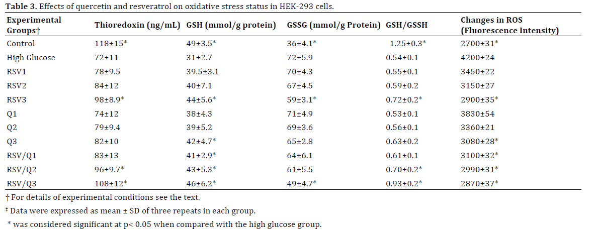
The results of the TBARS assay indicated that MDA levels as a marker of lipid peroxidation in the HEK-293 cell lysate increased markedly after treatment with 100 mM glucose compared to those in the control group. In contrast, the MDA levels in the cell lysate having different concentrations of RSV, Q, and RSV/Q decreased significantly in a dose-dependent manner. The combined therapy was more effective than the RSV and Q alone. In fact, at high doses of both antioxidants, the MDA levels decreased to control group level. Treatment with different doses of RSV, Q, and RSV/Q increased antioxidant capacity in the cell lysate of HEK-293 in a dose-dependent-fashion. As shown in Table 3 high doses of RSV/Q increased the antioxidant values of the treated cells to the level of the control group.
RSV And Q Inhibited the Gene Expression of NOX4 and RAGE and Improved Gene Expression of SMP30
The antioxidant effects of RSV, Q, and RVS/Q on NOX4 and RAGE transcript levels were examined using qRT-PCR analysis. Figures 2,3 show that the high glucose condition induced expression of NOX4 and RAGE, respectively, in the HEK-293 cell line that could be key factors in the progression of diabetic nephropathy. Treatment with different concentrations of antioxidants reduced expression of NOX4 and RAGE. Additionally, RSV/Q was more effective than RSV or Q alone. Expression of SMP30 anti-aging factor in high glucose medium decreased (Figure 4). Treatment with various concentrations of the antioxidants increased expression of SMP30.
Figure 2. Expression of NOX4 mRNA in HEK-293 cells under high glucose condition before and after treatment with RSV, Q and RSV/Q. Treatment with RSV and Q significantly attenuated high glucose-induced NOX4 mRNA expression. The combination effect of RSV/Q showed more effective than RSV and Q alone.
Figure 3. Effects of RSV, Q and RSV/Q on expression of RAGE mRNA in high glucose treated HEK-293 cells. Treatment with RSV and Q significantly attenuated high glucose-induced RAGE mRNA expression. The combination effect of RSV/Q showed more effective than RSV and Q alone.
Figure 4. Expression of SMP30 mRNA in HEK-293 cells under high glucose condition with or without treatment with RSV, Q and RSV/Q. Treatment with different concentrations of RSV, Q and notably RSV/Q induced the mRNA expression of SMP30 in HEK-293 cell.
DISCUSSION
Diabetic nephropathy is a leading cause of end-stage renal disease. Hyperglycemia through induction of ROS production [8] and by a decrease in antioxidant defense systems plays an important role in the pathogenesis of diabetic nephropathy. The present investigation showed that maintaining the high glucose condition for 48 h dramatically decreased the viability of HEK-293 cells. In line with these results, studies have shown that hyperglycemic condition has a negative effect on cell viability [18]. The current data demonstrates that cotreatment of hyperglycemic HEK-293 cells with different concentrations of RSV and Q alone and RSV/Q increased cell viability in a dose-dependent manner. Previous studies have confirmed the cytoprotective effect of RSV and Q under high glucose conditions in bEnd.3 cultured cells [12] and dorsal root ganglion neurons of rats [18], respectively.
According to the present study, antioxidant activity of thioredoxin decreased in the high glucose condition that is line with other studies results [19]. Studies have also shown that the antioxidant activity of thioredoxin decreases after hyperglycemia-mediated thioredoxin interacting protein (Txnip) induction as a critical mechanism to promote vascular oxidative stress in diabetes mellitus [19]. Improvement of thioredoxin level in RSV, Q and especially RSV/Q treated groups indicates beneficial effects of the flavonoids in kidney cells (Table 3). The glutathione level (as a predictor of cell redox status) and GSH/GSSG ratio dropped in the high glucose condition (Table 3). The reduction in the GSH/GSSG ratio and GSH in patients with type II diabetes has been reported elsewhere [20]. The present study recorded an increase in GSH level and the GSH/GSSG ratio in response to various concentrations of RSV, Q, and especially RSV/Q. MDA levels, as a marker of oxidative damage, increased under high glucose condition that confirmed by recent studies [21]. The results of the present study showed that MDA levels of HEK-293 lysate increased under hyperglycemic conditions and treatment with different concentrations of R, Q, and especially RVS/Q decreased MDA levels in a dose-dependent manner. High glucose condition also decreased total antioxidant capacity in kidney cells and different doses of RSV, Q and especially RSV/Q improved antioxidant status in the cells (Table 3). It seems reduced antioxidant capacity in hyperglycemic condition may be through elevated ROS production in the cells.
Treatment with RSV, Q, and RSV/Q downregulated ROS production in kidney cells resulting improved antioxidant capacity. In addition to direct ROS scavenging, studies indicate that RSV induced expression of antioxidants, including thioredoxin. In vivo studies have shown that RSV/Q has a greater pharmacological effect and more effectively improves brain antioxidant capacity [22]. In the present study, results obtained from biochemical analysis collectively confirmed beneficial effects of RSV, Q RSV/Q mixture.
The results of the gene expression assay in the present study revealed that RSV, Q, and RSV/Q decreased high glucose-induced RAGE and NADPH oxidase expressions in HEK-293 cells. It has recently been shown that the activity of NADPH oxidase increases under diabetic conditions in the kidney and can induce oxidative stress resulting in diabetic nephropathy [5].
Based on the literature, RSV inhibits NADPH oxidase activity and ROS generation and can prevent high glucoseinduced kidney damage in rat renal mesangial cells [3], which agrees with the results of the present study. Studies have shown that RSV can attenuate the expression of NADPH oxidase (NOX4) in endothelial cells [23]. RSV inhibits NADPH oxidase in high glucose-treated cells which protects against cell death [12]. Furthermore, the effect of flavonoids such as Q in down-regulation of NADPH oxidase gene expression in spontaneously hypertensive rats was reported by Sancheza et al. [24].
RAGE is another factor in oxidative stress induction and is activated under hyperglycemia conditions [7]. The interaction of RAGE and AGE induces signal transduction and increases cellular oxidative stress through activation of NADPH oxidase [25]. Moreover, RAGE plays a key role in the progression of diabetic nephropathy. Its expression is significantly reduced by RSV in renal tissue of diabetic rats [26]. Other results indicate that RSV and Q significantly reduce diabetes-induced renal damage in rats by attenuating histopathological changes [27].
It has been reported that SMP30 deficiency induces proximal tubule injury in diabetic mice. A reduction in the SMP30 level increases the risk of chronic kidney disease, including diabetic nephropathy, with age [7]. Studies have shown that NADPH oxidase activity and ROS production increase significantly in the brains of SMP30 knockout mice [9]. The decrease in SMP30 during aging has been related to elevated ROS generation in rat livers and kidneys [9]. It was found that RSV extends the lifespan of various organisms [28] and the activation of antiaging factor (SIRT-1) in HT29 cells extends their lifespan [29].
The cytoprotective effects of RSV and Q were found in the present study as well as the additive effects of RSV. Taken together, this data gives clues for identifying new formulations of antioxidants drugs for treatment of diabetic nephropathy. In the present study, biochemical and gene expression results collectively revealed that Q and especially RSV protect against hyperglycemiamediated oxidative stress in kidney cells. The protective effects of Q and RSV could occur by modification of RAGE downstream signaling pathways. To better understand the exact mechanisms of Q and RSV in kidney physiology, more genes in the RAGE and NOX4 related signaling pathways should be investigated.
In conclusion, the present study increases understand of the beneficial effects of Q and RSV at the molecular level to induce cytoprotective effects against diabetic nephropathy. The effect of RSV on oxidative stress-related gene expression was notable. Of the doses used, RSV/Q3 formulation provided the most protective effect with no cytotoxicity and should be considered for design of new drugs to increase the life span of the kidney.
Acknowledgments
This investigation was supported by Grant No. 773 from the office of Vice Chancellor for Research, Birjand University of Medical Sciences.
Conflict of interest
Authors declare that they have no conflict interests.
References
- Shaw JE, Sicree RA, Zimmet PZ. Global estimates of the prevalence of diabetes for 2010 and 2030. Diabetes Res Clin Pract 2010; 87:4-14. [PMID: 19896746]
- Kanwar YS, Sun L, Xie P, Liu Fy, Chen S. A glimpse of various pathogenetic mechanisms of diabetic nephropathy. Annu Rev Pathol 2011; 6:395-423. [PMID: 21261520]
- Zhang L, Pang S, Deng B, Qian L, Chen, J, Zou J, Zheng J,et al. High glucose induces renal mesangial cell proliferation and fibronectin expression through JNK/NF-κB/NADPH oxidase/ROS pathway, which is inhibited by resveratrol. Int J Biochem Cell Biol 2012; 44:629-638. [PMID: 22245600]
- Chabrashvili T, Tojo A, Onozato ML, Kitiyakara C, Quinn MT, Fujita T, Welch WJ,et al. Expression and cellular localization of classic NADPH oxidase subunits in the spontaneously hypertensive rat kidney. Hypertension 2002; 39:269-274. [PMID: 11847196]
- Paravicini TM, & Touyz RM. NADPH oxidases, reactive oxygen species, and hypertension clinical implications and therapeutic possibilities. Diabetes care 2008; 31:170-180.[PMID: 18227481]
- Soldatos G, & Cooper M. Diabetic nephropathy: important pathophysiologic mechanisms. Diabetes Res Clin Pract 2008; 82:75-79. [PMID: 18994672]
- Leu JG, Lin CY, Jian JH, Shih CY, & Liang YJ. Epigallocatechin-3-gallate combined with alpha lipoic acid attenuates high glucose-induced receptor for advanced glycation end products (RAGE) expression in human embryonic kidney cells. An Acad Bras Cienc 2013; 85:745-752. [PMID: 23780308]
- Marrazzo G, Barbagallo I, Galvano F, Malaguarnera M, Gazzolo D, Frigiola A, D'Orazio N,et al. Role of dietary and endogenous antioxidants in diabetes. Critical Reviews in Food Science and Nutrition 2014; 54:1599-1616.[PMID: 24580561]
- Mapanga R, Musabayane C. The renal effects of blood glucose-lowering plant-derived extracts in diabetes mellitus-an overview. Ren Fail 2010; 32:132-138.[PMID: 20113279]
- Kaul TN, Middleton E, Ogra PL. Antiviral effect of flavonoids on human viruses. J Med Virol 1985; 15:71-79. [PMID: 2981979]
- Vessal M, Hemmati M, Vasei M. Antidiabetic effects of quercetin in streptozocin-induced diabetic rats. Comp Biochem Physiol C Toxicol Pharmacol 2003; 135:357-364. [PMID: 12927910]
- Chen F, Qian LH, Deng B, Liu ZM, Zhao Y, Le YY. Resveratrol protects vascular endothelial cells from high glucose-induced apoptosis through inhibition of NADPH oxidase activation-driven oxidative stress. CNS Neurosci Ther 2013; 19:675-681. [PMID: 23731528]
- Palsamy P, Subramanian S. Ameliorative potential of resveratrol on proinflammatory cytokines, hyperglycemia mediated oxidative stress, and pancreatic βÃÆÃâÃââÃÆââ¬Å¡Ã¢ââ¬Å¡Ã¬ÃÆââ¬Å¡ÃâÃÂcell dysfunction in streptozotocinÃÆÃâÃââÃÆââ¬Å¡Ã¢ââ¬Å¡Ã¬ÃÆââ¬Å¡ÃâÃÂnicotinamideÃÆÃâÃââÃÆââ¬Å¡Ã¢ââ¬Å¡Ã¬ÃÆââ¬Å¡ÃâÃÂinduced diabetic rats. J Cell Physiol 2010; 224:423-432.[PMID: 20333650]
- Ji J, Zhang J, Huang G, Qian J, Wang X, Mei S. Over-expressed microRNA-27a and 27b influence fat accumulation and cell proliferation during rat hepatic stellate cell activation. FEBS Lett 2009; 583:759-766. [PMID: 19185571]
- Benzie IF, Strain J. The ferric reducing ability of plasma (FRAP) as a measure of “antioxidant power”: the FRAP assay. Anal Biochem 1996; 239:70-6. [PMID: 8660627]
- Yagi K. A simple fluorometric assay for lipoperoxide in blood plasma. Biochem Med 1976; 15:212-6.
- Ji J, Zhang J, Huang G, Qian J, Wang X, Mei S. Over-expressed microRNA-27a and 27b influence fat accumulation and cell proliferation during rat hepatic stellate cell activation. FEBS lett 2009; 583:759-66. [PMID: 19185571]
- Shi Y, Liang XC, Zhang H, Wu Ql, Qu L, & Sun Q. Quercetin protects rat dorsal root ganglion neurons against high glucose-induced injury in vitro through Nrf-2/HO-1 activation and NF-κB inhibition. Acta Pharmacol Sin 2013; 34: 1140-1148. [PMID: 23770986]
- Schulze Ch, Yoshioka J, Takahashi T, He Zh, King GL, Lee RT. Hyperglycemia promotes oxidative stress through inhibition of thioredoxin function by thioredoxin-interacting protein. J Biol Chem 2004; 279:30369-30374. [PMID: 15128745]
- Calabrese V, Cornelius C, Leso V, Trovato-Salinaro A, Ventimiglia B, Cavallaro M, Scuto M,et al. Oxidative stress, glutathione status, sirtuin and cellular stress response in type 2 diabetes. Biochim Biophys Acta 2012; 1822:729-736. [PMID: 22186191]
- Edremitlioglu M, Fatih Andic M, Beyza Sayin D, Korkut O, Kisa U. Quercetin, a powerful antioxidant bioflavonoid, attenuated renal dysfunction in long-term experimental diabetes mellitus. Marmara Medical Journal 2011; 24:88-99.
- Eddine BI, Hakima T, Abdelkrim T. Combined effect of quercetin and resveratrol induces anxiolytic-like behavior and improves brain antioxidant capacity in male wistar rat. Asia J Anim Sci 2015; 9:157-161.
- Ladurner A, Schachne D, Schueller K, Pignitter M, Heiss E H, Somoza V, Dirsch VM. Impact of trans-resveratrol-sulfates and–glucuronides on endothelial nitric oxide synthase activity, nitric oxide release and intracellular reactive oxygene species. Molecules 2015; 19:16724-16736. [PMID: 25329867]
- Sanchez M, Galisteo M, Vera R, Villar IC, Zarzuelo A, Tamargo J, Pérez-Vizcaíno F,et al. Quercetin downregulates NADPH oxidase, increases eNOS activity and prevents endothelial dysfunction in spontaneously hypertensive rats. J Hypertens 2006; 24:75-84. [PMID: 16331104]
- Kislinger T, Fu C, Huber B, Qu W, Taguchi A, Du Yan S, Hofmann M,et al. N ε-(carboxymethyl) lysine adducts of proteins are ligands for receptor for advanced glycation end products that activate cell signaling pathways and modulate gene expression. J Biol Chem 1999; 274:31740-31749. [PMID: 10531386]
- Moridi H, Karimi J, Sheikh N, Goodarzi MT, Saidijam M, yadegarazari R, Khazaei M,et al. Resveratrol-dependent down-regulation of receptor for advanced glycation end-products and oxidative stress in the kidney of rats with diabetes. Int J Endocrinol Metab 2015; 13:1-6.[PMID: 25892997]
- Elbe H, Vardi N, Esrefoglu M, Ates B, Yologlu S, Taskapan C. Amelioration of streptozotocin-induced diabetic nephropathy by melatonin, quercetin, and resveratrol in rats. Hum Exp Toxicol 2015; 34:100-113. [PMID: 24812155]
- Li H, Förstermann U. Resveratrol: a multifunctional compound improving endothelial function. Cardiovasc Drugs Ther 2009; 23:425-429. [PMID: 19937102]
- Boer VC, Goffau MC, Arts IC, Hollman PC, Keijer J. SIRT1 stimulation by polyphenols is affected by their stability and metabolism. Mech Ageing Dev 2006; 127:618-27.[PMID: 16603228]


