Keywords
Oxidative stress; Buccal cells; Shisha; S-glutathionylation; NOX2; iNOS; MAWI iSWAB tubes
Introduction
Oxidative stress is the disruption of the redox signaling due to an overproduction of reactive species, or an impairment of the antioxidant defense system resulting in the pathogenesis of many diseases such as cancer, cardiovascular, pulmonary and neurodegenerative diseases as well as aging processes [1,2]. The reactive species, divided into free and non-radicals, are classified into three groups: Reactive Oxygen Species (ROS), Reactive Nitrogen Species (RNS), and Reactive Chlorine Species (RCS) [3].
One of the reactive oxygen species, superoxide anion, is produced enzymatically by nicotinamide adenosine dinucleotide phosphate oxidase (NADPH oxidase) [2]. NADPH oxidase 2 (NOX2) or gp91phox is one of the family members of NADPH oxidase. It’s a membrane - embedded glycoprotein that needs p22phox protein for its stabilization. These proteins are inactive until they interact with other cytosolic components: p47phox, p67phox, p40phox and Rac. The activation consists of the phosphorylation of p47phox that enables the binding to p22phox and membrane lipids. It binds also to p67phox, promoting its interaction with gp91phox. For a better association of the subunits and the main constituents, p40phox binds to p67phox as well. In resting cells, Rac is maintained in an inactive cytoplasmic complex with guanine nucleotide dissociation inhibitor (GDI). Upon activation, they dissociate and Rac is translocated to the membrane, following a GDP to GTP exchange promoted by a guanine nucleotide exchange factors (GEFs) [4].
Also, hydrogen peroxide is one of the reactive oxygen species that has the ability of oxidizing specific thiols of cysteine residues within proteins in a basic environment. One result of the formation of cysteine-sulfenic acid, is the high reactivity with the glutathione sulfhydryl (GSH) resulting in the formation of protein S-glutathionylation [5] known as biomarkers of exposure to hydrogen peroxide [6].
Nitric oxide is a free radical produced by nitric oxide synthases (NOS) when converting L-Arginine to L-Citrulline with NADPH and oxygen as cofactors. NOS has three isoforms: endothelial NOS (eNOS), neuronal NOS (nNOS) and inducible NOS (iNOS). The constitutive calcium-dependent NOS: eNOS is present in the vascular endothelium and activated by arterial pressure changes, electric stimulation and several vasoactive substances such as bradykinin and angiotensin II; nNOS can be found in the postsynaptic terminals of the neuron [7]. The calciumindependent iNOS is induced by interleukins, cytokines and inflammations. Thus, if expressed, iNOS is active [8].
Once produced, NO could act directly or indirectly. The direct effect is done by activating guanyl cyclase (GC) (which produces GMPc and activates PKG) and promoting smooth muscles relaxation. The direct action of NO could also be on sulfhydryl of cysteine residues in proteins leading to S-nitrosylation, used as footprint of NO-mediated nitrosative stress [9,10]. The indirect roles are mainly by producing peroxynitrite through a reaction between NO and superoxide anion [2].
Peroxynitrite is a potent oxidant that can oxidize and destruct host cellular constituents (proteins, lipids, DNA), leading to the dysfunction of critical cellular processes and cell signaling pathways (Figure 1) [2]. The most predominant reaction of peroxynitrite is with tyrosine (known as nitration) and generates 3-nitrotyrosine. Due to peroxynitrite’s high affinity for tyrosine residues in proteins, 3-nitrotyrosine has been used as a protein oxidation biomarker of peroxynitrite [9].
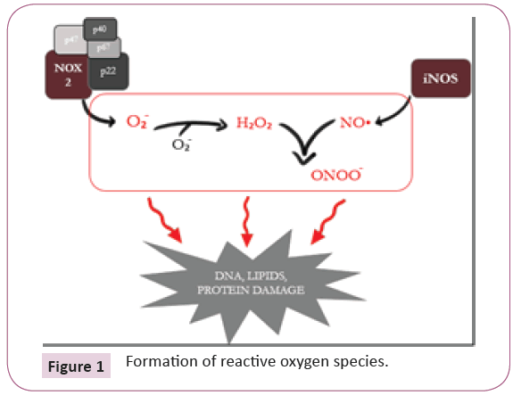
Figure 1: Formation of reactive oxygen species.
Oxidative stress has many endogenous and exogenous sources. One of the lasts, smoking or tobacco usage in all its form can cause oxidative stress. Water pipe, also known as shisha, involves the passage of charcoal-heated air through perforated aluminum foil which covers the tobacco placed in the head in order to generate smoke [11].
Waterpipe smoke is harmful. It causes impaired cellular growth and repair, inflammation, impaired vasodilation and oxidative stress [12]. The formation of free radicals, superoxide or nitric oxide result in lung oxidant toxicity through subsequent chain reactions resulting in uncontrolled destructive oxidation [13]. Hence, the damaging effects on cell function in lung epithelial cells and vascular endothelial cells are the two main factors in the development of chronic obstructive pulmonary disease (COPD), which is one of many respiratory diseases caused by tobacco smoke. Furthermore, oxidative stress and all these factors lead to cardiovascular diseases, including severe stenosis and ischemic heart disease. It affects also the heart rate variability, predictor of coronary artery disease. Besides, long history of waterpipe smoking increases the risk of having cardiovascular diseases [14].
Waterpipe smoking can also be a factor that increases the chance of having cancer due to genotoxic and clastogenic components in the water pipe smoke such as tar and polycyclic aromatic hydrocarbons [15].
However, the water pipe smoke effect on buccal cells has not been investigated yet, knowing that this type of cells represent an accessible pool of epithelial cells constituting a high diagnostic tool to evaluate the oxidative stress levels [16]. Besides, buccal cells collection is noninvasive and could be useful to provide information on host DNA, mRNA and proteins. In fact, these methods could be less time-consuming, and avoid the many limiting factors that could be faced such as inaccessibility and insufficiency of tissues. In addition, this collection method is practical compared to other methods since it does not need special expertise. Thus, buccal cells collection could be useful to investigate the molecular and cellular basis of many diseases [17].
The MAWI i-SWAB tubes Protein kit is a new buccal cells collection methods that allows the collection and stabilization of proteins from the point of collection. They enable the mechanistic release of buccal cells in a specific lysis and stabilization buffer. These tubes are suitable for any population and for any age. They allow self-collection, less time-consuming and facilitates sample concentration, transportation, extraction, and long term storage at room temperature [18].
The aim of our study was to evaluate the effect of water pipe smoke on oxidative stress levels in buccal cells, using MAWI i-SWAB tubes for the detection of different protein oxidation biomarkers (S-glutathionylation, S-nitrosylation and tyrosine nitration). Furthermore, these tubes were used for the revelation of iNOS and NOX2.
Material and Methods
Study population
Buccal cells samples were collected from ten non-shisha and heavy shisha smokers, aged between 20 and 50 years. Participants were also asked to fill a questionnaire after signing the informed consent, which provided us with more information about their age, work, health status, shisha or cigarette smoking and more.
Sample collection
Members were asked to rinse their mouth twice with water and wait thirty minutes before applying swabbing. They had to rotate a cotton swab twenty times on both cheeks, and repeat the action twice. Swabs were then released into a 1 mL iSWABTM Protein (iSWAB-P-1200) tube for sample collection.
Sample processing
Samples were kept overnight at room temperature before extraction. They were centrifuged at 50.3 g for one minute at 23°C. Supernatants were then removed for protein dosage by Bradford Assay (following Sigma Aldrich protocol, Catalog Number B6916). After the preparation of a standard curve starting from [BSA]=2 mg/mL, absorbance of the samples was read by spectrophotometer at 595 nm. Each tube was assayed in duplicates.
Laemmli reducing buffer (BioRAD) containing Tris-HCl, β-mercaptoethanol, SDS, glycerol, and bromophenol blue was then added to the samples. Then samples were boiled for three minutes before performing western blot.
Western blot
After three minutes of boiling, 15 μg of each sample were loaded in the 12% polyacrylamide gel’s wells, and then run at 80- 160 volts for two hours. Afterwards, gels were transferred to a nitrocellulose membrane (BioRad). Next, the membranes were stained with Ponceau Red for twenty minutes and then visualized by versa Doc imaging system (Bio-RAD).
Later, nitrocellulose membranes were blocked with 5% BSA (affymetrix) and incubated at 4°C overnight.
The day after, membranes were washed three times for five minutes with PBS 0.05% Tween 20 and incubated for 2 hours with primary antibody with gentle shaking at room temperature. Primary antibodies were: mouse monoclonal anti-nitrotyrosine (Abcam ab7048), mouse monoclonal anti-S-nitrosocysteine (Abcam ab94930), rabbit polyclonal anti-iNOS (Cell Signaling 2982), mouse monoclonal anti-glutathione (Abcam ab19534), rabbit polyclonal anti–NOX2 (Abcam ab31092) and anti-GAPDH.
Anti-glutathione gels were performed under non reducing conditions (Laemmli buffer without β-mercaptoethanol and boiling) and were then blocked with 5% milk and not BSA.
Anti-iNOS membranes were incubated overnight with the diluted antibody in 5% BSA, 1X PBS and 0.1% tween-20 at 4°C overnight.
After incubation with primary antibody, membranes were then washed three times with PBST for five minutes and incubated again with secondary antibodies for one hour at room temperature. The secondary antibodies were: HRP-conjugated rabbit anti-mouse polyclonal antibody (Abcam ab97046) and HRP-conjugated goat anti-rabbit antibodies.
TMB (Sigma Aldrich) was added to the membranes to be then visualized by Versa DOC imaging system.
Molecular weights of the bands obtained from the samples were compared to a broad range MW ladder (BioRAD, 161-0317) and to a pre-stained protein ladder-broad molecular weight (ab116028).
Results
The presence of the different protein oxidation biomarkers in nonsmokers and smokers samples were detected by western blots.
In Figure 2 bands of molecular weights ranging mainly between 30 KDa and 60 KDa revealed the presence of tyrosine nitration in buccal cells of non - shisha and heavy shisha smokers.
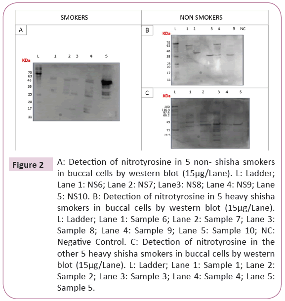
Figure 2: A: Detection of nitrotyrosine in 5 non- shisha smokers in buccal cells by western blot (15μg/Lane). L: Ladder; Lane 1: NS6; Lane 2: NS7; Lane3: NS8; Lane 4: NS9; Lane 5: NS10. B: Detection of nitrotyrosine in 5 heavy shisha smokers in buccal cells by western blot (15μg/Lane). L: Ladder; Lane 1: Sample 6; Lane 2: Sample 7; Lane 3: Sample 8; Lane 4: Sample 9; Lane 5: Sample 10; NC: Negative Control. C: Detection of nitrotyrosine in the other 5 heavy shisha smokers in buccal cells by western blot (15μg/Lane). L: Ladder; Lane 1: Sample 1; Lane 2: Sample 2; Lane 3: Sample 3; Lane 4: Sample 4; Lane 5: Sample 5.
In Figure 3, bands of molecular weights mainly between 30 KDa and 50 KDa revealed the presence of S-nitrosylated proteins in buccal cells in non-smokers and heavy shisha smokers samples.
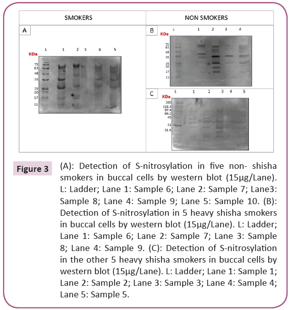
Figure 3: (A): Detection of S-nitrosylation in five non- shisha smokers in buccal cells by western blot (15μg/Lane). L: Ladder; Lane 1: Sample 6; Lane 2: Sample 7; Lane3: Sample 8; Lane 4: Sample 9; Lane 5: Sample 10. (B): Detection of S-nitrosylation in 5 heavy shisha smokers in buccal cells by western blot (15μg/Lane). L: Ladder; Lane 1: Sample 6; Lane 2: Sample 7; Lane 3: Sample 8; Lane 4: Sample 9. (C): Detection of S-nitrosylation in the other 5 heavy shisha smokers in buccal cells by western blot (15μg/Lane). L: Ladder; Lane 1: Sample 1; Lane 2: Sample 2; Lane 3: Sample 3; Lane 4: Sample 4; Lane 5: Sample 5.
In Figure 4, bands at variable molecular weights revealed the presence of S-glutathionylated proteins in all samples.
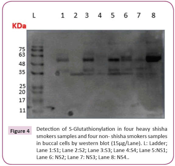
Figure 4: Detection of S-Glutathionylation in four heavy shisha smokers samples and four non- shisha smokers samples in buccal cells by western blot (15μg/Lane). L: Ladder; Lane 1:S1; Lane 2:S2; Lane 3:S3; Lane 4:S4; Lane 5:NS1; Lane 6: NS2; Lane 7: NS3; Lane 8: NS4.
In Figure 5, the anti-gp91phox antibody recognized proteins at 55 KDa, and bands had almost same intensities in all non-shisha and heavy shisha smokers samples.
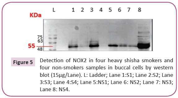
Figure 5: Detection of NOX2 in four heavy shisha smokers and four non-smokers samples in buccal cells by western blot (15μg/Lane). L: Ladder; Lane 1:S1; Lane 2:S2; Lane 3:S3; Lane 4:S4; Lane 5:NS1; Lane 6: NS2; Lane 7: NS3; Lane 8: NS4.
Figure 6 shows the GAPDH band at 37 KDa. GAPDH is one of the many housekeeping proteins. It was used as an internal control for Western blot analysis to normalize different proteins expressions [19] in order to compare the levels of protein oxidation biomarkers in non- smokers and heavy shisha smokers.
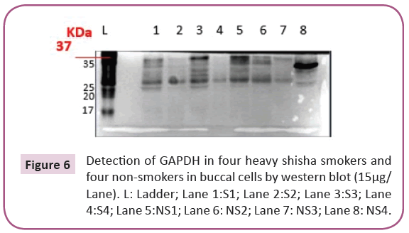
Figure 6: Detection of GAPDH in four heavy shisha smokers and four non-smokers in buccal cells by western blot (15μg/Lane). L: Ladder; Lane 1:S1; Lane 2:S2; Lane 3:S3; Lane 4:S4; Lane 5:NS1; Lane 6: NS2; Lane 7: NS3; Lane 8: NS4.
In Figure 7, the presence of iNOS at the level of 135 KDa was revealed in buccal cells of non-shisha and heavy shisha smokers which are almost equally expressed.
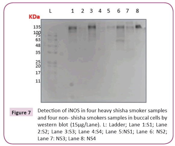
Figure 7: Detection of iNOS in four heavy shisha smoker samples and four non- shisha smokers samples in buccal cells by western blot (15μg/Lane). L: Ladder; Lane 1:S1; Lane 2:S2; Lane 3:S3; Lane 4:S4; Lane 5:NS1; Lane 6: NS2; Lane 7: NS3; Lane 8: NS4
Figure 8 shows the bands of GAPDH of 37 KDa in non-shisha smokers and heavy shisha smokers samples.
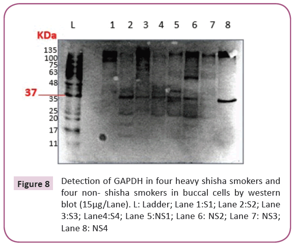
Figure 8: Detection of GAPDH in four heavy shisha smokers and four non- shisha smokers in buccal cells by western blot (15μg/Lane). L: Ladder; Lane 1:S1; Lane 2:S2; Lane 3:S3; Lane4:S4; Lane 5:NS1; Lane 6: NS2; Lane 7: NS3; Lane 8: NS4
Discussion
The overall difference between the intensity of the bands in Figures 1 and 2 is clear and shows a higher amount of tyrosine nitration in shisha smokers (Figure 1) compared to non-shisha smokers (Figure 2). However, housekeeping gene quantification such as GAPDH will be needed as internal loading control for further experiments.
Concerning the S-nitrosylated proteins obtained, our finding correlates with the previous ones of Bassil et al about the ability of MAWI tubes to preserve the protein oxidation biomarkers of nitric oxide and peroxynitrite: S-nitrosylation and tyrosine nitration [20]. The variation of the bands intensities could be correlated to the lifestyle and health condition of the participant.
It is important to note here that the appearance of oxidative stress biomarkers in non-shisha smokers is normal, because reactive oxygen species are always present. The damaging state is when these species overcome the antioxidant defense system [2].
The results of S-glutathionylation (Figure 4) show that the MAWI tubes can also be used for the detection of the protein oxidation biomarker of hydrogen peroxide in buccal cells: S-glutathionylation [6]. Thus, our results refer to the presence of hydrogen peroxide in both non-shisha and heavy shisha smoker samples. In addition to Bassil et al. MAWI protein tubes can also detect the presence of protein oxidation biomarker hydrogen peroxide “S-glutathionylation” in human buccal epithelial cells [20].
The bands of 91 KDa represent the mature form of NOX 2 (glycosylated) and those of 55 KDa represents the immature form of NOX 2 (unglycosylated) [21]. These results show that NOX 2 in all samples are immature (Figure 8), which means that in all non-shisha and heavy shisha smokers samples, NOX 2 is expressed in its immature form.
The first step of NOX maturation is the glycosylation on arginine residues in the endoplasmic reticulum ER, before passing by the Golgi apparatus to finally attend the trans-membrane [22]. Previous findings have shown that smoking and alcohol consumption induce cell death and ER stress in human lung epithelial cells and in pancreatic acinar cells [23]. ER stress induces dysfunction of the ER and disturbs its normal functions as folding, disulfide introduction and N-glycolysation and basically will not be able to glycosylate protein as NOX2 [24]. Based on that, these results could be correlated with our findings of unglycosylated NOX2 since all participants are alcohol consumers, heavy shisha smokers, and passive smokers.
In addition, our findings showed for the first time the presence of immature NOX2 in human buccal epithelial cells.
The comparison of iNOS expression Figure 8 was unclear due to many factors. It could be because of the lifestyle of participants, since buccal cells are exposed to food and other agents that could make the comparison inaccurate, as well as patient’s health status health status can highly affect the results [20]. Slomiany et al. findings about the increase of iNOS in buccal epithelial cells of alcohol consumers showed a positive correlation with our findings since most of the participants (non-shisha and heavy shisha smokers) were alcohol consumers [25].
Moreover, the small number of samples could also be a factor.
However, the groups of Hoyt have found that smoking decreases the expression of iNOS in lung epithelial cells by decreasing its mRNA transcription [26].
The difference between our results and previous results could be due to the different localization of epithelial cells.
In addition, buccal epithelial cells have a higher proliferation rate than other keratinized cells [27]. That might also be a difference factor between results, since the exposure of smoking might not be optimal due to the high proliferation rate.
Conclusion
In conclusion, our results confirmed previous findings about the ability of MAWI tubes to detect the protein oxidation biomarkers S-nitrosylation and tyrosine nitration in buccal cell. As well as that, it showed that these tubes are able to detect S-glutathionylation (presence of hydrogen peroxide) found in buccal cells of the non-shisha and heavy shisha smokers. Our findings also included the presence of iNOS protein expression equally in non-shisha and heavy shisha smokers in epithelial buccal cells. Finally, our results showed for the first time the presence of the immature NOX2 in buccal epithelial cells equally in smokers and non-smokers and was also detectable by the same tubes.
For more significant results, our future research will include first of all the increase of our sample size. We will need also to confirm the immaturity of NOX2 by more advanced molecular biology tools in human epithelial buccal cells and the process behind this immaturity.
Author’s Contribution
All authors participated in the analyses of the study and reviewed each drafts of the result article. MB and JM searched the databases and wrote the article. JAA contributed in experimental work. JM was responsible of sample collection.
Acknowledgments
I would like to acknowledge my supervisor Dr. Marcel Bassil for his continuous efforts, his professionalism and his encouragement all along the study. I would also like to emphasize on the fundamental role of the staff members of the Biotechnology Department at Benta Pharmaceutical Industries, especially Mrs. Joelle Abi Azar, who helped me use all required equipment and the necessary material to complete my study. My sincere thanks goes also to the donors and participants, without them it wouldn’t be possible to accomplish this research.
References
- Shintani H (2013) Determination of Xanthine Oxidase. Pharm Anal Acta 1: 1-27.
- Phaniendra A, Jestadi DB, Periyasamy L (2015) Free Radicals: Properties, Sources, Targets, and Their Implication in Various Diseases. Indian J Clin Biochem 30: 11-26.
- Halliwell B (2006) Free Radicals in Biology and Medicine 141: 312-322.
- Rada B, Leto TL (2008) Oxidative Innate Immune Defenses by Nox/Duox Family NADPH Oxidases, in Trends in Innate Immunity. Basel: KARGER 15: 164-187.
- Kiley PJ, Storz G (2004) Exploiting Thiol Modifications. PLoS Biol 2: e400.
- Grek CL, Reyes L, Townsend DM, Tew KD (2014) S-glutathionylation of buccal cell proteins as biomarkers of exposure to hydrogen peroxide. BBA Clin 2: 31-39.
- Jerca L, Jerca O, Manca? G, Constantinescu I, Lupu?oru R (2002) Mechanism of Action and Biochemical Effects of Nitric Oxide. J Prev Med 10: 35-45.
- Forstermann U, Sessa WC (2012) Nitric oxide synthases: Regulation and function. Eur Heart J 33: 829-837.
- Ahmad R, Ahsan H (2011) Contribution of peroxynitrite, a reactive nitrogen species, in the pathogenesis of autoimmunity. Autoimmune Disord 8: 1-18.
- Hidalgo C, Bull R, Behrens MI, Donoso P (2004) Redox regulation of RyR-mediated Ca2+ release in muscle and neurons. Biol Res 37: 539-552
- A.-N. Kb.-H. Ka.-A. A, E. Al (2007) Water-pipe (shisha) smoking influences total antioxidant capacity and oxidative stress of healthy Saudi males. J Food Agric Environ 5: 3-4.
- Rammah M, Dandachi F, Salman R, Shihadeh A, El-Sabban M (2013) In vitro effects of waterpipe smoke condensate on endothelial cell function: A potential risk factor for vascular disease. Toxicol Lett 219: 133-142.
- Kluchova Z, Tká?ová R (2006) The role of oxidative stress in lung injury induced by cigarette smoke. Sect Cell Mol Biol 6: 643-650.
- Jawad M, Ali M (2017) Health Effects of Waterpipe Tobacco Use: Getting the Public Health Message Just Right. Tob Use Insights 10.
- Waziry R, Jawad M, Ballout RA, Al Akel M, Akl EA (2017) The effects of waterpipe tobacco smoking on health outcomes: An updated systematic review and meta-analysis. Int J Epidemiol 46: 32-43.
- DeCoursey TE, Ligeti E (2005) Regulation and termination of NADPH oxidase activity. Cell Mol Life Sci 62: 19-20.
- Michalczyk A, Varigos G, Smith L, Ackland ML (2004) Fresh and cultured buccal cells as a source of mRNA and protein for molecular analysis. Biotechniques 37: 262-266.
- https://www.mawidna.com/the-mawi-story/.
- Nie X, Li C, Hu S, Xue F, Kang YJ, et al. (2017) An appropriate loading control for western blot analysis in animal models of myocardial ischemic infarction. Biochem Biophys Reports 12: 108-113.
- Bassil M, Atat AA, Ibragim M (2018) Detection of Oxidative Stress in Buccal Cells using iSWAB Tubes. J Mol Biomark Diagn 9: 387.
- Wang C (2013) Metabolic syndrome-induced tubulointerstitial injury: Role of oxidative stress and preventive effects of acetaminophen. Free Radic Biol Med 65: 1417-1426.
- Miyano K, Sumimoto H, N-linked glycosylation of the superoxide-producing NADPH oxidase Nox1. Biochem Biophys Res Commun 443: 1060-1065.
- Lugea A (2017) The Combination of Alcohol and Cigarette Smoke Induces Endoplasmic Reticulum Stress and Cell Death in Pancreatic Acinar Cells. Gastroenterology 153: 1674-1686.
- Laurindo FRM, Araujo TLC, Abrahão TB (2014) Nox NADPH oxidases and the endoplasmic reticulum. Antioxid Redox Signal 20: 2755-2775.
- Slomiany BL, Piotrowski J, Piotrowski E, Slominay A (1998) Activation of apoptotic caspase-3 and NOS2 in buccal mucosa with chronic alcohol ingestion. J Biochem Mol Biol Int 45: 1199-1209.
- Hoyt JC (2003) Cigarette Smoke Decreases Inducible Nitric Oxide Synthase In Lung Epithelial Cells. Exp Lung Res 29: 17-28.
- Rowat JS, Squier CA (1986) Rates of Epithelial Cell Proliferation in the Oral Mucosa and Skin of the Tamarin Monkey (Saguinus fuscicollis). J Dent Res 65: 1326-1331.

