Keywords
Stem cells; Dental cell implants; Enamel matrix derivatives; Tooth regeneration; Enamel matrix derivatives; Adhesion factors and biomaterials
Abbreviations
PDL: Periodontal Ligament; PDLSCs: Periodontal Ligament Stem Cells; EMD: Enamel Matrix Derivative; DPSC: Dental Pulp Stromal Cells; DMCs: Dermal Multipotent Cells; MSC: Mesenchymal Stem Cells; FBS: Fetal Bovine Serum; MDO: Mandibular Distraction in Osteogenesis; MSCT: Mesenchymal Stem Cell Transplantations; DBM: Demineralized Bone Matrix; DFDBA: Human Demineralized Freeze-Dried Bone Allograft; BMP: Bone Morphogenetic Proteins; BMMSCs: Bone Marrow Mesenchymal Stem Cells
Introduction
Periodontal diseases and tooth decay and enamel destruction including periodontal ligament (PDL), cementum, and bone are a major causes of tooth loss in adults. This is a major public-health problem in pediatric and adults groups worldwide. PDL is a specialized connective tissue that connects cementum and alveolar bone to maintain and support teeth in situ and preserve tissue homoeostasis. Recent advancements in implantation technology of dentally derived stem cells or human PDL stem cells, led to significant progress in the field of tooth regeneration [1]. PDLSCs in defined culture conditions differentiate into cementoblasts, adipocytes, and collagenforming cells. These cells when transplanted generate a cementum/PDL-like structure that contribute periodontal tissue repair. Today many advanced technologies are in use to support tooth enamel development and regeneration in vitro as well as in vivo. For successful tooth regeneration cultured stem cells, cementing adhesives and biological scaffold materials are used. More often, for transplantation purpose, stem cells are derived from an easily accessible tissue source and expanded ex vivo. These stem cell types are promising therapeutic tools which are used for reconstruction of enamel tissues that are destroyed due to periodontal diseases. In last three decades tissue engineering has emerged as a promising alternative approach to find solutions and clinical treatments for restoration of soft tissue defects mainly related to enamel and dental pulp. There have been made numerous rapid and exciting developments in tissue engineering technology which successfully regenerate and form a fully functional tooth in animal models, from a bioengineered tooth germ cell. In these methods bone-forming stem cells, new osteoinductive biomaterials, and growth factors are used. For growth and development of tissue engineered tooth, stem cells are implanted into different tissues that need suitable growing environment in vivo [2] (Figure 1). These have revolutionized the toot regeneration therapy and assisted the clinicians and enable them to conduct successful clinical trials for finding appropriate solutions of decayed and defective tooth [3] (Figure 2). Guided tissue regeneration is used for reconstructive osseous surgery [4].
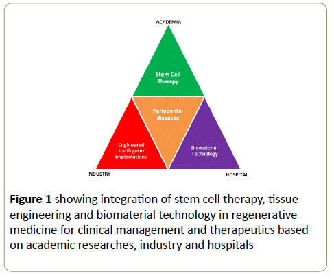
Figure 1: showing integration of stem cell therapy, tissue engineering and biomaterial technology in regenerative medicine for clinical management and therapeutics based on academic researches, industry and hospitals
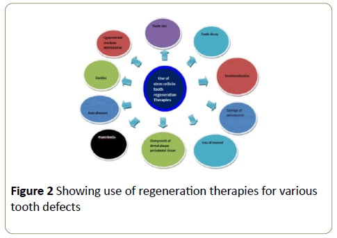
Figure 2: Showing use of regeneration therapies for various tooth defects
For development of bioengineered tooth important growth factors, stable and durable biodegradable polymer scaffolds materials are highly needful [5]. For framing, growth and regeneration of enamel and pulp tissue, cementum/ periodontal-ligament complex formation is highly essential [6]. This is also cooperatively regulated by the epithelial ameloblasts and mesenchymal odontoblasts [7]. For successful regeneration of tooth, formation of dentin complexes, removal of demineralized matrix, induction of enamel, vascularization of tooth need maintenance of microenvironment for responsiveness to cells and factors [8,9] (Figure 2). Similarly for successful replacement of tooth and cell implantation adhesion of cells to periodontal connective tissue amelogenin, bone sialoprotein and vimentin proteins is also important [10]. Similarly, enamel matrix derivative (EMD) also influence activities of cementoblasts and osteoblasts, which may able to regulate cell activities at a periodontal regenerative site. EMD and the type of cell populations present in the implant wound-healing environment may alter the implant-connective tissue interface [11] (Table 1). In addition, subrenal capsule implantation is used as a new alternative method for tissue-engineering in vivo [2].Moreover, tooth loss due to periodontal disease, dental caries, trauma, or a variety of genetic disorders continues to affect most adults adversely at some stage in their lives.
Hence, they essentially need a biological tooth substitute that could replace lost teeth and may provide a vital alternative to currently available clinical treatments. For this purpose dissociated porcine third molar tooth buds are used to make single-cell suspensions and seeded onto biodegradable polymers. The recent technology upholds the slow but highly effective regenerative therapy for successful treatment and replacement of tooth in pain. For tooth regeneration and healing of traumatic injuries niche factors, growth factors, and implantation of stem cells, are most essential requirements [12]. No doubt tooth bioengineering promises to be at the forefront of the next generation of dental treatments [3] (Table 1 and Figure 2).
More specifically, mammalian tooth root development is a long-term process during which root elongates along the apical direction and is accompanied with the formation of periodontium. In heterogeneous apical region of developing root as a functional entity, a developing apical complex is formed the root [13]. The developing apical complex shows sustainable development ability and functions as a growth center of tooth root. This is promising source of cells for tooth root and periodontal regeneration [13]. Similarly, periodontal ligament stem cells are considered as one of the best candidates for periodontal regeneration therapy [14]. Meanwhile, PDLSC cell pellet may be a promising alternative to promote periodontal defect repair for future clinical applications [15]. Still, there are limitations of conventional regeneration modalities which underscore the necessity of recapitulating development for periodontal tissue engineering [15]. No doubt stem cell derived cell types show enormous regenerative capacity but how these cells act in a inflammatory or toxic microenvironment. There is another question how stem cell origin affects optimal differentiation and regeneration is a major question [8].
Periodontal tissue engineering requires suitable biocompatible scaffold, growth factors, regenerative cells and instructional molecules. For clinical purposes an enamel matrix protein (EMP) is used to aid in hard tissue formation by postnatal mesenchymal stromal cells (MSCs) including bone marrow stromal cells (BMSCs) and periodontal ligament fibroblasts (PDLFs) [16]. Similarly, Straumann Bone Ceramic coated with Straumann Emdogain impose significant stimulatory effect in the commitment of mesenchymal cells to osteogenic differentiation in vitro while Emdogain inhibited AP activity and appeared not to induce ectopic bone formation. EMPs truly possess the capacity to induce the regeneration of bone or other components of the periodontium but it remains to be established. Similarly, new methods for restoration of crown, root, pulp, enamel, dentin, odontoblast, cementum, blood vessels, and periodontal ligaments to improve the indiscriminate shape of tooth are also highly needful [16]. In addition, tissue response after implantation determines the success of the healing process which is not only dependent on the chemical properties of the implant surface but also determines by the surface topography or roughness [17]. For better regeneration inducers of molecular pathways that drive tooth morphogenesis and enamel secretion are also needed to generate teeth from organ cultures for therapeutic implantation [18]. However, p38α MAPK is required for tooth morphogenesis and enamel secretion [19]. Similarly, CCN proteins and cell-associated molecules are also involved in several developmental processes are also needed [20]. Further, to induce complete tooth formation, in condition of tooth loss due to trauma or diseases, implantation of stem cells, morphogens and growth factors are needed to stimulate entire tooth development and progression [6] (Table 1). Epithelial ameloblasts in combination to mesenchymal odontoblasts cooperatively regulate tooth development and secrete enamel matrix, which is highly critical for enamel formation [7]. Formation of enamel knots is regulated by a cascade of gene activity where Fgf4, Shh, BMP4, Lef1 and p21 are the prime movers of the processes. Homeobox genes (Msx, Dlx) are the orchestrators of the framing and a series of proteins (adhesion molecules, extracellular matrix components) are the executors of tooth framing [6]. Formation of cementum/periodontal-ligament complex is an important field of tissue engineering (Table 1).
Certain questions, were raised i.e. how does enamel matrix derivatives (EMD) influence activities of cementoblasts and osteoblasts, and regulate cellular activities at a periodontal regenerative site. In addition, use of human periodontal ligament stem cells, mesenchymal stem cell (MSC) for tissue repairing and regeneration of periodontal tissues is highlighted [15,21]. Similarly, many thoughtful views have been given about use of broad spectrum of human cell types and tissues for transplantation purposes mainly in drug discovery and studying tooth disease mechanisms [22]. Moreover, for skeletal and muscular healing [23] dental pulp (DP) stem cells are used with plastic adherence and specific surface antigen. These cells show multipotent differentiation potential and provide faster healing of tooth enamel [24] (Table 1). These dental pulp stromal cells (DPSC) are considered to be a promising source of stem cells which are extensively used in regenerative therapy [25] (Figure 3) while PAFSCs are a distinctive cell population and are used as promising candidate for bio-root engineering [26].
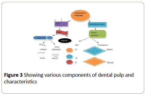
Figure 3: Showing various components of dental pulp and characteristics
Tooth Generation and Framing
Odontogenesis is a complex process in which a series of epithelial-mesenchymal interactions and odontogenic molecular cascades work together. For induction of regeneration after stem cell transplantation platelet-rich fibrin (PRF), growth factors and cytokines are mainly used [27]. In autogenic cell transplantation DBCs are seeded into fibrin glue- PRF could regenerate a complete tooth [27]. Orthodontic stem cell transplantation [28] needs cultured stem cells for periodontal regeneration [29]. Though, a donor is essential in each case and tooth extraction is required to obtain the periodontal ligament-derived cell. Transplantation of tissuecultured teeth decreased epithelial down growth and increased connective tissue attachment on the root-planed surface. The implantation of cultured dental cell reassociations allow for reproduction of complete functional differentiation at the cell, matrix, and mineral levels. In replantation of the tooth, the internal basal lamina kept at the surface of the enamel of the replanted tooth, that starts regeneration with junctional epithelium and form an attachment apparatus at the epithelium-tooth interface [30]. This new strategy might open new options to reconstruct extended periodontal defects and complete regeneration of the periodontium [31] (Figure 4). Similar experiments are also possible with muscle regeneration satellite cells, the skeletal muscle stem cells, become activated after trauma, proliferate, and migrate to the site of injury. These activated satellite cells form new multinucleated myofibers fuse to damaged myofibers [12] (Table 1).
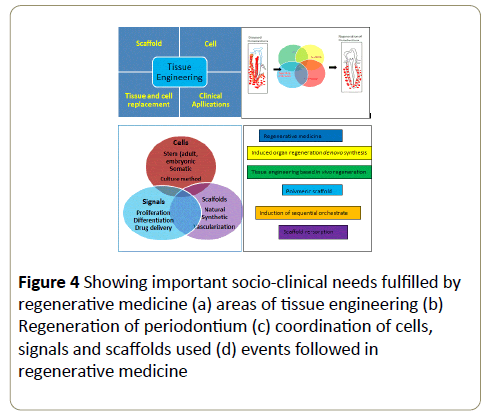
Figure 4: Showing important socio-clinical needs fulfilled by regenerative medicine (a) areas of tissue engineering (b) Regeneration of periodontium (c) coordination of cells, signals and scaffolds used (d) events followed in regenerative medicine
How to Maintain Microenvironment for Periodontal Development
In order to design predictable periodontal regenerative therapies, it is important to understand the responsiveness of cells within the local environment to factors used as important candidates [9] (Figure 5). This specific microenvironment of the satellite cells, acts as a functional niche that controls their behavior. The niche contains several components that maintain satellite cells quiescence until they are activated. In addition, a great diversity of stimulatory and inhibitory growth factors such as IGF-1 and TGF-beta1 regulate their activity [12]. Restoration of three-dimensional (3D) microenvironment of periodontal development elicits the intrinsic capacity of mesenchymal stem cells to proceed a redevelopment- like program that help cells to rapidly divide [31]. Similar to periodontal ligament stem cells (PDLSCs) in periodontal regeneration, bone marrow mesenchymal stem cells (BMMSCs) are very likely another cell source of physiological repair of periodontal tissues. Selfassembly approach of stem cells a physiological phenomenon occurs during organogenesis that enhance complete reconstruction of functional complex periodontium-organ systems. However, for filling the periodontal defects monodispersed cells are allowed to self-assemble into a microtissue such as a 3D spheroid. In addition, bilayered cell pellet constructs comprising calcified boneforming cell pellets (i.e. BMMSCs) and cementum/PDLforming cell pellets (i.e. PDLSCs) are to be fabricated in vitro in a tissue-mimicking way. This novel strategy could be used for complete regeneration of the periodontium to reconstruct extended periodontal defects [31] (Figure 5 and Table 1).
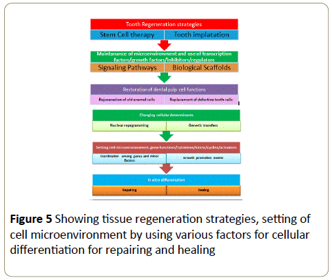
Figure 5: Showing tissue regeneration strategies, setting of cell microenvironment by using various factors for cellular differentiation for repairing and healing
The Induction of Enamel and Dentin Complexes
The combination of bone replacement graft materials is used for treatment of periodontal osseous defects. Bone morphogenetic proteins (BMPs) produced by Hertwig’s epithelial root sheath or present in enamel matrix derivatives (EMD) seem to be involved in the control of dental follicle (DF) cell differentiation, but their precise function remains largely unknown. The DF surrounding the developing tooth germ is an ectomesenchymal tissue composed of various cell populations derived from the cranial neural crest. These dental follicle cells (HDFC) are believed to contain precursor cells for cementoblasts, periodontal ligament cells, and osteoblasts. Similarly, both EMD combined with a bovine-derived xenograft (BDX) are used for treatment of intraosseous defects in patients with periodontitis. For transplantation of human tooth germ components, heterologously recombined mouse dental epithelia and xenogenic graft tissue are used to make reconstructed transplants. Further, differentiation of mouse dental epithelia is restrained by putative suppressive factors derived from human dental papilla until they are separated by mineralized dentin layers that serve as a diffusion barrier. The mouse enamel organ nevertheless retains its own phenotypic characteristics and intrinsic timing of cell differentiation and function [32].
ds and enamel matrix derivative
Human demineralized freeze-dried bone allograft (DFDBA) and enamel matrix derivative (EMD) are used for treatment of bone and periodontal defects [33]. Tbx1 expression in epithelium requires mesenchyme-derived signals because dental mesenchyme induces expression of Tbx1 in recombined dental and non-dentalepithelia. Forced expression of Tbx1 in dental explants activates amelogenin expression. Thus, Tbx1 expression in developing teeth takes place under direct control of FGF signaling that correlates withs determination of the ameloblast lineage [34]. Sonic hedgehog (Shh), one of the essential molecules for embryogenesis and organogenesis, is strongly expressed in the enamel knot, which represents the signaling center for odontogenesis due to the presence of essential secretory molecules [35]. There are methods which are used for formation of adipose tissue in vitro and in vivo by using human adipose-derived stromal cells (ADSCs) utilizing a gelatin sponge (Gelform) as a scaffold. These tissue-engineered constructs are exposed to adipogenic differentiation medium for in vitro and implanted in the backs of severe combined immunodeficient (SCID) in animal models for in vivo adipose regeneration [36]. Enamel matrix derivative (EMD) influence activities of cementoblasts and osteoblasts, and thus may be able to regulate cell activities at a periodontal regenerative site (Table 1).
Biodegradable polymer scaffolds
Dissociated tooth tissues contain both dentin and enamel, show presence of epithelial and mesenchymal dental stem cells in porcine third molar tissues. For bio-engineering, complex tooth structures are obtained from pig tooth bud tissues which show potential for the regeneration of mammalian dental tissues. These cultured rat tooth bud cells are obtained from 3 to 7 day post-natal (dpn) rats and cellseeded biodegradable scaffolds are grown in the omenta of adult rat hosts for 12 wks, and then harvested. Thus, bioengineer tooth structures derived from cultured tooth bud cells dental epithelial and mesenchymal stem cells can be maintained in vitro in culture medium. Similarly, bone constructs are grown in vitro with use of isolated cells, biodegradable polymer scaffolds, and bioreactors [5]. Culturing bone marrow mesenchymal stem cells (BMSCs) on ceramic bovine bone scaffolds in different environments in vitro are used to induce proliferation, differentiation, and maturation of BMSCs [5]. Regeneration of extensive bone defects is done using stem cells mainly by injecting adipose derived stem cells and demineralized bone matrix (DBM) into areas of bone defect [8] (Table 1).
Vascularization of engineered teeth
The implantation of cultured dental cell-cell re-associations are allowed for the reproduction of fully formed teeth, crown morphogenesis, epithelial histogenesis. It also needs mineralized dentin and enamel deposition for rootperiodontium development. Vascularization is critical for organogenesis and tissue engineering generated tooth because it assists in blood vessel formation during tooth development. It is also essential for cell re-associations, in vivo implantation. Ex vivo, blood vessels are developed in the dental mesenchyme from the cap to bell stages and in the enamel organ, shortly before ameloblast differentiation. In cultured teeth and cell re-associations, blood-vessel-like structures are still remained in the peridental mesenchyme, but these never developed into dental tissues. After implantation, both teeth and re-associations get revascularized and these newly formed blood vessels originated from the host, are allowed for their survival, in affording conditions for fast organ growth, mineralization, and enamel secretion [37] (Table 1).
Factors Necessary for Periodontal Connective Tissue Attachment Formation on Dental Implants
There are important growth factors and adhesion molecules which are necessary for periodontal connective tissue attachment after formation of cellular associations on dental implants. TGF-beta, CCN proteins and cell-associated molecules, p38α MAPK are required for tooth morphogenesis and enamel secretion [19] (Figure 6). However for quick graft establishment and attachment amelogenin, bone sialoprotein and vimentin protein are used [10]. Recently, Cbfa1 was found to be a critical transcriptional regulator of osteoblast differentiation [38]. After periodontium transplantation of PL cells on wounded surface these cells secrete their own factors which do faster regeneration than ES cells, possibly because of PL cell plasticity and the capacity to undergo effective differentiation in the periodontal cellular microenvironment [39] (Figure 6). Besides, orthodontic tooth movement promotes the differentiation of transplanted cells, and the differentiation predominantly in the paravascular areas of the periodontium while in autogenous periodontal cell grafts, enamel matrix derivative (EMD), play important role on the implant-connective tissue interface. More specifically, implants that received GCT cell grafts were found surrounded by fibrous connective tissue. In contrast, implants that received PDL cells without the application of EMD demonstrated good bone contact, but strands of epithelium were observed in the implant-connective tissue interface (Table 1). Both amelogenins and soluble dentin proteins showed bone induction activity like bone morphogenetic protein and induce differentiation of mesenchymal cell into chondrocyte and osteocyte [40]. For dental root development conventional pediatric anticancer therapy is also used by using stem cell transplantation [41].
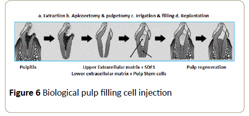
Figure 6: Biological pulp filling cell injection
Orthodontics and Stem Cell Transplantation
Implantation of cultured stem cells
Stem cells are used for finding cures for numerous diseases including skin wound healing through transplantation medicine. Implantations of cultured cells are applied for periodontal regeneration in which a donor is essential for tooth extraction and to obtain the periodontal ligament-derived cell. There are advanced regenerative techniques available combining tissue culture and transplantation of teeth [29]. Transplantation of tissuecultured teeth decreases epithelial down growth and increase connective tissue attachment on the root-planed surface. Furthermore, EMD could remarkably increase the new connective tissue attachment in this periodontal regenerative technique [29]. More specifically, implantation of cultured cellcell re-associations led to crown morphogenesis, epithelial histogenesis, organ vascularization, and root and periodontium development [37]. The implantation of cultured dental cell re-associations allow for reproduction of complete functional differentiation at the cell, matrix, and mineral levels. A specific microenvironment and beta-catenin signaling is required for embryonic tooth morphogenesis and promotion of continuous tooth development are embryonic stage [42] (Table 1 and Figure 7).
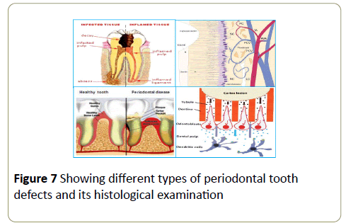
Figure 7: Showing different types of periodontal tooth defects and its histological examination
For regeneration of transplanted tooth the junctional epithelium attachment is highly essential [30]. The sizes of furcating defects and the coverage of gingival flap influence the outcome of the treatment using autogenous periodontal ligament cells with or without enamel matrix derivatives [43]. Atelocollagen membrane inhibits apical migration of regenerating epithelium and accelerates connective tissue reattachment in part by inhibiting the mitotic function of basal epithelial cells in early stages of wound healing [44]. However, implantation of bio-materials such as atelocollagen induce dentinogenesis of dental pulp tissue, but it also need basal growth factors to induce regeneration and utilization of biomaterials [45]. For determining success rate of implantation periodontal regeneration procedures are followed after finding connective tissue attachment level [46] (Table 1 and Figure 7). Mesenchymal stem cell (MSC)- mediated tissue regeneration methods are used for regeneration a bio-root and its associated periodontal tissues to restore tooth loss [21]. MSC increases bony regeneration in calvarial critical-size defects in rabbits, and provide a new promising therapeutic strategy to aid skeletal healing [23].
Bone marrow mesenchymal stem cells (MSCs) comprise a heterogeneous population of postnatal progenitor cells with profound immunomodulatory properties, such as upregulation of Foxp3 (+) regulatory T cells (Tregs) and downregulation of Th17 cells. These MSC subpopulations possess the range of immunomodulatory function [47]. Similarly, human umbilical cord-derived mesenchymal stem cells (UC-MSCs) are used to facilitate osteogenic differentiation in bone regeneration. These cells could be achieved by over expression of osterix (Osx) [48]. Due to their high proliferation property UC-MSCs could play important role in bone tissue engineering. More often, stem cells cultured in conditioned medium are used for transplantation purpose as a promising alternative to skin wound healing treatment [49] (Table 1). More specifically, for clinical management of soft tissues mixed population of bone marrow-derived autologous stem and progenitor cells are seeded onto β-tricalcium phosphate (β-TCP), which serve as a scaffold to deliver cells directly to the defect [50]. This is also used for vascularization, mineralization of bone tissues. Thus both bone marrow and adipose-derived stem cells were found are considered best choice for regeneration of the defect. These adipose-derived stem cells are easily accessible and abundantly proliferate into mesenchymal cells which form an effective bone regeneration material (Table 1).
Mesenchymal stem cell
Mesenchymal stem cell transplantations (MSCT) are used to treat human diseases and in orthodontics. These cells divide rapidly and differentiate within the injured tissue into specialized cells post transplantation [49]. Similarly, mesenchymal progenitor stem cells exist within the bone marrow stroma in form of subset of nonhematopoietic cells can expand ex vivo and induce either in vitro or in vivo. These cells terminally differentiate into osteoblasts, chondrocytes, adipocytes, tenocytes, myotubes, neural cells, and hematopoietic-supporting stroma. These mesenchymal progenitor cells require suitable microenvironment for cellular proliferation and differentiation with a wide range of growth factors, cytokines, chemokines, proteins and enzymes. Mesenchymal progenitors are used for reconstruction and regeneration of many organs and tissues [21]. These cells have many clinical applications in cell and gene therapies. For regeneration of various tissues, cryo-preserved MSCs (7 days in a -150°C deep freezer) are also used. After normalization in medium, cells these maintain high survival and proliferation rate and retain their adipogenic and osteogenic differentiation abilities in culture medium containing essential growth promoting factors [51] that could be obtained from the conditioned medium or spent media harvested from cultured cells (Table 1).
Bone regeneration through distraction osteogenesis (DO) is promising but remarkably slow. However, to accelerate bone regeneration, autologous mesenchymal stem cells are directly injected to the distraction site. In comparison to direct injection, scaffold-based method provides earlier cell delivery with potentially better controlled cell distribution and retention [52]. It is technically feasible and biologically sound to deliver autologous BM-MSCs to the distraction site immediately after osteotomy using a gelfoam scaffold to enhance mandibular DO (MDO) [52]. It is clear only a small percentage of transplanted cells integrate and survive in host tissues because of availability of trophic factors such as miR-29b levels and Fas. Fas deficiency causes failure of miR-29b release, thereby elevating intracellular miR-29b levels, and down regulates DNA methyltransferase 1 (Dnmt1) expression in MRL/lpr BMMSCs. This results in hypomethylation of the Notch1 promoter and activation of Notch signaling, in turn leading to impaired osteogenic differentiation [51]. It also causes MDO assisted by MSCT [53] (Table 1).
Embryonic stem cells
Embryonic stem cells possess unlimited self-renewal and differentiation capacity and show wider applications in biomedical research and regenerative medicine. Embryonic stem cell-associated antigens are expressed in a variety of adult stem cells as well as embryonic stem cells. Stage-specific embryonic antigen (SSEA)-4 are used to isolate dental pulp (DP) stem cells which show plastic adherence, specific surface antigen expression, and multipotent differentiation potential, similar to MSC. Embryonic stem (ES) cells, derived from the inner cell mass of mammalian blastocysts, show the ability to grow indefinitely while maintaining pluripotency [54,55]. Moreover, human ES cells are highly useful to understand disease mechanisms, to screen effective and safe drugs, and to treat patients of various diseases and injuries [56] but it is very difficult to generate patient- or disease-specific ES cells, which are required for their effective application [57]. SSEA-4+ DP cells possess osteogenic potential, and the SSEA-4+ clonal DP cells show multilineage differentiation potential toward osteoblasts, chondrocytes, and neurons in vitro [58]. More specifically, for removal of disturbances in dental development or impairment in children hematopoietic stem cell transplantation (HSCT) are done [24] (Table 1).
Dental pulp stromal cells (DPSC) are a promising source of stem cells for clinical regenerative therapy (Figure 3). A wider usage of DPSC used in transplantation requires largescale expansion in vitro to equalize the supply and demand of cell mass according to clinical requirement without compromising current good manufacturing practice. For DPSC cell culture fetal bovine serum (FBS) is used as a nutritional supplement, but it is undesirable additive to cells because it carries the risk of transmitting viral and prion diseases. Human platelet lysate (HPL) is also used as a substitute for FBS in a large-scale expansion of DPSC in a shorter time period under cGMP conditions [25]. Adult dental pulp cells (DPCs) are isolated from third molars have the capability to differentiate into keratocytes, cells of the corneal stoma (Figure 8). After inducing differentiation in vitro, DPCs expressed molecules characteristic of keratocytes, keratocan, and keratan sulfate proteoglycans at both the gene and the protein levels. DPCs cultured on aligned nanofiber substrates generate tissueengineered, corneal stromal-like constructs, recapitulating the tightly packed, aligned, parallel fibrillar collagen of native stromal tissue. Similarly, bone marrow-derived cells are known to contribute to wound healing, and are able to differentiate in many different tissue-specific cell types [59] (Table 1). The increasing clinical demand for bone substitutes appropriate cell source and three-dimensional (3D) scaffolds that support cell growth and enhance osteogenic potential are highly required [60]. Mainly for wound healing of defective tooth cell-scaffold constructs are implanted intraperitoneally in experimental animals [61]. However, for in vivo implantation of the HDPSC 3D 45S5 Bioglass scaffolds having a sporadic woven bone-like spicules and calcified tissue are implanted. These promote bone-like tissue formation in vitro and in vivo and were found promising candidate for clinical bone repair and regeneration [60] (Table 1).
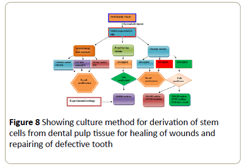
Figure 8: Showing culture method for derivation of stem cells from dental pulp tissue for healing of wounds and repairing of defective tooth
| Problem |
Orthodontic reason/defect |
Solution |
Reference |
| Erupted tooth |
Expressed cytokeratin 14, dentin matrix protein-1, vascular endothelial growth factor, and osteopontin |
DBC-fibrin glue-PRF composite was autografted back into the original alveolar sockets |
Yang, et al.[27] |
| Structural erosion or fragile tooth |
Induced morphological changes occur due to increased alkaline phosphatase (ALP) activity, runt-related transcription factor 2 (RUNX2), osteocalcin (OCN), and bone sialoprotein (BSP) expression in PDLSCs |
Correction of microRNAs (miRNAs) in human periodontal ligamentstem cells(PDLSCs |
Wei,et al.(2015) |
| Periodontal trauma |
Structural and functional integrity of the periodontium following periodontal trauma such as orthodontic tooth movement |
High mobility group box protein-1 (HMGB1) |
Wolf,et al.(2015) |
| Traumatic injuries |
Loss of structural/compositional sensitivity of enamel tissue |
A mixed population of bone marrow-derived autologousstemand progenitorcellsare seeded onto β-tricalcium phosphate (β-TCP) |
Rajan,et al.[50] |
| Osteoporosis |
Osteocalcium disorder, elemental loss and pulp and enamel destruction due to loss of calcium and phosphates |
FasL pathways mediated differentiation of ERK and GSK-3β-catenin pathway |
Ming, et al.(2014) |
| Osteoporosis |
intermittent mechanical strain (IMS) |
Promoted osteogenic differentiation of OVX BMSCs by activating Runt-related transcription factor 2 |
Zhang, et al.(2015) |
| Tumor progression and metastasis/ Tumor blood vessels |
Upregulation of vascular endothelial growth factor (VEGF) and VEGF receptor 2 |
Antitumor therapy, radioactive and laser based destruction |
Ohmura-Kakutani, et al.(2014) |
| Craniofacial problems |
Implantation of stem cells |
Use of cultured stem cells from source tissue and organ from whichstem cellscan be derived |
Mohanty, et al.(2015) |
| Fibrodysplasiaossificansprogressiva |
Low level of protein expression in receptor cells |
Bone morphogenetic protein (BMP) receptor ALK2, R206H are used |
Fujimoto,et al.(2014) |
| Bone and tooth pain |
Limited capacityto induce bone formation |
Restoration by using rhBMP-2, DFDBA and EMD |
Intini, et al.[33] |
| Demineralization |
embryonic tooth morphogenesis and promotes continuous tooth development |
beta-catenin signaling |
Liu, et al.(2010) |
| Faulty and defective tooth arrangment |
Organization of predentin/dentin,enamel, and cementum |
Mineralization afterimplantation |
Lechguer, et al.[37] |
| Hard tissue formation |
Post-natal mesenchymal stromalcells(MSCs) including bone marrow stromalcells(BMSCs) and periodontal ligament fibroblasts (PDLFs). |
Straumann Bone Ceramic coated with Straumann Emdogain |
Mrozik,et al.(16) |
| Loss of dental pulp and non clinical damage |
FGF molecules are able to maintain epithelial Tbx1 expression during odontogenesis,Expression of Tbx1 indentalepithelium of FGF receptor 2b(-/-) |
Mesenchyme-derived signals |
Mitsiadis, et al.[34] |
| Odontogenesis |
Forced expression of Gli1, a major transcription factor in Shh signaling |
Amelogenin and ameloblastin. |
Takahashi, et al.[35,57] |
| Odontogenesis |
Sonic hedgehog (Shh) inenamelknot |
Use of Sonic hedgehog (Shh) |
Takahashi, et al.[35,57] |
| Low osteogenic differentiation |
miR-26a potentially targeted on GSK3β and Smad1 to regulate Wnt and BMP signaling pathway |
MicroRNAs (miRNAs) used as important regulators ofstemcell |
Su, et al.(2015) |
| Orthodontic tooth movement (OTM) |
induces local inflammation in periodontium |
OTM induced a significant elevation of type 1 T helper cell (Th1) cytokines tumor necrosis factor-α (TNF-α) and interferon-γ (IFN-γ) around periodontal tissue in WT |
Yan, et al.(2015) |
| Untimely enamel loss |
Obstruction in muscle formation and decreasing scar tissue contraction |
Scaffold-freecellsand mesenchymalstem cellsscaffold |
Zhou, et al.(2015) |
| Loss of osteogenic capability |
Mitogen-activated protein kinase (MAPK) signaling pathways |
Use of BMMSCs and PBMSCs cells,regenerative medicine |
Zheng, et al.(63) |
| Enamel cell and dental pulp infection |
von Willebrand factor (vWF) and CD31 immunofluorescent staining,different energy densities of infrared LED on the cell viability |
Vascular endothelial growth factor (VEGF) and basic fibroblast growth factor (bFGF)are used for pulp and enamel repairing |
Feng, et al.(2015) |
| Fragiledeciduous teeth |
Enamel cell destruction and low calcium level |
Human exfoliated deciduous teeth (SHED) and adiposestem cells(ASC) |
Loo, et al.(2014) |
| Root cancer |
Inhibition of fucosylation may be used to block CSCs and metastatic spread. |
Oral squamous cell carcinoma (OSCC) |
Desiderio, et al.(2015) |
| Traumatic tooth and jaw injury |
Skeletal regenerative medicine,mesenchymalstem cells(MSC) |
Bone tissue engineering |
Asatrian, et al.(2015) |
| Large bone defects |
CACB/ADSCs compos |
Therapeutic potential of ERK signaling pathway |
Wei, et al.(2015) |
| Defects in growing bones |
Loss of majority of osteoblasts, Cxcl12 (chemokine (C-X-C motif) ligand 12)-abundant stromalcells and bone marrow stromal/mesenchymal progenitorcellsin postnatal life |
Implantation of periodontal human mesenchyme cells |
Ono, et al.(2014) |
| Bone breakage |
Osteoinductive signals are used as potent tool for bone regeneration,fabricated poly(L-lactic acid) (PLLA) electrospunnanofibers with random and aligned morphology immobilized with bone morphogenic protein-2 (BMP-2 |
Engineering bone tissue |
Perikamana, et al.(2015) |
| Skeletal deformities |
Mediates p53/miR-17/Smurf1 pathway |
Engineering bone tissue |
Liu, et al.(2015) |
| Delayed growth of deciduous tissues |
Kruppel-like factors (KLFs) are evolutionarily conserved zinc finger-containing transcription factors |
Differentiation, proliferation, embryogenesis and pluripotency. |
Ding, et al.(2015) |
| Tooth decay and damage |
Organic matrix mediated mineralization paves a way for formation of synthetic enamel |
Bone marrow-derived mesenchymalstemand stromalcells(MSCs) |
Jayasudha, et al.(2014) |
| Tooth decay |
Toll-like receptor 3, ankylosis-progressive homolog, decorin, osteocalcin, and runt-related transcription factor-2 |
Increasing cellular resistance and RCT |
Ramis, et al.(17) |
| Thermal hyper-sensitivity |
Impairs the development of the tooth root in lactational rats andalters the function of apical papilla-derivedstem cell |
Exposure to a continuous low dose of tetrachlorodibenzo-p-dioxin |
Guo,et al.(2015) |
Table 1: Different types of tooth related problems and solutions based on available technology and methods
Human pluripotent stem cells
Human pluripotent stem cells exhibit the essential characteristics of embryonic stem cells. These possess normal karyotypes, express telomerase activity, bear cell surface markers and genes that characterize human ES cells. These cells possess development potential to differentiate into advanced derivatives of all three primary germ layers and are proved highly useful in the production of new disease models for drug development, and transplantation medicine. Recently bone-resorbing osteoclasts were generated from human embryonic and induced pluripotent stem cells. Further, derivation of hematopoietic and osteoclast populations from human embryonic and induced pluripotent stem cells is invaluable for understanding embryonic bone development and postnatal bone disease [22]. Due to successful reprogramming of differentiated human somatic cells into a pluripotent state has made creation of patient- and diseasespecific stem cells. However, generation of induced pluripotent stem (iPS) cells is capable of germline transmission, from mouse somatic cells by transduction of four defined transcription factors. Similarly, generation of iPS cells from adult human dermal fibroblasts need Oct3/4, Klf4, and c-Myc factors. These cells were found similar to human ES cells in morphology, proliferation, surface antigens, gene expression, epigenetic status of pluripotent cell-specific genes, and telomerase activity. No doubt these cells could differentiate into various cell types in vitro from adult human fibroblasts (Table 1).
Periodontal ligament stem cells
Stem cell-based therapy represents a novel and more advantageous modality of treatment for tooth defect or loss. But it is true that tooth-derived stem cells are not readily accessible, instead dermal multipotent cells (DMCs) are easily available from skin tissue for odontogenic induction. However, by exposure to conditioned medium of embryonic and neonatal tooth germ cells in culture, the proliferation and mineralization activity of DMCs could be elevated, while the embryonic tooth germ cell-conditioned medium (ETGC-CM) produce more significant effects. DMCs are used as an alternative cell source for tooth regeneration and therapeutics [61]. Similarly, periodontal ligament stem cells (PDLSCs) are considered as potential MSC sources for clinical applications in periodontal regeneration therapy [62]. But there occurs a significant difference in proliferation and differentiation capacity of PDLSCs according to age. PDLSCs obtained from aged donors exhibit decreased proliferation and differentiation capacity when compared with those from young donors. Young PLC-CM show enhanced cell proliferation and differentiation capacity of PDLSCs than aged donors. There is another fact that aged PDLSCs induced by young PLC-CM showed enhanced tissue-regenerative capacity to produce cementum/periodontal ligament-like structures, whereas young PDLSCs induced by aged PLC-CM transplants form connective tissues. PDLSCs are modulated by the extrinsic microenvironment [63] but upon transplantation into immunocompromised mice, a regular aligned cementum/PDLlike complex is formed [15]. Moreover, the combination of apical tooth germ cell-conditioned medium and endogenous extracellular matrix could maximally mimic the microenvironment of root/periodontal tissue development and enhance the reconstruction of physiological architecture of a cementum/PDL-like complex in a tissue-mimicking way. This is the main reason that periodontal ligament stem cells (PDLSCs) are one of the best candidates for periodontal regeneration. Their function can be impaired in periodontitis microenvironment. PDLSCs are promising alternative to promote periodontal defect repair for future clinical applications in regenerative medicine and tissue engineering [15] (Figure 4). However, successful regeneration of periodontium, coordination of cells, signals and scaffolds is highly needful. Thus, dental follicle cells (DFCs), serving as precursor cells and mesenchymal stem cells show intimate association with PDLSCs. DFCs could provide a favorable microenvironment to improve the proliferation and differentiation capacity of PDLSCs from healthy subjects (HPDLSCs) and patients diagnosed with periodontitis (PPDLSCs) [14]. Dental follicle cells rescue the regenerative capacity of periodontal ligament stem cells in an inflammatory microenvironment.
Odontogenic epithelial cells from nonodontogenic
For successful regeneration there should be a clear identification of odontogenic and non odontogenic epithelial cells. Moreover, induction of dental epithelial cell differentiation marker gene expression in nonodontogenic HaCaT cells by TMSB4X is established [64] (Table 1). The upregulation of odontogenesis-related genes, such as runt-related transcription factor 2 (RUNX2), Amelogenin (AMELX), Ameloblastin (AMBN) and Enamelin (ENAM) is also required. Thymosin beta 4 (Tmsb4x) is closely related to the initiation and development of the tooth germ. Coculture of PBCD34+ cells and MSC increases bony regeneration and used as therapeutic strategy for skeletal healing [14] (Table 1). It is fact that recombination of cells liberated from developing tooth germs develop into teeth. Guided tissue regeneration is used for reconstructive osseous surgery [4]. It could regenerate alveolar bone in conjunction with the placement of titanium dental implants. Meanwhile complete osseointegration of an implant could be achieved by the placement of a Teflon membrane over an implant that had been inserted into an alveolus immediately following tooth extraction (Table 1).
Artificial Dental Implants
Osseointegrated oral implants are available in different materials, body shapes, diameters, lengths, platforms, surface properties and coatings. Implant surface modifications and coatings provide market value to them, and produce competitive superiority on the basis of surface material over the others. In implant dentistry there are common codes to identify good implant as turned, milled or polished surface. A machined good surface can be produced by a machine and polish, ground, honed and sand blasting surfaces [65]. Numerous surface modifications including turned, blasted, acid-etched, porous-sintered, oxidized, plasma-sprayed, hydroxyapatite coated surfaces, or a combination of these procedures have been developed. There are more than 1300 implant types that vary in form, material; dimension, surface properties and interface geometry are used by dentists in different countries [66]. Good implant surfaces are also prepared by embedding in polymethyl methacrylate resin. Surface modification of implant shapes or particular materials is highly important because it can improve clinical results of commercially available implants. Un-decalcified sections are prepared with the sectioning-grinding technique. The percentage of bone contacting the implant surface can be measured with a self-designed histomorphometry method using a millimeter grid in a stereomicroscope.
Titanium-surfaced implant contains significantly higher percentage of bone along the hydroxyapatite-coated material. Implant failure is a consequence of prosthetic loading in which healing is poorly developed. It could be resolved by measuring the bone response around implants placed in the mandible that supported prostheses exhibiting two levels of fit and not loaded exclusively. A misfitting prostheses with high dynamic functional loads are superimposed with misfit loads always provides unsatisfactory results and is not suitable for clinical applications, because of fit does not alter the osseointegrated interface. Finite element analysis (FEA) is considered a precise and applicable method for evaluating dental implant systems. By means of FEA, a parasaggital model could be digitized with addition of computed tomography (CT)-generated patient data set, and various single-teeth, osseointegrated, twodimensional dental implant models could be simulated for finding a best fit. For successful implantation it is necessary to examine the effect of implant diameter variation (3.8 mm-6.5 mm) of both a press-fit, stepped cylindrical implant type and a press-fit, straight cylindrical implant type as osseointegrated in the posterior mandible. In addition a comparison of stressdissipating characteristics of the stepped implant versus the straight implant design should be made to analyze bite force direction (vertical, horizontal, and oblique 45°) on implant types. It is true that using the widest diameter implant is not necessarily the best choice when considering stress distribution to surrounding bone, but within certain morphological limits, for implant, an optimum dental implant exists for decreasing the stress magnitudes at the boneimplant interface. It could manage the stress and dissipate it throughout the stepped cylindrical implant in comparison to the straight implant. Therefore, it is highly important in FEA of dental implants to consider not only axial forces (vertical loading) and horizontal forces (moment-causing loads), but also to consider a combined load (oblique bite force). As it is true that there is more realistic bite directions which can spread given force that will cause the highest localized stress in cortical bone.
Conclusion
Recent advancements made in regenerative techniques by combining tissue culture and transplantation of stem cells for wound healing and replacement of defective teeth. Moreover, human periodontal ligament stem cells are considered as good source of physiological repair of periodontal tissues. MSCs are used in skeletal healing and are considered as promising candidate for bio-root engineering. Mesenchymal stem cells and SSEA)-4 show plastic adherence, specific surface antigen expression, and multipotent differentiation potential. In addition due to high proliferation property genetically engineered UC-MSCs are also used in bone tissue engineering. For successful development, framing, growth and regeneration of tooth stable and durable biodegradable polymer scaffolds materials and cementum/periodontal-ligament complex formation are highly important. In addition,, induction of enamel and formation of dentin complexes and removal of demineralized matrix, vascularization of engineered teeth, maintenance of microenvironment are important issues. For successful wound healing of tooth implantation of cultured cells, adhesion and cementing molecules/factors are also needed for stable attachment formation on dental implants. For adhesion of cells to periodontal connective tissue amelogenin, bone sialoprotein and vimentin proteins are also needed. Osseointegrated oral implants are available in different materials, body shapes, diameters, lengths, platforms, surface properties and coatings.
Acknowledgements: Author is thankful to Prof. Ashok Kumar, Vice Chancellor, DDU Gorakhpur University, Gorakhpur for his kind support.
References
- Wang Y, Preston B, Guan G (2012) Tooth bioengineering leads the next generation of dentistry.Int J Paediatr Dent 22: 406-418.
- Nie X, Jin Y, Long J, Wu L, Jing W, et al. (2008) Experimental study on the development of tissue-engineered tooth germ with heterotopic allotransplantation.Sichuan Da XueXueBao Yi Xue Ban 39: 279-282.
- Scott MA, Levi B, Askarinam A, Nguyen A, Rackohn T, et al. (2012) Brief review of models of ectopic bone formation.Stem Cells Dev 21: 655-667.
- Nyman S, Lang NP, Buser D, Bragger U (1990) Bone regeneration adjacent to titanium dental implants using guided tissue regeneration: a report of two cases. The International Journal of Oral & Maxillofacial Implants 5: 9-14.
- Jin F, Zhang Y, Xuan K, He D, Deng T, et al. (2010) Establishment of three-dimensional tissue-engineered bone constructs under microgravity-simulated conditions. Artif Organs 34: 118-25.
- D'Antò V, Cantile M, Cillo C, Valletta A (2003) The cellular and molecular bases of "tooth framing".Minerva Stomatol 52: 489-506.
- Nakata A, Kameda T, Nagai H, Ikegami K, Duan Y, et al. (2003) Establishment and characterization of a spontaneously immortalized mouse ameloblast-lineage cell line.Biochem Biophys Res Commun 308: 834-839.
- Han DS, Chang HK, Kim KR, Woo SM (2014) Consideration of bone regeneration effect of stem cells: comparison of bone regeneration between bone marrow stem cells and adipose-derived stem cells.J Craniofac Surg 25: 196-201.
- Tokiyasu Y, Takata T, Saygin E, Somerman M (2000) Enamel factors regulate expression of genes associated with cementoblasts.J Periodontol 71: 1829-1839.
- Honda MJ, Shinohara Y, Sumita Y, Tonomura A, Kagami H, et al. (2006) Shear stress facilitates tissue-engineered odontogenesis.Bone 39: 125-133.
- Craig RG, Kamer AR, Kallur SP, Inoue M, Tarnow DP (2006) Effects of periodontal cell grafts and enamel matrix proteins on the implant-connective tissue interface: a pilot study in the minipig.J Oral Implantol 32: 228-236.
- Ten Broek RW, Grefte S, von den Hoff JW (2010) Regulatory factors and cell populations involved in skeletal muscle regeneration.J Cell Physiol 224: 7-16.
- Xu L, Tang L, Jin F, Liu XH, Yu JH, et al. (2009) The apical region of developing tooth root constitutes a complex and maintains the ability to generate root and periodontium-like tissues.J Periodontal Res 44: 275-282.
- Liu J, Wang L, Liu W, Li Q, Jin Z, et al. (2014) Dental follicle cells rescue the regenerative capacity of periodontal ligament stem cells in an inflammatory microenvironment.PLoS One 9: e108752.
- Yang Z, Jin F, Zhang X, Ma D, Han C, et al. (2009) Tissue engineering of cementum/periodontal-ligament complex using a novel three-dimensional pellet cultivation system for human periodontal ligament stem cells. Tissue Eng Part C Methods 15: 571-81.
- Mrozik KM, Gronthos S, Menicanin D, Marino V, Bartold PM (2012) Effect of coating Straumann Bone Ceramic with Emdogain on mesenchymal stromal cell hard tissue formation.Clin Oral Investig 16: 867-878.
- Ramis JM, Taxt-Lamolle SF, Lyngstadaas SP, Reseland JE, Ellingsen JE, et al. (2012) Identification of early response genes to roughness and fluoride modification of titanium implants in human osteoblasts. Implant Dent 21: 141-9.
- Abdollah S, Macias-Silva M, Tsukazaki T, Hayashi H, Attisano L, et al. (1997) J. Biol. Chem. 272.
- Greenblatt MB, Kim JM, Oh H, Park KH, Choo MK, et al. (2015) p38a MAPK is required for tooth morphogenesis and enamel secretion.J Biol Chem 290: 284-295.
- Kanyama M, Shimo T, Sugito H, Nagayama M, Kuboki T, et al. (2013) Regulation of CCN2 gene expression and possible roles in developing tooth germs.Arch Oral Biol 58: 1659-1666.
- Kaku M, Tai M, Kawata T, Fujita T, Motokawa M, et al. (2011) Mesenchymal stem cell-induced cranial suture-like gap in rats.PlastReconstr Surg 127: 69-77.
- Grigoriadis AE, Kennedy M, Bozec A, Brunton F, Stenbeck G, et al. (2010) Directed differentiation of hematopoietic precursors and functional osteoclasts from human ES and iPS cells.Blood 115: 2769-2776.
- Li G, Wang X, Cao J, Ju Z, Ma D, et al. (2014) Coculture of peripheral blood CD34+ cell and mesenchymal stem cell sheets increase the formation of bone in calvarial critical-size defects in rabbits.Br J Oral Maxillofac Surg 52: 134-139.
- Vesterbacka M, Ringdén O, Remberger M, Huggare J, Dahllöf G (2012) Disturbances in dental development and craniofacial growth in children treated with hematopoietic stem cell transplantation.OrthodCraniofac Res 15: 21-29.
- Govindasamy V, Ronald VS, Abdullah AN, Ganesan Nathan KR, Aziz ZA, et al. (2011) Human platelet lysate permits scale-up of dental pulp stromal cells for clinical applications. Cytotherapy 13: 1221-33.
- Han C, Yang Z, Zhou W, Jin F, Song Y, et al. (2010) Periapical follicle stem cell: a promising candidate for cementum/periodontal ligament regeneration and bio-root engineering.Stem Cells Dev 19: 1405-1415.
- Yang KC, Wang CH, Chang HH, Chan WP, Chi CH, et al. (2012) Fibrin glue mixed with platelet-rich fibrin as a scaffold seeded with dental bud cells for tooth regeneration.J Tissue Eng Regen Med 6: 777-785.
- Liu Y, Jiang M, Hao W, Liu W, Tang L, et al. (2013) Skin epithelial cells as possible substitutes for ameloblasts during tooth regeneration.J Tissue Eng Regen Med 7: 934-943.
- Saito A, Saito E, Yoshimura Y, Takahashi D, Handa R, et al. (2011) Attachment formation after transplantation of teeth cultured with enamel matrix derivative in dogs. J Periodontol 82: 1462-8.
- Shioya K, Sawada T, Miake Y, Inoue S, Yanagisawa T (2009) Ultrastructural study of tissues surrounding replanted teeth and dental implants.Clin Oral Implants Res 20: 299-305.
- Yang ZH, Jin F, Zhang XJ, Liu X, Zhang YF, et al. (2010) A novel possible strategy based on self-assembly approach to achieve complete periodontal regeneration. Artif Organs 34: 603-9.
- Isogawa N, Terashima T, Nakano Y, Kindaichi J, Takagi Y, et al. (2004) The induction of enamel and dentin complexes by subcutaneous implantation of reconstructed human and murine tooth germ elements.Arch HistolCytol 67: 65-77.
- Intini G, Andreana S, Buhite RJ, Bobek LA (2008) A comparative analysis of bone formation induced by human demineralized freeze-dried bone and enamel matrix derivative in rat calvaria critical-size bone defects. J Periodontol 79: 1217-24.
- Mitsiadis TA, Tucker AS, De Bari C, Cobourne MT, Rice DP (2008) A regulatory relationship between Tbx1 and FGF signaling during tooth morphogenesis and ameloblast lineage determination.Dev Biol 320: 39-48.
- Takahashi S, Kawashima N, Sakamoto K, Nakata A, Kameda T, et al. (2007) Differentiation of an ameloblast-lineage cell line (ALC) is induced by Sonic hedgehog signaling.Biochem Biophys Res Commun 353: 405-411.
- Hong L, Peptan IA, Colpan A, Daw JL (2006) Adipose tissue engineering by human adipose-derived stromal cells.Cells Tissues Organs 183: 133-140.
- NaitLechguer A, Couble ML, Labert N, Kuchler-Bopp S, Keller L, et al. (2011) Cell differentiation and matrix organization in engineered teeth.J Dent Res 90: 583-589.
- D'Souza RN, Aberg T, Gaikwad J, Cavender A, Owen M, et al. (1999) Cbfa1 is required for epithelial-mesenchymal interactions regulating tooth development in mice.Development 126: 2911-2920.
- Nayak BN, Wiltshire WA, Ganss B, Tenenbaum H, McCulloch CA, et al. (2008) Healing of periodontal tissues following transplantation of cells in a rat orthodontic tooth movement model. Angle Orthod 78: 826-31.
- Wang W (1993) Ectopic bone induction by human fetal enamel proteins.Zhonghua Kou Qiang Yi XueZaZhi 28: 362-364, 384.
- Hölttä P, Hovi L, Saarinen-Pihkala UM, Peltola J, Alaluusua S (2005) Disturbed root development of permanent teeth after pediatric stem cell transplantation. Dental root development after SCT.Cancer 103: 1484-1493.
- Liu J, Bian Z, Kuijpers-Jagtman AM, von den Hoff JW (2010) Skin and oral mucosa equivalents: construction and performance.OrthodCraniofac Res 13: 11-20.
- Zhu WD, Hou JX, Liu KN, Meng HX, Tang XL. (2009) Clinical and radiographic evaluation of class III furcation defects in the treatment using autogenous periodontal ligament cells with or without enamel matrix derivatives. Beijing Da XueXueBao 18;41: 56-61.
- Numabe Y, Ito H, Hayashi H, Ryder MI, Kamoi K (1993) Epithelial cell kinetics with atelocollagen membranes: a study in rats.J Periodontol 64: 706-712.
- Inoue T, Shimono M (1992) Repair dentinogenesis following transplantation into normal and germ-free animals.Proc Finn Dent Soc 88 Suppl 1: 183-194.
- Caton J, Nyman S, Zander H (1980) Histometric evaluation of periodontal surgery. II. Connective tissue attachment levels after four regenerative procedures.J Clin Periodontol 7: 224-231.
- Yang R, Liu Y, Kelk P, Qu C, Akiyama K, et al. (2013) A subset of IL-17(+) mesenchymal stem cells possesses anti-Candida albicans effect.Cell Res 23: 107-121.
- Wang B, Huang S, Pan L, Jia S (2013) Enhancement of bone formation by genetically engineered human umbilical cord-derived mesenchymal stem cells expressing osterix.Oral Surg Oral Med Oral Pathol Oral Radiol 116: e221-229.
- Jayaraman P, Nathan P, Vasanthan P, Musa S, Govindasamy V (2013) Stem cells conditioned medium: a new approach to skin wound healing management.Cell Biol Int 37: 1122-1128.
- Rajan A, Eubanks E, Edwards S, Aronovich S, Travan S, et al. (2014) Optimized cell survival and seeding efficiency for craniofacial tissue engineering using clinical stemcell therapy. Stem Cells Transl Med 3: 1495-503.
- Kojima SI, Kaku M, Kawata T, Motokawa M, Sumi H, et al. (2015) Cranial suture-like gap and bone regeneration after transplantation of cryopreserved MSCs by use of a programmed freezer with magnetic field in rats. Cryobiology 70: 262-8.
- Sun Z, Tee BC, Kennedy KS, Kennedy PM, Kim DG, et al. (2013) Fields, H.W. Scaffold-based delivery of autologous mesenchymal stem cells for mandibular distraction osteogenesis: preliminary studies in a porcine model. PLoS One 8: e74672.
- Tee BC, Sun Z (2015) Mandibular distraction osteogenesis assisted by cell-based tissue engineering: a systematic review.OrthodCraniofac Res 18 Suppl 1: 39-49.
- Evans MJ, Kaufman MH. (1981) Establishment in culture of pluripotential cells from mouse embryos. Nature 292: 154-156.
- Martin GR (1981) Isolation of a pluripotent cell line from early mouse embryos cultured in medium conditioned by teratocarcinoma stem cells.Proc Natl Acad Sci USA 78: 7634-7638.
- Thomson JA, Itskovitz-Eldor J, Shapiro SS, Waknitz MA, Swiergiel JJ, et al. (1998) Embryonic stem cell lines derived from human blastocysts.Science 282: 1145-1147.
- Takahashi K, Tanabe K, Ohnuki M, Narita M, Ichisaka T, et al. (2007) Induction of pluripotent stem cells from adult human fibroblasts by defined factors.Cell 131: 861-872.
- Kawanabe N, Murata S, Fukushima H, Ishihara Y, Yanagita T, et al. (2012) Stage-specific embryonic antigen-4 identifies human dental pulp stem cells.Exp Cell Res 318: 453-463.
- Verstappen J, van Rheden RE, Katsaros C, Torensma R, Von den Hoff JW (2012) Preferential recruitment of bone marrow-derived cells to rat palatal wounds but not to skin wounds.Arch Oral Biol 57: 102-108.
- El-Gendy R, Yang XB, Newby PJ, Boccaccini AR, Kirkham J (2013) Osteogenic differentiation of human dental pulp stromal cells on 45S5 Bioglass® based scaffolds in vitro and in vivo.Tissue Eng Part A 19: 707-715.
- Huo N, Tang L, Yang Z, Qian H, Wang Y, et al. (2010) Differentiation of dermal multipotent cells into odontogenic lineage induced by embryonic and neonatal tooth germ cell-conditioned medium.Stem Cells Dev 19: 93-104.
- Zhao BJ, Liu YH (2014) Simvastatin induces the osteogenic differentiation of human periodontal ligament stem cells.Fundam Clin Pharmacol 28: 583-592.
- Zheng W, Wang S, Wang J, Jin F (2015) Periodontitis promotes the proliferation and suppresses the differentiation potential of human periodontal ligament stem cells.Int J Mol Med 36: 915-922.
- Kiyoshima T, Fujiwara H, Nagata K, Wada H, Ookuma YF, et al. (2014) Induction of dental epithelial cell differentiation marker gene expression in non-odontogenic human keratinocytes by transfection with thymosin beta 4.Stem Cell Res 12: 309-322.
- Stout KJ, Davis EJ, Sullivan P (1990)Atlas of Machined Surfaces, eBook ISBN 978-94-011-7772-6. Publisher Springer Netherlands.
- Binon PP (2000) Implants and components: entering the new millennium.Int J Oral Maxillofac Implants 15: 76-94.