Research Article - (2025) Volume 10, Issue 1
Topographic Evaluation of Periapical Inflammatory Lesions in the First Molar’s Region using CBCT
Maryam Kazemipoor1,
Fatemeh Foroughipour2* and
Yaser Safi3
1Department of Endodontics, Shahid Sadoughi University of Medical Sciences, Yazd, Iran
2Department of Endodontics, Dental School, Yazd, Iran
3Department of Oral and Maxillofacial Radiology, Shahid Beheshti University of Medical Sciences, Tehran, Iran
*Correspondence:
Fatemeh Foroughipour, Department of Endodontics, Dental School, Yazd,
Iran,
Email:
Received: 08-May-2024, Manuscript No. IPJHCC-24-19790;
Editor assigned: 11-May-2024, Pre QC No. IPJHCC-24-19790 (PQ);
Reviewed: 26-May-2024, QC No. IPJHCC-24-19790;
Revised: 07-Feb-2025, Manuscript No. IPJHCC-24-19790 (R);
Published:
14-Feb-2025, DOI: 10.36846/2472-1654-10.1.64
Abstract
Background: Investigating the pattern of extension in the periapical inflammatory lesions is important in the treatment plan and prognosis of treatment.
Introduction: This study would assess the topography of periapical inflammatory lesions in the first molars using Cone Beam Computed Tomography (CBCT).
Methods: In this descriptive study, 197 CBCT images about patients in the age group of 14-77 years were analyzed. The maximum extension of the periapical lesion in the three orthogonal planes related to the regions of maxillary and mandibular first molars was measured and reported in millimeters. Measurements were compared based on age, gender‚ dental arch and root type. Statistical analysis was performed using percentages, repeated measure ANOVA, paired t-tests and Pearson correlation coefficient. The significant level was set at 0.05.
Results: The highest total mean lesion extensions were in the vertical plane followed by the buccolingual and mesiodistal plane. There was a statistically significant difference between the extension of the periapical lesion in the vertical and mesiodistal (P=0.000), vertical and buccolingual (P=0.001), as well as the mesiodistal and buccolingual planes (P=0.027). In the maxilla and mandible, the highest mean lesion extension was in the vertical, buccolingual and mesiodistal plane respectively. According to the root type, there was only a statistically significant difference in lesion extension in the buccolingual plane and between the mesial and distobuccal roots (P=0.030).
Conclusion: Given the limitations of the present study, regarding the extension of the periapical lesion in the first molar region, the bone structure of the maxilla and mandible follows a precise and delicate pattern. In this regard, future studies in different communities and races should be designed to address this issue in different communities. In addition, the CBCT is a reliable imaging method to evaluate the extension of the periapical lesion both morphologically and morphometrically.
Keywords
Cone-beam computed tomography; Diagnosis; Endodontics; Molars; Periapical disease; Periapical periodontitis
Introduction
The dental pulp is a sterile environment that is protected by enamel and cementum. Serious damage to these structures leads to pulpitis and if tooth inflammation continues, it causes necrosis [1]. The effect of these inflammatory processes on the root canal system and periapical tissue causes periapical bone lesions which account for 75% of primary endodontic lesions [2-4]. Although most of these lesions are without clinical symptoms [5], they can affect the treatment and prognosis of Root Canal Therapy (RCT) and future tooth replacement approaches [1-6]. Histological evaluation can be used to diagnose periapical lesions, which is considered the gold standard; yet, this method is invasive. Therefore, the diagnosis and evaluation of these lesions are carried out through radiography and clinical examination [2]. Intraoral periapical radiographs (PA) has been used for many years to diagnose and evaluate periapical lesions. However, these lesions are detected in PA radiographs when 30% to 50% of the bone mineral content has been lost during the progression of the disease [5,6]. In the diagnosis of these lesions in radiographic images, the density of the surrounding bone, the location of the tooth, the three-dimensional shape of the lesion, the projection angle of the X-ray and the contrast of the image are effective [6]. PA images are twodimensional and this somewhat limits information about the size, extension and location of the lesion. Furthermore, in these radiographs, the important structures in root canal therapy may be covered by anatomical structures [5-7]. Cone Beam Computed Tomography (CBCT) is a relatively new method in the diagnosis of periapical lesions; today, it is even considered the gold standard for diagnosis [2,6]. In this type of imaging, the superimposition seen in regular radiographs does not occur and the anatomical structures are seen more clearly. CBCT images with their 3D view can show us the relationship of the lesion with important anatomical structures such as the maxillary sinus and the mandibular canal more precisely [7]. In a comparison between periapical radiographs, CBCT images and histological evaluation, it has been shown that bone lesions in periapical radiographs were not detectable in 22% of cases, while this value was 9% in CBCT [2]. Molars are the most difficult teeth for interpreting periapical lesions in periapical graphs; but when CBCT images were used to diagnose these lesions, the number of detected periapical lesions increased by 63% [6]. In the posterior region of the mandible, the molars tend to be lingually inclined and this causes the roots of these teeth to be in the vicinity of the lingual nerve. Besides, the extension of periapical inflammatory lesions in this region has a more tendency to the lingual area [8]. The proximity of the root tip of the mandibular molar teeth to the Inferior Alveolar Nerve (IAN) canal causes the nerve to be damaged during non-surgical root canal therapy due to filling behind the canal or during endodontic surgery. In addition, the extension of local infections such as periapical inflammatory lesions in this region can cause nerve paresthesia [9,10]. In maxillary molars, the maxillary sinus is in the vicinity of the molar roots and this proximity can cause the extension of periapical inflammatory lesions to the sinus space [11]. Considering the proximity of molars to important anatomical structures and the inefficacy of two-dimensional periapical radiographs in determining the pattern of periapical lesion extension in this region, the investigation of the periapical lesion and the pattern of bone destruction with three-dimensional imaging methods is of utmost importance. Hence, the present study aimed to investigate the pattern of extension in periapical inflammatory lesions attributed to maxillary and mandibular first molars in the three spatial planes of axial, coronal and sagittal using CBCT.
Materials and Methods
The protocol of this study was approved by the Shahid Sadoughi University of Medical Sciences Ethics Committee Yazd, Iran (IR.SSU.REC.1400.004) before data analysis. The inclusion criteria for the study were specified as maxillary and mandibular molar teeth with periapical lesions, the age of the patients in the range of 14-77 years, and teeth with fully formed apices. Each subject had buccal or lingual-palatal plates perforation and open apices were excluded from the analysis.
In this descriptive-correlational study, the research samples were 197 CBCT images stored in the archives of a private radiology center in Tehran. CBCT scans were captured by Scanora 3D (Soredex, Tuusula, Finland) with exposure settings of 13 mA, 90 kvp, scan/exposure time of 16/3.75 sec, a voxel size of 0.20 mm and 0.5 mm slice thickness. Information about each patient including sex, age, dental arch and root type recorded in special tables designed for the present study.
The samples included 84 (43.2%) men and 113 (56.8%) women aged 14 to 77 years who were randomly selected from the available data set. The 197 images included maxillary and mandibular first molars with periapical lesions selected from 812 CBCTs. Samples were categorized into three age groups: A (14-44 yrs), B (45-54 yrs) and C (55-77 yrs).
CBCT images were evaluated using a computer (Lenovo IdeaPad 310-15IKB, IBM co., USA) with a screen size of 15.40 and a resolution of 1280 × 800 and RadiAnt software (RadiAnt DICOM Viewer, Radiant Software Solution Inc., Maharashtra, India).
A manual magnifier with a magnification of 2.5X and the internal zoom of the software was applied for magnifying, increasing the accuracy and visibility of the lesions’ border. The lesions’ border was determined by the naked eye.
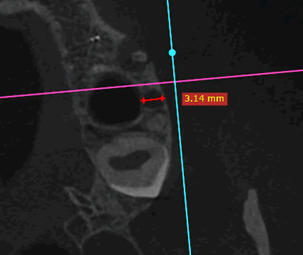
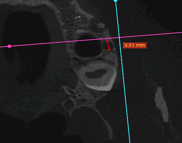
In the axial plane, maximum lesion extensions in the horizontal buccolingual (buccopalatal) and horizontal mesiodistal dimensions were measured in millimeters using a software ruler (Figures 1 and 2).

Figure 1: Periapical lesion extension in axial view, buccolingual dimension.

Figure 2: Periapical lesion extension in the axial view, mesiodistal dimension.
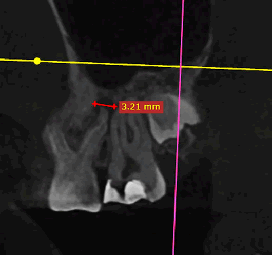
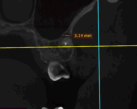
In the sagittal plane, the lesion's maximum vertical (occlusoapical) and horizontal mesiodistal extensions were measured (Figure 3). In the same manner, in the coronal plane, the maximum vertical (occluso-apical) and horizontal buccolingual (buccopalatal) extensions of the lesion were recorded (Figure 4). The highest rate of lesion extension was reported for each root of the first molars.

Figure 3: Periapical lesion extension in the sagittal view.

Figure 4: Periapical lesion extension in the coronal view.
Data were recorded in SPSS software (SPSS version 24, Chicago, IL, USA). Statistical analysis was performed using percentages, repeated measures ANOVA, paired t-test, and Pearson's correlation coefficient. The significant level was set at 0.05.
Results
Repeated measures ANOVA showed that the extensions of the lesion were not similar in all directions and there were significant differences between the lesion expansions in the three-dimensional planes (p-value<0.001). Generally, in the examined population, the highest average of lesion extension was reported in the vertical dimension (2.04 ± 1.61), followed by horizontal buccolingual (1.84 ± 1.28) and horizontal mesiodistal dimension (1.74 ± 1.19) respectively (Table 1).
| Lesion extension aspect |
14-44 year-old group |
45-54 year-old group |
55-77 year-old group |
Total |
P-value |
| Frequency |
Mean (SD) mm |
Frequency |
Mean (SD) mm |
Frequency |
Mean (SD) mm |
Frequency |
Mean (SD) mm |
| Occlusoapical aspect |
97 |
2.25 (1.79) |
51 |
2.2 (1.53) |
49 |
1.46 (1.15) |
197 |
2.04 (1.61) |
0.015 |
| Mesiodistal aspect |
97 |
1.91 (1.32) |
51 |
1.77 (1.07) |
49 |
1.38 (0.97) |
197 |
1.74 (1.19) |
0.044 |
| Buccolingual aspect |
97 |
2.00 (1.43) |
51 |
1.87 (1.12) |
49 |
1.50 (1.04) |
197 |
1.84 (1.28) |
0.082 |
Table 1: Mean (SD) lesion extension in three aspects of the samples under study in terms of age.
A statistically significant difference was reported between the lesion extension in vertical and buccolingual (P=0.001), vertical and mesiodistal (P=0.000) and between two horizontal planes (P=0.027). In the age group of 14-44 years, the highest mean lesion extension was observed in the vertical, buccolingual and mesiodistal planes, respectively. In the age group of 45-54 years, the highest mean lesion extension was observed in the vertical, mesiodistal and buccolingual planes respectively. Also, in the age group of 55-77 years, the highest mean lesion extension was observed in the buccolingual aspect, vertical aspect and mesiodistal planes respectively. Regarding the effect of age, a statistically significant difference was reported in lesion extension in the vertical (P=0.015) and mesiodistal (P=0.034) planes between the age groups of 14-44 and 55-77 years (Table 1). A different pattern of lesion extension was reported between men (n=84) and women (n=113). In the male patients, the highest mean lesion extension was observed in the vertical, buccolingual and mesiodistal planes, respectively. In contrast in the female patients, the highest mean lesion extension was observed in the vertical, mesiodistal and buccolingual aspects, respectively. No statistically significant difference was reported between men and women with regard to lesion extension (Table 2). According to the dental arch, 61.42% of the samples were related to the maxilla and 38.57% were related to the mandible. A similar extension pattern was reported for both the maxilla and mandible. The highest mean lesion extension in either maxilla or mandible was reported in the vertical, buccolingual and mesiodistal, respectively. No statistically significant difference was reported between the two jaws with regard to lesion extension (Table 3). 289 lesions were reported in the present study, which included 13.84% distal roots, 22.14% mesial roots, 13.84% distobuccal roots, 32.87% mesiobuccal roots and 17.3% palatal roots. The highest mean lesion extension in all the molars’ roots was reported in the vertical plane. In the distal, distobuccal and mesiobuccal roots, the highest mean lesion extension was related to the vertical, buccolingual and mesiodistal, respectively. In the mesial and palatal roots, the highest mean lesion extension was related to the vertical, mesiodistal and buccolingual planes. Considering the effect of root type, there was only a statistically significant difference in the buccolingual plane (P=0.024). A pairwise comparison of the two groups revealed that there was only a statistically significant difference between the buccolingual extension of the mesial root of mandibular molars and the distobuccal root of the maxillary molars (P=0.030) (Table 4).
| Lesion extension aspect |
Male |
Female |
Total |
P-value |
| Frequency |
Mean (SD) mm |
Frequency |
Mean (SD) mm |
Frequency |
Mean (SD) mm |
| Occlusoapical aspect |
84 |
2.14 (1.68) |
113 |
1.96 (1.57) |
197 |
2.04 (1.61) |
0.439 |
| Mesiodistal aspect |
84 |
1.94 (1.31) |
113 |
1.77 (1.25) |
197 |
1.84 (1.28) |
0.721 |
| Buccolingual aspect |
84 |
1.77 (1.16) |
113 |
1.71 (1.22) |
197 |
1.74 (1.19) |
0.353 |
Table 2: Mean (SD) lesion extension in three aspects of the samples under study in terms of gender.
| Lesion extension aspect |
Maxilla |
Mandible |
Total |
P-value |
| Frequency |
Mean (SD) mm |
Frequency |
Mean (SD) mm |
Frequency |
Mean (SD) mm |
| Occlusoapical aspect |
121 |
1.91 (1.53) |
76 |
2.25 (1.73) |
197 |
2.04 (1.61) |
0.166 |
| Mesiodistal aspect |
121 |
1.67 (1.20) |
76 |
1.85 (1.18) |
197 |
1.74 (1.19) |
0.284 |
| Buccolingual aspect |
121 |
1.72 (1.22) |
76 |
2.03 (1.35) |
197 |
1.84 (1.28) |
0.11 |
Table 3: Mean (SD) lesion extension in three aspects of the samples under study in terms of the dental arch.
| Lesion extension aspect |
Distal root |
Mesial root |
Distobuccal root |
Mesiobuccal root |
Palatal root |
Total |
P-value |
| Frequency |
Mean (SD) mm |
Frequency |
Mean (SD) mm |
Frequency |
Mean (SD) mm |
Frequency |
Mean (SD) mm |
Frequency |
Mean (SD) mm |
Frequency |
Mean (SD) mm |
| Occlusoapical aspect |
40 |
3.16 (2.36) |
64 |
3.63 (2.27) |
40 |
2.99 (1.76) |
95 |
3.85 (2.16) |
50 |
3.11 (2.19) |
289 |
3.52 (2.18) |
0.356 |
| Mesiodistal aspect |
40 |
3.17 (1.74) |
64 |
3.04 (1.72) |
40 |
2.48 (1.58) |
95 |
3.17 (1.72) |
50 |
2.93 (1.70) |
289 |
3.01 (1.70) |
0.645 |
| Buccolingual aspect |
40 |
3.22 (1.52) |
64 |
3.50 (1.80) |
40 |
2.99 (1.76) |
95 |
3.40 (1.80) |
50 |
2.85 (1.90) |
289 |
3.16 (1.75) |
0.024 |
Table 4: Mean lesion extension in three aspects in the samples under study in terms of tooth root type.
Discussion
Microorganisms and their secondary byproducts play an important role in the development of pulpal diseases and their extension to the periapical tissue [12]. The success of root canal therapy is achieved when microorganisms can be removed from the root canal system [12]. Routinely, the success of root canal therapy is mainly evaluated by intraoral periapical radiographs [13]. In follow-up studies, one of the key factors determining the success of root canal therapy is the healing or extension of the periapical lesion related to RCT [1]. Because intraoral periapical radiographs, which create a two-dimensional view, are routinely used to evaluate the recovery process and the success of the treatment, it is recommended to take at least two radiographs with two projection angles so that the third aspect of the lesion can be examined [14]. CBCT imaging is highly accurate for the early diagnosis of periapical diseases and the actual location of the lesion can be evaluated with CBCT images [15]. CBCT can be prescribed in difficult cases where two-dimensional imaging cannot provide full diagnostic information to the therapist. These data can be integrated with the information obtained from intraoral scanners to design a complete treatment guide [14]. The floor of the maxillary sinus extends from the first premolar to the maxillary tuberosity. The roots of the maxillary posterior teeth may have close relations to the sinus floor so that odontogenic infections in the periapical region can extend to the maxillary sinus and cause maxillary sinusitis [16]. Previous studies have concluded that CBCT images detect periapical lesions in the upper jaw 34% more than periapical images. Besides, CBCT images can show the extension of the lesion to the sinus [16]. In a study aimed at the anatomical analysis of the periapical bone of the maxillary posterior teeth with CBCT, Hu et al. [16] concluded that in the upper posterior teeth, the mesiobuccal root of the upper second molar is located at the closest distance to the sinus floor and the greatest vertical distance can be seen in the buccal roots of the first maxillary premolar. They also demonstrated that this distance is also related to age and sex and this gap was reported more in people over 40 years old and women [16]. During research conducted in 2020, Sakir et al. concluded that the thickness of the sinus mucosa can be evaluated in three-dimensional images such as CBCT compared to two-dimensional images [17]. There is also a relationship between the thickness of the maxillary sinus mucosa and the anatomical and periodontal conditions of the maxillary posterior teeth [17]. The thickness of the sinus mucosa is greater when the roots of the molars are closer to the sinus floor and have a periapical lesion [17]. Since CBCT images can determine the thickness and density of bone, the inclination of roots and their relationship with anatomical structures, thus, it is necessary to measure the real topography of the periapical lesion with CBCT images before surgical procedures [18]. Meirinhos et al. [19] investigated the relationship between the prevalence of periapical lesions with previous root canal therapy and the type of crown restoration with the help of CBCT during a cross-sectional study. They concluded that maxillary first molars have a higher prevalence of periapical lesions. In terms of roots, the mesiobuccal roots of maxillary first molars had the highest prevalence of lesions [19]. In a study aimed at determining the prevalence of periapical lesions in root-treated teeth using CBCT images, Nascimento et al., [20] concluded that the prevalence of periapical lesions was higher in maxillary molars and anterior teeth [20]. In the present study, the topography of the periapical lesion related to maxillary and mandibular first molars was evaluated in three orthogonal planes of CBCT slices. Based on the results of the present study, in general, the highest mean lesion extension in this dental group was reported in the vertical, buccolingual and finally mesiodistal aspects, respectively. In the present study, considering the effect of gender on the pattern of lesion extension, no statistically significant difference was found between males and females. The results of the study by Jamshidi et al., to investigate the prevalence of periapical lesions in extracted teeth in the Iranian population reported that the prevalence of periapical lesions was equal in males and females. Kazemipoor et al., conducted a study on the pattern of lesion extension in anterior teeth. Based on the results of this study, there was a statistically significant difference between males and females in the extension pattern, so in men, the highest mean extension was reported in the vertical aspect and women in the buccolingual aspect. Bone density decreases with age and this decrease in density is higher in women due to the effect of female hormones, low bone mass and longer mean age compared to men. Pablo Florenzano et al., stated that the amount of bone regeneration is related to age and its quantity and quality change with age. Based on the results of the present study, a statistically significant difference was reported between the 14-44 year-old group and the 55-77 year-old group in the extension of the vertical and mesiodistal aspects. The thickness of the spongy bone in the maxilla and mandible is different and this plays an important role in lesion extension in different regions of the maxilla and mandible. In the present study, no statistically significant difference was found between the upper and lower jaws in lesion extension in three planes. Also, in terms of root type, the highest mean lesion extension in the vertical aspect was related to the mesiobuccal root of the first molar. Based on the results of the present study, only lesion extension in the buccolingual aspect in the distobuccal roots of the maxillary first molar and the mesial root of the mandibular first molar showed a statistically significant difference.
CONCLUSION
Given the limitations of the present study, the extension of the periapical lesion in the first molar region of the bony structure of the maxilla and mandible follows a precise pattern. In this regard, future studies should be designed in different communities and races, as well as in terms of examining the volume. Moreover, the CBCT method is a reliable imaging method to evaluate the extension and topography of the periapical lesion in terms of morphology and morphometric.
Ethics Approval
All the experimental procedures were approved by the ethics committee of the Shahid Sadoughi University of Medical Sciences Ethics Committee Yazd, Iran (IR.SSU.REC.1400.004) before data analysis.
Human and Animal Rights
All the images that have been analyzed in the present study were anonymous and non-identifiable. Each CBCT image had a code number and patient age and gender were recorded for every code.
Consent to Participate
We have applied an archive from a private radiology clinic and all the personal information including patients' names, initials or hospital numbers is not mentioned anywhere in the manuscript (including figures).
Consent for Publication
The explanation has been made in the consent to publication section.
Availability of Data and Materials
The data supporting the findings of the article is from an archive of a private radiology clinic and is not public.
Acknowledgement
We would like to thank the Vice-chancellor of Research and Technology, Shahid Sadoughi University of Medical Sciences, who approved this study.
Conflict of Interest
The authors declare no conflict of interest.
Funding
The present study was supported by the Vice-chancellor of Research and Technology, Shahid Sadoughi University of Medical Sciences (ID: IR.SSU.REC.1400.004).
References
- Karamifar K, Tondari A, Saghiri MA (2020) Endodontic periapical lesion: An overview on the etiology, diagnosis and current treatment modalities. Eur Endod J. 5(2):54-67.
[Crossref] [Google Scholar] [PubMed]
- Ketabi A-R, Ketabi S, Nabli MB, Lauer H-C, Brenner M (2019) Detection and measurements of apical lesions in the upper jaw by cone beam computed tomography and panoramic radiography as a function of cortical bone thickness. Clin Oral Investig. 23(11):4067-4073.
[Crossref] [Google Scholar] [PubMed]
- Kruse C, Spin-Neto R, Wenzel A, Kirkevang LL (2015) Cone beam computed tomography and periapical lesions: A systematic review analysing studies on diagnostic efficacy by a hierarchical model. Int Endod J. 48(9):815-828.
[Crossref] [Google Scholar] [PubMed]
- Setzer FC, Shi KJ, Zhang Z, Yan H, Yoon H, et al. (2020) Artificial intelligence for the computer-aided detection of periapical lesions in cone-beam computed tomographic images. J Endod. 46(7):987-993.
[Crossref] [Google Scholar] [PubMed]
- Antony DP, Thomas T, Nivedhitha MS (2020) Two-dimensional periapical, panoramic radiography versus three-dimensional cone-beam computed tomography in the detection of periapical lesion after endodontic treatment: A systematic review. Cureus. 12(4):e7736.
[Crossref] [Google Scholar] [PubMed]
- Venskutonis T, Daugela P, Strazdas M, Juodzbalys G (2014) Accuracy of digital radiography and cone beam computed tomography on periapical radiolucency detection in endodontically treated teeth. J Oral Maxillofac Res. 5(2):12-19.
[Crossref] [Google Scholar] [PubMed]
- Huumonen S, Kvist T, Gröndahl K, Molander A (2006) Diagnostic value of computed tomography in reâ?treatment of root fillings in maxillary molars. Int Endod J. 39(10):827-833.
[Crossref] [Google Scholar] [PubMed]
- Aksoy U, Orhan K (2018) Risk factor in endodontic treatment: Topographic evaluation of mandibular posterior teeth and lingual cortical plate using cone beam Computed Tomography (CT). Med Sci Monit. 24:75-88.
[Crossref] [Google Scholar] [PubMed]
- Aljarbou FA, Aldosimani MA, Althumairy RI, Alhezam AA, Aldawsari AI (2019) An analysis of the first and second mandibular molar roots proximity to the inferior alveolar canal and cortical plates using cone beam computed tomography among the Saudi population. Saudi Med J. 40(2):189.
[Crossref] [Google Scholar] [PubMed]
- Ozkan BT, Celik S, Durmus E (2008) Paresthesia of the mental nerve stem from periapical infection of mandibular canine tooth: A case report. Oral Surg Oral Med Oral Pathol Oral Radiol Endod. 105(5):e28-e31.
[Crossref] [Google Scholar] [PubMed]
- Goller-Bulut D, Sekerci A-E, Köse E, Sisman Y (2015) Cone beam computed tomographic analysis of maxillary premolars and molars to detect the relationship between periapical and marginal bone loss and mucosal thickness of maxillary sinus. Med Oral Patol Oral Cir Bucal. 20(5):e572.
[Crossref] [Google Scholar] [PubMed]
- Bordea IR, Hanna R, Chiniforush N, GrÄ?dinaru E, Câmpian RS, et al. (2020) Evaluation of the outcome of various laser therapy applications in root canal disinfection: A systematic review. Photodiagn Photodyn Ther. 29(10):11-19.
[Crossref] [Google Scholar] [PubMed]
- Liang Y-H, Li G, Shemesh H, Wesselink PR, Wu M-K (2012) The association between complete absence of post-treatment periapical lesion and quality of root canal filling. Clin Oral Investig. 16(6):1619-1626.
[Crossref] [Google Scholar] [PubMed]
- Moreno-Rabié C, Torres A, Lambrechts P, Jacobs R (2020) Clinical applications, accuracy and limitations of guided endodontics: a systematic review. Int Endod J. 53(2):214-231.
[Crossref] [Google Scholar] [PubMed]
- Jindal G, Batra H, Kaur S, Vashist D (2015) Dentigerous cyst associated with mandibular 2nd molar: An unusual entity. J Maxillofac Oral Surg. 14(Suppl 1):154-157.
[Crossref] [Google Scholar] [PubMed]
- Hu X, Lei L, Cui M, Huang Z, Zhang X (2019) Anatomical analysis of periapical bone of maxillary posterior teeth: A cone beam computed tomography study. J Int Med Res. 47(10):4701-4710.
[Crossref] [Google Scholar] [PubMed]
- Sakir M, Ercalik Yalcinkaya S (2020) Associations between periapical health of maxillary molars and mucosal thickening of maxillary sinuses in cone-beam computed tomographic images: A retrospective study. J Endod. 46(3):397-403.
[Crossref] [Google Scholar] [PubMed]
- Patel S, Dawood A, Ford TP, Whaites E (2007) The potential applications of cone beam computed tomography in the management of endodontic problems. Int Endod J. 40(10):818-30.
[Crossref] [Google Scholar] [PubMed]
- Meirinhos J, Martins JNR, Pereira B, Baruwa A, Gouveia J, et al. (2020) Prevalence of apical periodontitis and its association with previous root canal treatment, root canal filling length and type of coronal restoration-a cross-sectional study. Int Endod J. 53(4):573-584.
[Crossref] [Google Scholar] [PubMed]
- Nascimento EHL, Gaêta-Araujo H, Andrade MFS, Freitas DQ (2018) Prevalence of technical errors and periapical lesions in a sample of endodontically treated teeth: A CBCT analysis. Clin Oral Investig. 22(7):2495-2503.
[Crossref] [Google Scholar] [PubMed]
Citation: Kazemipoor M, Foroughipour F, Sa i Y (2025) Topographic Evaluation of Periapical In lammatory Lesions in the First
Molar’s Region using CBCT. J Healthc Commun. 10:64.
Copyright: © 2025 Kazemipoor M, et al. This is an open-access article distributed under the terms of the Creative
Commons Attribution License, which permits unrestricted use, distribution, and reproduction in any medium, provided the
original author and source are credited.


