Keywords
Biomarkers; Pepsinogen I; Pepsinogen II; Gastrin-17; Helicobacter pylori; Serological testing; Marker panel; Gastropanel; Gastric cancer risk; Atrophic gastritis; Antrum; Corpus; Dyspepsia; Diagnosis; Screening; Primary prevention; Limitations; False positive; False negative; Hp tests
Introduction
Gastric Cancer (GC) continues to be one of the most common cancers and causes of global cancer mortality; nearly one million new cases and 736.000 annual deaths worldwide [1]. In many Western countries, GC incidence has been steadily declining, however, attributed to major changes in the exposure to the risk factors of GC [2]. These known risk factors for GC include smoking, use of alcohol, dietary factors, occupational exposures, radiation and/or radiotherapy, as well as genetic predisposition in certain rare syndromes. According to the current concepts, a different global distribution of these risk factors explains the large geographic variation in the incidence of GC. It is estimated that nearly 80% of GC cases among males and 70% in women are due to different life-style and environmental factors [1,3,4] including a protective effect of the Mediterranean type of diet [5].
In addition to those common-type of risk factors, there are two specific risk factors that far exceed in importance of all the others in pathogenesis of GC: Helicobacter pylori (HP) infection and Atrophic Gastritis (AG). As early as in 1994, the International Agency for Research on Cancer (IARC, Lyon) stated that the scientific evidence is sufficient to declare HP as a human carcinogen. This bacterium primarily infects the gastric mucosa, and if uneradicated, develops AG in about half of the affected patients. Although HP itself is not directly carcinogenic, AG is the single most potent risk factor of GC. In fact, the risk of GC increases in parallel with the severity of AG: Compared with healthy stomach, the risk is 2-5x in those with only chronic HP gastritis but up to 90x in patients with severe AG both in the corpus and antrum (Atrophic Pan-Gastritis; AGpan) [3,6-10].
The other main histological type of GC (intestinal type) develops in atrophic mucosa through various degrees of dysplasia (mild, moderate, and severe), which are often accompanied by Intestinal Metaplasia (IM) This pathogenetic chain is known as the correa cascade [3]. It is important to recall that this cascade can often (but not invariably) be interrupted by appropriate early treatment of HP infection [3,4,9,11]. Based on the Updated Sydney System Classification (USS), AG is classified by its topographic location (antrum, corpus, or both) as AGA, AGC or AGpan, respectively [12-14]. In addition to being the key risk factor of GC, HP-infection also plays a causative role in the development of peptic ulcer disease [8-10]. Similarly, both AG and HP can be responsible for the symptoms known as dyspepsia; organic or functional [15] Debate still continues, however, on the value of systematic HP-eradication in relieving the dyspeptic symptoms [9,10,15].
The diagnosis of AG has traditionally been made using histological biopsies on gastroscopy. However, gastroscopy is an invasive diagnostic tool, which requires expensive equipment and considerable professional skills. Similarly, gastroscopy (like any endoscopies) is a subjective diagnostic method, which is not suitable for population-based screening of GC. As to the diagnosis of HP-infections, attempts have been made to standardize the management (diagnosis included) since 1996 by the Maastricht Consensus Conferences publishing their consensus statements at regular intervals. Unfortunately, it happens far too often in daily practice that only the merits of the commonly used HP tests are being emphasized, and the limitations of their use in special clinical settings are ignored, although clearly described in the European Consensus Reports [9,10]. This applies to both of the two most widely used HP tests; the 13C-Urea Breath Test (UBT) and Stool Antigen test (SAT), the limitations of which were recently discussed in timely reviews [16-18].
GastroPanel® Test (Serological Biopsy)
Because of these obstacles in diagnosis of both AG and HPinfection, the need to develop a reliable diagnostic blood test has increased in parallel with the increasing understanding of the importance of HP and AG as the key risk factors of GC, as a result of long-term prospective studies conducted e.g. in Finland [7,13]. To meet such an increasing demand, the GastroPanel® test was designed in the late 1990’s by Biohit Oyj (Helsinki, Finland), representing the first non-invasive diagnostic test for stomach health and disease [19-22]. With GastroPanel®, both the key risk factors of GC can be identified in a simple blood test, which is based on the simultaneous measurement of four serum biomarkers of gastric mucosal function and its structure. This biomarker panel is equally applicable as the first-line diagnostic test for patients with dyspeptic symptoms, with potential to replace the invasive gastroscopy in this diagnostic and management algorithm [21,22].
Introduction to GastroPanel® test
GastroPanel® is the first-line diagnostic test for HP-infection (5-80% of the world population affected), for the examination of all patients with dyspepsia (20-40% of the Western population), as well as for the screening of AG with related risks, such as stomach and esophageal cancer [22-24]. As well known, AG also enhances the risk of malabsorption of vitamin- B12, calcium, iron, magnesium, zinc, and some medicines [20,21].
GastroPanel® consists of key stomach-specific biomarkers representing the key regulators of normal stomach physiology. These biomarkers include: Pepsinogen I (PGI), Pepsinogen II (PGII), Amidated Gastrin-17 (G-17), and HP Antibodies (HPAb), designed to give information on both the structure and function of the stomach mucosa [20-27]. Most importantly, this panel gives accurate estimates of the capacity of the corpus and antrum mucosa to produce gastric acid and G-17, respectively, as well as of important gastric pathologies, e.g. inflammation, topography and grade of AG [28-30].
Normal plasma levels of all four biomarkers indicate that the stomach mucosa has normal structure and function, whereas abnormal levels are signs of a non-healthy stomach, reflecting disturbances in the feedback mechanisms regulating the acid output of the corpus, PGs and G-17. For G-17 assessment, there are two options; G-17 basal (G-17b) values and G-17 stimulated (G-17s) values, the latter being particularly important in distinguishing between functional disturbance of the antrum (G-17s normal) and AGA (G-17s does not increase upon protein stimulation) [31,32].
GastroPanel® is unique in that the results are interpreted by a software application (GastroSoft®) (https://www.GastroPanel.com), specifically designed for this purpose. GastroPanel® results are classified into one of five possible diagnostic categories related to stomach morphology: 1) Normal mucosa, 2) Superficial or non-atrophic (HP) gastritis, 3) AG in the corpus, 4) AG in the antrum, and 5) AG in both antrum and corpus (pan-gastritis) [13,32]. Thus, GastroPanel® is optimized for use together with the USS classification of gastritis, which is based on these same five diagnostic categories [33]. In addition, there are three other marker profiles specific to functional disturbances of the stomach where morphology is normal (details to follow) (Figure 1).
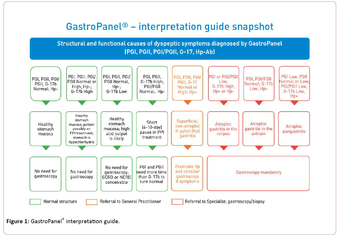
Figure 1: GastroPanel® interpretation guide.
GastroPanel® has been validated in several large trials based on biopsy-confirmed gastroscopies [23,34,35], all included in a recent meta-analysis [36]. These studies have been used for validating the reference (cut-off) values for each biomarker of the panel against the gold standard histological endpoints. These studies also confirm the high accuracy of GastroPanel® in detecting the most important endpoint, moderate-to-severe AG (AG2+) [36]. Thus, normal values of PGI, PGII and their ratio (PGI/PGII) preclude AGC with a NPV of over 95% [23]. In turn, the values of PGI and PGII as well as their ratio below the established cut-off levels predict AGC2+ with area under ROC curve (AUC) values >0.950 in adequately-powered, USS-validated series [34,36].
In brief, the levels of PGI decrease in AGC and in AGpan, but remain within a normal range in all other conditions. Elevated PGII levels reflect mucosal inflammation, the highest values being detected in HP-associated non-AG. The G-17b values are highest in AGC, because of the missing negative feedback by the acid output from an atrophic corpus, resulting in uninhibited secretion of G-17b by the normal antral mucosa.
The same applies to the situation where acid output is inhibited by prolonged use of PPI-medication. By definition, when antral mucosa is atrophic and the G cells disappear; G-17 secretion remains very low even after protein stimulation (G-17s) [19] HP IgG antibodies (HPAb) provide significant added diagnostic value to the three biomarkers. HPAb levels measure two potentially different conditions: 1) An ongoing HP-infection, or 2) A previous exposure to HP. As the only abnormal marker, HPAb implicates an [16-18] [HP-associated superficial gastritis (non-AG)], while associated with abnormalities in the other three markers, elevated HPAb levels confirm the diagnosis of HP-associated AG (AGA or AGC) [37,38].
Biomarkers of the GastroPanel® Test
Pepsinogen I (PG I)
This biomarker is included in GastroPanel® to identify patients who have mucosal atrophy in the gastric corpus (AGC), for which the plasma PGI is a highly specific biomarker [36,38-42]. Pepsinogen I (PGI) is a precursor enzyme (zymogen) of pepsin, synthesized by the chief cells and the neck cells of the gastric corpus (in oxyntic glands) as a pepsin precursor, the major part of PGI is secreted into the gastric lumen but a minor fraction is excreted into the blood. The circulating PGI concentration closely correlates with the quantity of the chief cells in the corpus mucosa, and any loss of these cells due to mucosal atrophy results in a linear decrease in plasma levels of PGI [38-42].
For as yet unknown reasons, AG increases the risk of GC [3,6,7,9,10]. Compared with a healthy stomach, this risk is 5- fold among patients with advanced AGC, but up to 90-fold in patients with advanced AG in both the antrum and corpus (AGpan) [7]. In the screening of middle-aged (50-69 years) males in Finland, the circulating PGI level was low (<25 μg/l) in 9.8% of the subjects, of whom 4.7% revealed either a GC or a precursor lesion on endoscopy [13]. Similar results have been published in several previous studies, included in a recent meta-analysis [43].
Pepsinogen II (PG II)
Pepsinogen II is produced by the chief cells and the mucous neck cells of the gastric corpus, in pyloric glands of the gastric antrum, and in Brunner’s glands of the proximal duodenum. The ratio of PGI to PGII plasma levels in normal subjects is between 3 and 20 [30]. The PGI/PGII ratio decreases linearly with increasing grade of AGC [36,38,39,44]. The ratio falls below 3.0 when AGC is advanced (AGC2+) [39]. It has been shown that the risk of GC is increased (5-fold) when the PGI/ PGII ratio is low [22,26,29,42,45-50]. This test is intended as an additional diagnostic tool for AGC. Decreased PGI, PGII and the PGI/PGII ratio, along with elevated G-17 confirm the diagnosis of AGC. An elevated PGII level alone reflects mucosal inflammation, the highest values being detected in HP-associated non-AG. Since HPAb levels can remain elevated for several months after successful eradication, PGII is a useful marker for the confirmation of positive HP-eradication results [22,36,38].
Gastrin-17 (G-17)
Gastrins are linear peptide hormones produced by the G cells in the duodenum, in the pyloric part of the gastric antrum, as well as in the pancreas [22]. The main function of gastrins is to stimulate the secretion of gastric acid (HCl) by the parietal cells of the corpus, as well as to increase the motility of antrum [51]. In addition, gastrins are known to stimulate gastric chief cells to secrete pepsinogens and also induce the contraction of the Lower Esophageal Sphincter (LES) like most of the peptide hormones, different molecular weight gastrins are synthesized as a result of post-translational modifications from preprogastrin. Thus, a mixture of different molecular weight gastrins is released from the G cells into the circulation, including gastrin-71, -52, -34, -17, -14, and -6, all of which are carboxy-amidated and circulate in an O-sulfated and non-sulfated form [52]. In healthy humans, the dominant forms of gastrin in plasma/serum are amidated gastrin-34 (G-34) and G-17 [53].
In healthy antral tissue, G-17 is the most potent form of all gastrins, almost exclusively produced by the antral G cells. Thus, the G-17 included in GastroPanel® test is a specific biomarker of antral structure and function, and through a negative feedback loop, an indirect biomarker of gastric corpus as well. G-17 plasma levels within the normal range implicate a normal structure and function of the antrum, whereas low or high values of G-17 also reflect abnormal functions of the corpus. The maximum information is obtained when G-17 testing is done separately for fasting (G-17b) and stimulated (G-17s) levels, accompanied by PGI, PGII and HPAb in the full GastroPanel testing [7,19,44,54-57].
The measurement of G-17b may also be used for the monitoring of the patients who have undergone gastric surgery; secretion of G-17b is practically zero after successful radical antral resection. In HP-negative subjects, a low fasting level of G-17 can indicate high acid output. This in turn may increase the risk of gastroesophageal reflux disease (GERD) and Barrett’s esophagus (up to 3-4-fold), whereas a normal or elevated G-17b excludes the presence of Barrett’s esophagus with high probability [57,58].
Helicobacter pylori antibody (IgG)
Helicobacter pylori (HP) infection is the most important cause of chronic gastritis leading to mucosal atrophy. A much more uncommon cause of AG is an autoimmune disease [59,60]. In GastroPanel® test, ELISA technique is used to detect in the plasma levels of HP IgG antibody levels.
HP is a spiral-shaped, gram-negative bacterium that colonizes in human stomach [61]. The organism is found within the mucous layer overlying the gastric epithelium, and also within the mucosal glands, but it does not appear to invade the epithelial cells. However, the mucosa underneath and surrounding the areas of the HP colonization is invariably inflamed; this condition is referred to as chronic superficial or non-atrophic gastritis which, if untreated, persists for life [22,60,61]. Without adequate eradication of the bacteria, this chronic inflammatory process leads to AG [9,10]. Peptic ulceration is another important sequel of HP-infection [62-65]. The epidemiological evidence indicates a link between HP-infection and gastric adenocarcinoma, as well as a Mucosa- Associated Lymphatic Tissue (MALT) lymphoma [9,17-19,66-68].
Interpretation of the GastroPanel® Results
GastroPanel® is optimized for use in context with the USS classification of gastritis [14,38]. Both the USS and the GastroSoft® use five diagnostic categories to classify the biopsies and GastroPanel® results, respectively. These include: 1) Normal mucosa, 2) Superficial (HP) gastritis, 3) AGA, 4) AGC, and 5) AG in both antrum and corpus (AGpan) [14,69]. In addition to these five categories related to stomach morphology, three other marker profiles are possible in GastroPanel® , being defined as functional disturbances with normal stomach morphology. All eight diagnostic categories are depicted in Table 1, and explained in the following.
| GastroPanel® Biomarkers |
| Marker Profile |
Pepsinogen I (30-160 µg/l)@ |
Pepsinogen II (3-15 µg/l) |
PGI/PGII Ratio (3-20) |
Gastrin-17b (1-7 pmol/l) |
Gastrin-17s (3-30 pmol/l) |
Helicobacter pylori IgG Antibody titer (<30 EIU) |
Interpretation |
| 1 |
N |
N |
N |
N |
N |
N |
Healthy mucosa (no atrophy, no HP-infection) |
| 2 |
N |
N |
N |
L* |
N |
N |
Healthy mucosa. High acid output in the corpus |
| 3 |
N or H^ |
N or H^ |
N |
H** |
N |
N |
Healthy mucosa. Low acid output due to e.g. PPI medication |
| 4a |
N or H^ |
N or H^ |
N |
N or H^ |
ND |
N or H† |
Active HP-infection, not treated |
| 4b |
N |
N |
N |
N |
ND |
H |
HP-infection successfully eradicated |
| 4c |
N |
H |
N |
H |
ND |
H |
HP eradication failed |
| 5 |
L |
L |
L |
H |
ND |
N^^ or H |
Atrophic gastritis in the corpus (AGC) |
| 6 |
N |
N |
N |
L |
L |
H |
Atrophic gastritis in the antrum (AGA) |
| 7 |
L |
L |
L |
L |
L |
N^^ or H |
Atrophic gastritis in the antrum and corpus (AGpan) |
| 8 |
H |
H |
N |
H |
ND |
N |
Short (4-10d) break in PPI treatment |
| N=normal; L=low; H=high; *Test PPI medication for two weeks, G17b should normalize;**Stop PPI medication, G-17b should normalize within two weeks; ND, no need for testing;^PGI, PGII and G-17 can be elevated due to mucosal inflammation; ^^HP antibodies can disappear in mucosal atrophy with protracted clinical course; @Pepsinogen I cut-off value 30 µg/l is consonant with moderate/severe AG; †HP antibody levels can remain elevated for months after successful eradication of HP. |
Table 1: The diagnostic categories of GastroPanel® test results.
Normal biomarker profile
With all four biomarkers within the normal reference range, gastric mucosa functions normally. Given that the function of gastric mucosa is critically dependent on the specific cells responsible for the output of gastric acid (parietal cells), pepsinogens (chief cells) and G-17 (G cells), normal function necessitates the presence of these cells in normal quantities [22,24,29,32,38]. Thus, stomach function and mucosal structure go hand-in-hand, and by definition, a normal GastroPanel® result is a surrogate marker of a healthy stomach. As we know now, however, a normal marker profile does not exclude minor abnormalities like non-specific inflammation, mild irritation or micro-erosions that do not impact on the marker profiles [24,38].
High acid output
Gastric acid (HCl) is produced by the highly specialized parietal cells in the corpus. Acid output is controlled, among other things, by the output of G-17 in the antrum as a result of a positive feedback loop stimulating acid secretion after a meal [22,38,51,52]. Acid output results in progressively lower pH in the stomach contents, and the threshold of pH 2.5 triggers a negative feedback to antral G cells, signaling them to down-regulate the secretion of G-17 [51-54]. As a result, G-17 output decreases in parallel with the increasing acid output of the corpus [19,22,24,34]. When, due to any reason (e.g. other stimulatory mechanisms), acid content in the corpus remains abnormally high, the end result is abnormally low G-17b secretion from the antral G cells. Using GastroPanel®, this condition is best diagnosed after a test medication with PPI, when the G-17b should be normalized within approximately 2 weeks of therapy. In this highly acidic milieu with low G-17b, however, the levels of stimulated G-17s will remain within normal limits, because the G cells are intact and capable of increasing their G-17 secretion upon protein stimulation (e.g. by a protein powder; Biohit Cat. No. 601038) [38].
Low acid output due to proton pump inhibitor (PPI) medication
This regulation also works in the other way round. When acid output in the corpus is reduced (for any reason), the positive feedback loop triggers antral G cells to increase their G-17b secretion, resulting in elevated serum levels of G-17b [19,24,38]. The two prime conditions leading to low acid output are: 1) AGC, and 2) Long-term use of PPI-medication (or to a lesser extent, H2-receptor blockers). The former is excluded by the normal (or even elevated) values of PGI, PGII, and normal PGI/PGII ratio [24,38], while the latter is best diagnosed by discontinuing the PPI medication. In that case, the antral G-17b should be normalized within two weeks [24,38,51-54].
Superficial (non-atrophic), Helicobacter pylori associated gastritis
Like all bacteria, HP will also induce acute inflammation in the gastric mucosa, with the usual onset in the antrum [9,10,22,24,28,61,67,68]. Three different marker profiles can be encountered in association with HP-infection (Table 1)
Active HP-infection: In an active HP-infection, HPAb levels are raised above the cut-off value (30 EIU), which can be the only abnormal finding in GastroPanel® test, with all other markers falling within a normal range. Not infrequently, however, an active ongoing HP-infection causes a severe inflammatory reaction which, due to increased cell permeability, can lead to increased leakage of PGI, PGII and even G-17 from the secretory cells and result in elevated serum levels of any or all of these three biomarkers, as depicted in Table 1 (4a) [9,10,24,28,38].
Successful HP-eradication: Successful HP-eradication by active treatment should result in normalized values of HPAbs as well as the three (“inflammatory”) markers (PGI, PGII, G-17) (Table 1 (4b)) For the latter, this is known to take place with a delay of some weeks [24,38] In contrast, HPAb levels can remain elevated for a longer period of time which is subject to individual variation and limits the usefulness of GastroPanel® in the immediate post-treatment control of HP-eradication. Because a marked individual variation exists in the dynamics of these marker profiles, an accurate record of timing of the HP-eradication is mandatory while making the re-testing with GastroPanel® [2,21,38,56].
Failed HP-eradication: In cases where HP-eradication attempt fails, HPAb levels remain elevated (usually slightly), PGI and PGI/PGII ratio usually fall within a normal range, whereas PGII and/or G-17b may remain slightly elevated as a sign of an ongoing inflammatory process (Table 1, profile 4c) The result can be confirmed after 5-6 months, followed by a new treatment attempt if indicated [9,10,24]. An option is to use another test for the control of HP-eradication, e.g. the Helicobacter pylori Quick Test (fast) or Helicobacter pylori UFT 300 Quick test (ultrafast) [69,70].
Atrophic gastritis of the corpus (AGC)
By definition, the loss of specific cells (chief cells) in the oxyntic glands of the corpus mucosa as a result of mucosal atrophy will lead to a progressively reduced output of PGI and (to a lesser extent) PGII, which is also produced by the same cells in the antral mucosa [24,38]. This disproportionate reduction of these two markers will result in a reduced PGI/ PGII ratio, which is another excellent signature of AGC [22,24,26-28,30,34,36,38,43]. This reduction in the PGI and PGI/PGII ratio is progressive and closely correlates with the severity of AGC, with total atrophy and acid-free stomach as the endpoint [56,59]. In the case of intact (normal) antral mucosa, this leads to markedly increased output and serum levels of G-17b [19,24,38] (Table 1, profile 5) There is no need to test G-17s in such a situation. In chronic AGC cases with a protracted course over decades, HP itself may disappear from the stomach, resulting in gradual normalization of the HPAb levels [71-73].
Atrophic gastritis of the antrum (AGA)
When the mucosal atrophy only affects the antrum, all corpus-specific markers will remain within the normal range (Table 1) by definition, AGA is caused by HP-infection, and HPAbs are invariably elevated in the GastroPanel® testing. As a result of AGA, the G cells are reduced in number and finally disappear, leading to progressively reduced plasma levels of G-17b. In severe AGA, there is no response in G-17 output to protein stimulation (G-17s), because of the lack of (target) G cells in the antral mucosa (Table 1, profile 6) [19,22,24,34,35,37,38,40,56,58]. Thus, the distinction between the two potential causes of low G-17b: i) High acid output (profile 2) and ii) AGA (profile 6), is neatly done by using the G-17s testing after protein stimulation [24,38]. As pointed out, G-17s will react normally only in the former, but fails to react in severe AGA.
Atrophic gastritis of the antrum and corpus (AGpan)
The most severe form of AG is known as atrophic pangastritis (AGpan), affecting both the antrum and corpus [24,38]. As an end result, the specified cells (chief cells) in the corpus and antrum (G cells) disappear, leading to a biomarker expression profile where both pepsinogens (PGI, PGII) and G-17 are substantially reduced (Table 1, profile 7) [19,22,24,34,35,37,38,40,59,60]. This applies to both G-17b and G-17s, which remain low even after protein stimulation because of the missing G cells. Like in AGC (profile 5), HPAb levels can be within a normal range or elevated. This is because in chronic AG, HP itself can disappear from an atrophic mucosa, and in the absence of antigen stimulus, a normal decay of IgG antibodies will revert the HPAb levels below the 30 EIU cut-off [71-73].
Panel profile in context of PPI medication
Any gastric acid suppressive medication (PPI, H2 blockers) will inevitably interfere with the profile of the GastroPanel® markers because of an altered acid output, as explained above. To enable the assessment of the biomarker profile without such an interference, the manufacturer recommends that the patient discontinues any acid-suppressive treatment 7 days before the sampling [24,38]. It is appreciated that because of severe symptoms, this withdrawal of PPI-or H2-blocker medication is not always possible. Because of this fact, the new version of the GastroSoft® was designed to take into account also the continued use of these drugs. Import is an accurate record of the PPI/H2-medication, the fact whether or not discontinued, and if so, for how many days before the sampling. With this information accurately recorded, the GastroSoft® is capable of interpreting the test results correctly, defined as profile 8 in Table 1, based on the following rational.
PPI and H2-blockers effectively reduce gastric acid production in the parietal cells of the corpus [24]. This increases the production of G-17 and also the output of pepsinogens. Once the PPI/H2-treatment is discontinued, it takes approximately 4-10 days for HCl production and G-17 levels to normalize. However, pepsinogens respond more slowly, and PGI and PGII levels may remain above the cut-off values for up to 2-3 weeks [24,38]. Furthermore, an abrupt termination of a long-term PPI-medication is typically followed by rebound acid hypersecretion, frequently accompanied by heartburn (and other) symptoms and extremely low levels of G-17b [19,22,24,32,38].
Clinical Performance Confirmed in a Formal Meta-Analysis
To provide an unbiased estimate of the accumulated evidence, we recently performed a systematic review and meta-analysis of all studies published on GastroPanel® test since its introduction in the early 2000’s [36]. Altogether, 27 studies were eligible, comprising 8.654 tested patients from different geographic regions. GastroPanel® was shown to perform better in diagnosis of AGC than AGA, with 70.2% vs. 51.6% pooled SE, and 93.9% vs. 84.1% pooled SP, respectively [36]. Limited number of studies erodes the Q test’s power to detect true heterogeneity in meta-analysis stratified by geographic origin of the studies. The results of this first metaanalysis of GastroPanel® literature corroborates the above cited statement of the international experts [22]. Due to its high specificity for AGA and AGC [36] as well as its extremely high longitudinal negative predictive value [34], GastroPanel® is truly a test for stomach health and disease. In other words, testing GastroPanel-negative at any time point during one’s life-time precludes (with >95% probability) a significant gastric pathology for several years ahead [34]. Meantime, however, abnormal GastroPanel® profiles implicating AGC are powerful independent predictors of an incident GC, as recently shown in a long-term longitudinal setting [74].
GastroPanel® Test is Devoid of the Caveats of Conventional HP Tests
Management of HP (including diagnosis and therapy) has been exhaustively reviewed in several reports. The message is unanimous in that several clinical conditions seriously hamper the diagnostic value of the most commonly used HP tests: 13C-Urea Breath Test (UBT) and Stool Antigen Test (SAT), both false-negative and false-positive results being not uncommon [9,10,16-18,71-73,75-80]. Basically, these false-negative results are due to decreased bacterial loads in the stomach mucosa, and include the following clinical conditions: 1) Use of PPI medication; 2) Use of antibiotics; 3) Bleeding peptic ulcer; 4) Atrophic Gastritis (AG; with or without intestinal metaplasia); 5) Gastric cancer; 6) MALT lymphoma, and 7) Partial gastrectomy. Since the late 1990’s, it has been well established that UBT also gives false-positive results in cases where urease-producing bacterial species are colonizing an acid-free stomach due to AG or a long-term use of PPI medication [16-18,81-91]. It is to be emphasized that neither UBT nor SAT (or HP serology) is capable of diagnosing AG (of HP-or autoimmune origin), thus missing the patients at high risk for the potentially serious clinical sequels of AG: i) GC), ii) Esophageal cancer, iii) Vitamin-B12 deficiency, and iv) Malabsorption of calcium, iron, magnesium and certain medicines. It is mandatory that these serious limitations (i.e., false-negative results in true disease, false-positives with no HP infection, and failure to diagnose AG) in use of UBT and SAT are properly acknowledged when these two tests are promoted for HP diagnosis [9,10,16-18,24,28,38].
As might be appreciated from the above, GastroPanel® test is devoid of these diagnostic caveats of the UBT and SAT tests [16-18,24,28,38]. In fact, GastroPanel® is the most comprehensive HP test, with its HPAb measurement being complemented by the 3 other biomarkers (PGI, PGII, G-17) The latter are sensitive indicators of mucosal inflammation, which is important because like all bacteria, also HP will induce acute inflammation in the gastric mucosa, with a usual onset in the antrum [9,10,22].
Depending on the different phase of infection, three different marker profiles can be encountered in association with HP-infection, as explained before (Table 1). First, in an active HP-infection, HPAb titers are raised, which can be the only abnormal finding in GastroPanel, with all other markers falling within a normal range. Frequently, however, an active ongoing HP-infection causes an inflammatory reaction severe enough to increase the levels of the inflammation markers: PGI, PGII and even G-17 [34,38,92]. Second, a successful HP-eradication by active treatment should result in normalized values of all three markers, with a delay of some weeks to months. This delay should be taken into account while interpreting the GastroPanel® results in samples taken soon after HP-eradication [22,34,38]. Third, in cases where HPeradication fails, HP-antibody titers remain elevated, accompanied by PGII and/or G-17b values that are slightly elevated due to a persistent inflammatory reaction.
Conclusion
GastroPanel® test has been on the market for roughly 10 years by now. The test design exploits the increased understanding of the natural history data on gastritis provided by long-term cohort studies run since the 1960’s [7,13,14,19,39,40,44,55,58-60,65,71-73]. This test is the first non-invasive diagnostic tool based on physiology of 3 stomach-specific biomarkers of structure and function, complemented by ELISA (IgG) testing for HP, the key etiological factor of peptic ulcer disease and GC [9,10]. In its current version, the Unified GastroPanel® test is fully automated, all 4 biomarkers being processed under identical conditions. The test will be soon available as the quick test version as well, particularly suitable for the POC (point-of-care) testing at doctors’ offices with meager facilities for blood sample processing. With the refined diagnostic algorithm of the GastroSoft®, the results are classified into 8 specific marker profiles [38,92], of which 4 represent functional disturbances (in acid output), 3 indicate AG (and its topographic location), and 1 is specific for HP-infection.
With all these sophisticated diagnostic properties, this panel of 4 biomarkers makes GastroPanel® test also the most comprehensive HP test, devoid of the known shortcomings of the conventional HP tests [16-18]. In 2017, the International Helicobacter pylori Study Group stated in their Maastricht V Consensus Conference (2016), that the blood biomarker tests are a reliable means to identify and screen for gastric diseases and their risk status [10]. In 2012, 16 experts from 12 countries in the HSI (Healthy Stomach Initiative, http:\\www.hsinitiative.org) published a position paper with a set of recommendations implicating that this biomarker test is suitable for both screening of asymptomatic patients and for diagnosis of dyspeptic patients [22]. Given that this bacteria is the single most important risk factor of GC, it is time to move a step forward towards a comprehensive diagnosis of Helicobacter pylori infections, using the test that is: i) Free from the shortcoming of the conventional HP tests, and ii) Provides an added value by detecting also the other key risk factor of GC, i.e., atrophic gastritis, with a high precision [36].
References
- https://globocan.iarc.fr/
- https://www.cancer.fi/syoparekisteri/tilastot/ajantasaiset-perustaulukot/koko-maa/
- Correa P, Haenszel W, Cuello C (1990) Gastric precancerous process in a high risk population: cohort follow-up. Cancer Res 50: 4737-4740.
- Filipe MI, Munoz N, Matko I (1994) Intestinal metaplasia types and the risk of gastric cancer: a cohort study in Slovenia. Int J Cancer 57: 324-329.
- Buckland G, Agudo A, Lujan L (2010) Adherence to a mediterranean diet and risk of gastric adenocarcinoma within the european prospective investigation into cancer and nutrition (epic) cohort study. Am J Clin Nutr 91: 381-390.
- Wong BC, Lam SK, Wong WM (2004) Helicobacter pylori eradication to prevent gastric cancer in a high-risk region of China: a randomized controlled trial. JAMA 291: 187-194.
- Sipponen P, Kekki M, Haapakoski J, Ihamäki T, Siurala M (1985) Gastric cancer risk in chronic atrophic gastritis: statistical calculations of cross-sectional data. Int J Cancer 35: 173-177.
- International Agency for Research on Cancer, World Health Organization Schistosomes, liver flukes and Helicobacter pylori. IARC working group on the evaluation of carcinogenic risks to human (1994) IARC Monogr Eval Carcinog Risks Hum 61: 218-220.
- Malfertheiner P, Sipponen P, Naumann M. H (2005) pylori-Gastric cancer Task Force. Helicobacter pylori eradication has the potential to prevent gastric cancer: a state-of-the-art critique. Am J Gastroenterol 100: 2100-2115.
- Malfertheiner P, Megraud F, O’Morain CA, et al. (2017) on behalf of the European Helicobacter and Microbiota Study Group and Consensus panel. Management of Helicobacter pylori infection-the Maastricht V/Florence Consensus Report. Gut 66: 6-30.
- Uemura N, Okamoto S, Yamamoto S (2001) Helicobacter pylori infection and the development of gastric cancer. N Engl J Med 345: 784-789.
- Ohata H, Kitauchi S, Yoshimura N (2004) Progression of chronic atrophic gastritis associated with Helicobacter pylori infection increases risk of gastric cancer. Int J Cancer 109: 138-143.
- Varis K, Sipponen P, Laxen F (2000) The Helsinki Gastritis Study Group. Implications of serum pepsinogen I in early endoscopic diagnosis of gastric cancer and dysplasia. Scand J Gastroenterol 35: 950-956.
- Sipponen P, Price AB (2011) The Sydney system for classification of gastritis 20 years ago. J Gastroenterol Hepatol 26: 31-34.
- Moayyedi P, Talley NJ, Fennerty MB, Vakil N (2006) Can the clinical history distinguish between organic and functional dyspepsia? JAMA 295: 1566-1576.
- Syrjänen K, Eronen K (2016) Serological testing in management of dyspeptic patients and in screening of gastric cancer risks. J Gastrointest Disord Liver Funct 2: 1-5.
- Syrjänen K (2016) Caveats in diagnosis of Helicobacter pylori infection can be avoided by a panel of serum biomarkers (GastroPanel® ) J Carcinog Mutagen 7: 123.
- Syrjänen K (2017) False negative and false positive results in diagnosis of Helicobacter pylori infections can be avoided by a panel of serum biomarkers (GastroPanel) M J Gast 1: 007-014.
- Sipponen P, Ranta P, Helske T, et al (2002) Serum levels of amidated gastrin-17 and pepsinogen I in atrophic gastritis: an observational case-control study. Scand J Gastroenterol 37: 785-791.
- Sipponen P, Härkönen M, Salaspuro M (2008) Atrophic gastritis often gets little attention. The Finnish Medical Journal 63: 1428-1430.
- Suovaniemi O (2007) GastroPanel research into the treatment of dyspepsia practice. General practitioner 4: 104-106.
- Agreus L, Kuipers EJ, Kupcinskas L (2012) Rationale in diagnosis and screening of atrophic gastritis with stomach-specific plasma biomarkers. Scand J Gastroenterol 47: 136-147.
- Storskrubb T, Aro P, Ronkainen J (2008) Serum biomarkers provide an accurate method for diagnosis of atrophic gastritis in a general population: the Kalixanda study. Scand J Gastroenterol 43: 448-1455.
- Wikström M (2012) Assessment of stomach health by “chemical gastroscopy”. Eur Gastroenterol Rev 1-6.
- Lomba-Viana R, Dinis-Ribeiro M, Fonseca F (2012) Serum pepsinogen test for early detection of gastric cancer in a European country. Eur J Gastroenterol Hepatol 24: 37-41.
- Miki K (2006) Gastric cancer screening using the serum pepsinogen test method. Gastric Cancer 9: 245-253.
- Bornschein J, Selgrad M, Wex T, Kuester D, Malfertheiner P (2012) Serological assessment of gastric mucosal atrophy in gastric cancer. BMC Gastroenterol 12: 10.
- Germaná B, Di Mario F, Cavallaro LG (2005) Clinical usefulness of serum pepsinogens I and II, gastrin-17 and anti-Helicobacter pylori antibodies in the management of dyspeptic patients in primary care. Dig Liver Dis 37: 501-508.
- Miki K, Ichinose M, Shimizu A (1987) Serum pepsinogens as a screening test of extensive chronic gastritis. Gastroenterol Jpn 22: 133-141.
- Samloff IM, Varis K, Ihamaki T, Siurala M, Rotter JI (1982) Relationships among serum pepsinogen I, serum pepsinogen II, and gastric mucosal histology. A study in relatives of patients with pernicious anemia. Gastroenterol 83: 204-209.
- Korstanje A, den Hartog G, Biemond I, Lamers CB (2002) The serological gastric biopsy: a non-endoscopical diagnostic approach in management of the dyspeptic patient: significance for primary care based on a survey of the literature. Scand J Gastroenterol 236: 22-26.
- Oksanen A, Sipponen P, Miettinen A, Sarna S, Rautelin H (2000) Evaluation of blood tests to normal gastric mucosa. Scand J Gastroenterol 35: 791-795.
- Dixon MF, Genta RM, Yardley JH, Correa P (1996) Classification and grading of gastritis. The updated Sydney System. International Workshop on the Histopathology of Gastritis, Houston 1994. Am J Surg Pathol 20: 1161-1181.
- Väänänen H, Vauhkonen M, Helske T (2003) Non-endoscopic diagnosis of atrophic gastritis with a blood test. Correlation between gastric histology and serum levels of gastrin-17 and pepsinogen I: a multicenter study. Eur J Gastroenterol Hepatol 15: 885-891.
- Telaranta-Keerie A, Kara R, Paloheimo L, Härkönen M, Sipponen P (2010) Prevalence of undiagnosed advanced atrophic corpus gastritis in Finland: an observational study among 4,256 volunteers without specific complaints. Scand J Gastroenterol 45: 1036-1041.
- Syrjänen K (2016) A Panel of serum biomarkers (GastroPanel® ) in non-invasive diagnosis of atrophic gastritis. Systematic review and meta-analysis. Anticancer Res 36: 5133-5144.
- Benberin V, Bektayeva R, Karabayeva R (2013) Prevalence of H.pylori infection and atrophic gastritis among symptomatic and dyspeptic adults in Kazakhstan. A hospital-based screening with a panel of serum biomarkers. Anticancer Res 33: 4595-4602.
- Syrjänen KJ, Sipponen P, Härkönen M, Peetsalu A, Korpela S (2015) Accuracy of GastroPanel testing in detection of atrophic gastritis. Eur J Gastroenterol Hepatol 27: 102-104.
- Varis K, Samloff IM, Ihamäki T, Siurala M (1979) An appraisal of tests for severe atrophic gastritis in relatives of patients with pernicious anemia. Dig Dis Sci 24: 187-191.
- Varis K, Kekki M, Härkönen M, Sipponen P, Samloff IM (1991) Serum pepsinogen I and serum gastrin in screening of atrophic pangastritis with high risk of gastric cancer. Scand J Gastroenterol 186: 117-123.
- Knight T, Wyatt J, Wilson A, et al. (1996) Helicobacter pylori gastritis and serum pepsinogen levels in a healthy population: development of a biomarker strategy for gastric atrophy in high groups. Br J Cancer 73: 819-824.
- Borch K, Axelsson CK, Halgreen H, et al. (1989) The ratio of pepsinogen A to pepsinogen C: a sensitive test for atrophic gastritis. Scand J Gastroenterol 24: 870-876.
- Dinis-Ribeiro M, Yamaki G, Miki K, et al. (2004) Meta-analysis on the validity of pepsinogen test for gastric carcinoma, dysplasia or chronic atrophic gastritis screening. J Med Screen 11: 141-147.
- Scherlock P, Morson PC, Barbara L (1983) Surveillance of pernicious anemia. In Precancerous Lesions of the Gastrointestinal Tract. Veronesi U (eds) 189-194.
- Hattori Y, Tashiro H, Kawamoto T, Kodama Y (1995) Sensitivity and specificity of mass screening for gastric cancer using the measurement of serum pepsinogens. Jpn J Cancer Res 86: 1210-1215.
- Yoshihara M, Sumii K, Haruma K, et al.(1997) The usefulness of gastric mass screening using serum pepsinogen levels compared with photofluorography. Hiroshima J Med Sci 46: 81-86.
- Kodoi A, Yoshihara M, Sumii K, Haruma K, Kajiyama G (1995) Serum pepsinogen in screening for gastric cancer. J Gastroenterol 30: 452-460.
- Aoki K, Misumi J, Kimura T, Zhao W, Xie T (1997) Evaluation of cut-off levels for screening of gastric cancer using serum pepsinogens and distribution of levels of serum pepsinogen I, II and of PG1/PGII ratios in a gastric cancer case-control study. J Epidemiol 7: 143-151.
- Kikuchi S, Wada O, Miki K, et al (1994) Serum pepsinogen as a new marker for gastric carcinoma among young adults. Research group on prevention of gastric carcinoma among young adults. Cancer 73: 2695-2702.
- Farinati F, Di Mario F, Plebani M, et al.(1991) Pepsinogen A/Pepsinogen C or Pepsinogen A multiplied by gastrin in the diagnosis of gastric cancer. Ital J Gastroenterol 23: 194-206.
- Rozengurt E, Walsh JH, Gastrin CCK (2001) signaling, and cancer. Annu Rev Physiol 63: 49-76.
- Sawada M, Dickinson CJ (1997) The G cell. Annu Rev Physiol 59: 273-98.
- Rehfeld JF (1998) The new biology of gastrointestinal hormones. Physiol Rev 78: 1087-108.
- Hallissey MT, Dunn JA, Fielding JW (1994) Evaluation of pepsinogen A and gastrin-17 as markers of gastric cancer and high-risk pathologic conditions. Scand J Gastroenterol 1: 1129-1134.
- Sipponen P (2002) Gastric cancer: Pathogenesis, risks, and prevention. J Gastroenterol 37: 39-44.
- Sipponen P, Härkönen M, Alanko A, Suovaniemi O (2002) Diagnosis of atrophic gastritis from a serum sample. Clin Lab 48: 505-515.
- Sipponen P, Vauhkonen M, Helske T, Kääriäinen I, Härkönen M (2004) Serum fasting level of gastrin-17 is low with long-segment Barrett’s esophagus. Gastroenterology 126: A-177
- Sipponen P, Valle J, Varis K (1990) Fasting levels of serum gastrin in different functional and morphological states of the antro-fundal mucosa. An analysis of 860 subjects. Scand J Gastroenterol 25: 513-519.
- Varis K, Sipponen P. Gastritis (1994) In: Principles and Practice of Gastroenterology and Hepatology. Gitnick G (ed.) Appleton & Lange, Connecticut 85: 197.
- Sipponen P. Helicobacter pylori gastritis-epidemiology (1997) J Gastroenterol 32: 273-277
- Marshall BJ, Warren JR (1984) Unidentified curved bacilli in the stomach of patients with gastritis and peptic ulceration. Lancet 1: 1311-1315.
- Sipponen P, Marshall BJ (2000) Gastritis and gastric cancer. Western countries. Gastroenterol Clin North Am 29: 579-592.
- Wadström T (1995) An update on Helicobacter pylori Current Opinion in Gastroenterology 11: 69-75.
- Northfield TC, Mendall M, Goggin PC (1994) Helicobacter pylori infection, pathophysiology, epidemiology and management. Kluwer Academic Press; Dordrecht.
- Sipponen P (2001) Update on the pathologic approach to the diagnosis of gastritis, gastric atrophy, and Helicobacter pylori and its sequelae. J Clin Gastroenterol 32: 96-202.
- Sande N, Nikulin M, Nilson I (2001) Increased Risk of Developing Atrophic Gastritis in Patients Infected with CagA+ Helicobacter pylori. Scand. J Gastroenterol 36: 928-933.
- Parsonnet J, Friedman GD, Vandersteen DP (1991) Helicobacter pylori infection and the risk of gastric carcinoma. N Engl J Med 325: 1127-1131.
- Parsonnet J, Hansen S, Rodriguez L (1994) Helicobacter pylori infection and gastric lymphoma. N Engl J Med 330: 1267-1271.
- Dixon MF, Genta RM, Yardley JH (1996) Correa P. Classification and grading of gastritis. The updated Sydney System. International Workshop on the Histopathology of Gastritis, Houston 1994. Am J Surg Pathol 20: 1161-1181.
- https://www.biohithealthcare.com/products
- Kokkola A, Rautelin H, Puolakkainen P (1998) Positive result by serology indicates active Helicobacter pylori infection in patients with atrophic gastritis. J Clin Microbiol 36: 1808-1810.
- 72.Kokkola A, Rautelin H, Puolakkainen P, et al. (2000) Diagnosis of Helicobacter pylori infection in patients with atrophic gastritis: comparison of histology, 13C-urea breath test, and serology. Scand J Gastroenterol 35: 138-141.
- Kokkola A, Kosunen TU, Puolakkainen P, et al. (2003) Spontaneous disappearance of Helicobacter pylori antibodies in patients with advanced atrophic corpus gastritis. APMIS 111: 619-624.
- Kurilovich SA, Belkovets AV, Reshetnikov OV, et al. (2016) Stomach-specific biomarkers (GastroPanel) can predict the development of gastric cancer in Caucasian population: A longitudinal nested case-control study in Siberia. Anticancer Res 36: 247-254.
- Ferwana M, Abdulmajeed I, Alhajiahmed A (2015) Accuracy of urea breath test in Helicobacter pylori infection: meta-analysis. World J Gastroenterol 2: 1305-1314.
- Levine A, Shevah O, Shabat SV (2004) Masking of13C-urea breath test by proton pump inhibitors is dependent on type of medication: comparison between omeprazole, pantoprazole, lansoprazole and esomeprazole. Aliment Pharmacol Therapeut 20: 117-122.
- Asfeldt AM, Lochen ML, Straume B (2004) Accuracy of monoclonal antibody-based stool antigen test in the diagnosis of Helicobacter pylori infection. Scand J Gastroenterol 39: 1073-1077.
- Vaira D, Gatta L, Ricci C (2001) Helicobacter pylori: diseases, tests and treatment. Digest Liver Dis 33: 788-794.
- Sanchez DJ, Gene E, Suarez D (2011) Has HP prevalence in bleeding peptic ulcer been underestimated? A meta-regression. Am J Gastroenterol 398-405.
- Lehours P, Ruskone-Fourmestraux A, Lavergne A (2003) Which test to use to detect Helicobacter pylori infection in patients with low-grade gastric mucosa-associated lymphoid tissue lymphoma? Am J Gastroenterol 98: 291-295.
- Lahner E, Vaira D, Figura N, et al. (2004) Role of noninvasive tests (13C-Urea Breath Test and Stool Antigen Test) as additional tools in diagnosis of Helicobacter pylori infection in patients with atrophic body gastritis. Helicobacter. 9: 436-442.
- 82.Franco M, Rugge M, D'Andrea E, et al. (2005) Gastric mucosa-associated lymphoid tissue lymphoma and Helicobacter pylori: Scratch and win. Scand J Gastroenterol 40: 115-119.
- Garza GE, Perez PGI, Maldonado GHJ, Bosques PFJ (2014) A review of Helicobacter pylori diagnosis, treatment, and methods to detect eradication. World J Gastroenterol 20: 1438-1449.
- Gurbuz AK, Ozel AM, Narin Y, et al. (2005) Is the remarkable contradiction between histology and 14C urea breath test in the detection of Helicobacter pylori due to false-negative histology or false-positive 14C urea breath test? J Int Med Res 33: 632-640.
- Brandi G, Biavati B, Calabrese C, et al. (2006) Urease-positive bacteria other than Helicobacter pylori in human gastric juice and mucosa. Am J Gastroenterol 101: 1756-1761.
- Osaki T, Mabe K, Hanawa T, Kamiya S (2008) Urease-positive bacteria in the stomach induce a false-positive reaction in a urea breath test for diagnosis of Helicobacter pylori infection. J Med Microbiol 57: 814-819.
- Michaud L, Gottrand F, Ganga ZPS (1998) Gastric bacterial overgrowth is a cause of false positive diagnosis of Helicobacter pylori infection using 13C urea breath test. Gut 42: 594-595.
- Brandi G, Biasco G, Biavati B (1995) Bacterial colonization in juice and biopsies of the achlorhydric stomach. Gastroenterol 108: A787.
- Brandi G, Pisi A, Biasco G (1996) Bacteria in biopsies in humans hypochlorhydric stomach: A scanning electron microscopy study. Ultrastruct Pathol 20: 203-209.
- Gurbuz AK, Ozel AM, Narin Y, et al. (2005) Is the remarkable contradiction between histology and 14C urea breath test in the detection of Helicobacter pylori due to false-negative histology or false-positive 14C urea breath test? J Int Med Res 33: 632-640.
- Brandi G, Biavati B, Calabrese C, et al. (2006) Urease-positive bacteria other than Helicobacter pylori in human gastric juice and mucosa. Am J Gastroenterol 101: 1756-1761.
- https://www.biohithealthcare.com/products/diagnostics-tests/products/1/gastropanel

