Kelbich P1,2,3*, HejÄÂÂÂÂl A4,5,6,Stanek I7, Svítilova E8,Hanuljakova E1,2 andSames M4,9
1Biomedical Centre, Masaryk Hospital Ústí nad Labem, Ústí nad Labem, Czech Republic
2Laboratory for Cerebrospinal Fluid Analysis, Neuroimmunology, Pathology and Special Diagnostics, Topelex, Prague, Czech Republic
3Faculty of Chemical Technology, University of Pardubice, Pardubice, Czech Republic
4Department of Neurosurgery, Masaryk Hospital Ústí nad Labem, J. E. Purkinje University, Ústí nad Labem, Czech Republic
5St. Anne’s University Hospital, International Clinical Research Center, Brno, Czech Republic
6Academy of Sciences of the Czech Republic, Institute of Experimental Medicine, Prague, Czech Republic
7Department of Thoracic Surgery, Masaryk Hospital Ústí nad Labem, Ústí nad Labem, Czech Republic
8Department of Nephrology and Haemodialysis, Masaryk Hospital Ústí nad Labem, Czech Republic
9Department of Neurosurgery, Faculty of Medicine, Charles University, Prague, Czech Republic
Corresponding Author:
Petr Kelbich
Biomedical Centre, Masaryk Hospital
Ústí Nad Labem, Usti Nad Labem, Czech
Republic.
Tel: 420607118725
E-mail: petr.kelbich@kzcr.eu
Received date: April 20, 2017; Accepted date: April 25, 2017; Published date: April 28, 2017
Citation: Kelbich P, HejÄÂÂÂÂl A, Stanek I, et al. Principles of the Cytological-Energy Analysis of the Extravascular Body Fluids. Biochem Mol Biol J. 2017, 3:1.
Introduction
We have investigated development of organ impairment via detection of local inflammatory response in certain extravascular body fluids, for example in the cerebrospinal fluid, in the intraocular fluid, in the pleural effusion in the abdominal effusion, in the peritoneal dialysate, in the synovial fluid etc. We have used the so called cytological-energy principle to analyse these fluids. An impairment of particular organs is usually associated with a local inflammatory response. Its carriers are immunocompetent cells. Therefore the first step is their detection via cytological investigation of certain extravascular fluid.
The further step is an evaluation of immunocompetent cells activation in according to their energy requirement in certain locality [1,2]. Therefore we derived an equation for identifying the theoretical average number of molecules of adenosine triphosphate (ATP) produced from one molecule of glucose under set conditions in certain compartment. We call this parameter the coefficient of energy balance (KEB; in Czech Koeficient Energetické Bilance) [3-5].
Derivation of the KEB (Koeficient Energetické Bilance)
Calculating the KEB originates from three axioms describing glucose metabolism [6,7]:
1. The aerobic production of 38 molecules of ATP from 1 molecule of glucose.
2. The anaerobic production of 2 molecules of ATP from 1 molecule of glucose.
3. The anaerobic production of 2 molecules of lactate from 1 molecule of glucose.
x = [glucose] [mMol.l−1]
y = [lactate] [mMol.l−1]
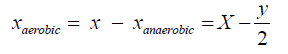
The number of molecules of glucose metabolized under anaerobic conditions (6 carbon atoms) corresponds to half of the amount of lactate produced (3 carbon atoms):

The calculation above shows that from the total amount of glucose in the extravascular fluid, double the amount of lactate is produced under anaerobic conditions, which is identical to the amount of produced ATP:
[ATPanaerobic ] = y
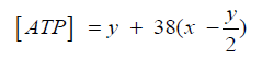
The remaining amount of glucose in the extravascular fluid is exchanged aerobically, i.e., 38 ATPs per 1 molecule of glucose:

The total production of ATP results from the sum of aerobic and anaerobic production:
[ATP] = [ ATPanaerobic] +[ ATP aerobic ]
The average production of ATP from 1 molecule of glucose represents the resulting KEB value:


Two models of energy relationships in the extravascular body fluids
The KEB suggests a foundation for two models of the extravascular fluids [4,5]:
Oxygen is diluted in the extravascular body fluids under normal conditions, enabling predominantly aerobic metabolism in the extravascular fluid compartment. This is associated with a relatively high production of ATP, which is expressed as a high KEB value (Figure 1).
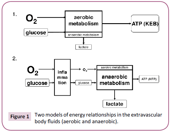
Figure 1: Two models of energy relationships in the extravascular body fluids (aerobic and anaerobic).
Pathological changes in the particular organs are usually associated with an immune system reaction (Figure 1). Activated immunocompetent cells in the extravascular fluid require increased energy; therefore they consume higher amounts of glucose along with more oxygen. This leads to a decrease in extravascular fluid oxygen and results in anaerobic metabolism with an overproduction of lactate. Anaerobic metabolism is less energy efficient, resulting in decreased ATP production. This is reflected in decreased KEB values.
1. Normal condition.
2. Local inflammatory response.
Cytological-energy findings
1. KEB values >28.0 in the extravascular fluid indicate normal condition, but cannot rule out a possible slight serous inflammation in particular organ [4,5].
2. KEB values from 15.0 to 28.0 indicate increased anaerobic metabolism in the extravascular fluid, which may be associated with serous inflammation in particular organ.
3. A significant number of neutrophil granulocytes and a normal energy relationships (with KEB values >28.0) or higher degree of anaerobic metabolism (with KEB values from 15.0 to 28.0) in the extravascular fluid we call “preventive protection” with an increased risk of purulent inflammation in the particular organ.
4. A significant number of neutrophil granulocytes and a high degree of anaerobic metabolism (with KEB values < 10.0) in the extravascular fluid is typical of purulent inflammation in the particular organ, usually involving extracellular bacteria.
5. The significant number of lymphocytes or monocytemacrophages elements and a high degree of anaerobic metabolism in the extravascular fluid (with KEB values < 10.0) may signify an intense inflammatory response in the particular organ with an oxidative burst of macrophages, involving intracellular bacteria, mycotic agents or cancer.
References
- Horejsi V, Bartunkova J (2009) Zakladyimunologie.(4th edn). Triton, Prague.
- Krejsek J, Andrys C, Krcmova I (2016) Imunologiecloveka.(1st edn). Hradec Králové: Garamonsro.
- Kelbich P, Slavík S, Jasanská J, Adam P, Hanuljaková E, et al. (1998) Evaluations of the energy relations in the CSF compartment by investigation of selected parameters of the glucose metabolism in the CSF.KlinBiochemMetab6: 213-225.
- Kelbich P, HejÄÂÂÂÂl A, Selke-Krulichová I, Procházka J, Hanuljaková E, et al. (2014) Coefficient of energy balance, a new parameter for basic investigation of the cerebrospinal fluid. ClinChem Lab Med. 52: 1009-1017.
- Kelbich P, HejÄÂÂÂÂl A, Procházka J, Hanuljaková E, Staněk I, et al. (2016) Cytological-energetic examination of the extra-vascular body fluids, including the cerebrospinal fluid. Biochem Anal Biochem5: Suppl: 4.
- Karlson P, KurzesP (1977) Lehrbuch der BiochemiefürMediziner und Naturwissenschaftler. (10th edn). Georg ThiemeVerlag, Stuttgart.
- Murray RK, Granner DK, Mayes PA, Rodwell VW (1998) Harper’s biochemistry. (23th edn). H&H, Prague.

