Keywords
Viral haemorrhagic fevers; Disseminated intravascular coagulation (DIC) Endotheliopathy; Thrombocytopenia; Multi-organ dysfunction syndrome (MODS); Thrombotic thrombocytopenic purpura (TTP) TTPlike syndrome
Introduction
Ebola viral haemorrhagic fever is a rare but life-threatening haemorrhagic disorder occurring in Ebola viral sepsis. Ebola viruses are found in several African countries. Ebola was first discovered in 1976 near the Ebola River in what is now the Democratic Republic of the Congo. Since then, outbreaks have appeared sporadically in Africa according to the Centers for Disease Control and Prevention (CDC) [1].
Clinical features of Ebola viral haemorrhagic fever include inflammatory symptoms such as fever, myalgia, arthralgia, malaise and weakness. Haemorrhagic signs are petechiae, bleeding in internal organs and from bodily orifices like the mouth, eyes, or ears. Some patients develop bloody diarrhea. Eventually, critically ill patients could progress to more serious conditions, including seizures, delirium, shock, and multi-organ dysfunction.
Thrombocytopenia in critically ill patients (TCIP)
Potential causes of Ebola haemorrhagic disease include: 1) thrombocytopenia related to bone marrow suppression from viral sepsis [2], 2) disseminated intravascular coagulation (DIC) [3,4], and 3) hepatic coagulopathy associated with virus-induced hepatitis/hepatic necrosis [5,6]. However, no credible clinical and laboratory data have been documented to explain the underlying coagulopathy.
Although Ebola haemorrhagic fever occurs with thrombocytopenia [7,8], its relationship to bleeding is not clearly determined because thrombocytopenia is typically mild to moderately severe and it alone can’t be accountable for the severe haemorrhagic disorder. Thus, thrombocytopenia has not entered as a serious issue in caring of Ebola infection other than platelet transfusion to maintain it at a safe level.
Just like other sepsis from bacteria, viruses, fungi or parasites, Ebola sepsis is often associated with TCIP [9]. This term has been applied to etiology-undetermined thrombocytopenia after exclusion of known causes of acute thrombocytopenia (e.g., heparin-induced, drug or transfusion-associated, DIC-associated, hypersplenism-related). An interesting finding is that TCIP not only occurs in sepsis/septic shock, but also occurs in other critical illnesses (e.g. severe trauma, complications of surgery, pregnancy and transplant, and immunologic and collagen vascular diseases).
Recently, significant correlation was noted between the degree of thrombocytopenia and severity of the critical illness as well as prognosis and the likelihood of recovery [10,11]. Severer thrombocytopenia has been associated with systemic inflammatory response syndrome (SIRS) and multi-organ dysfunction syndrome (MODS) [12,13]. These observations support TCIP is an important participant in the pathogenesis of the critical illness.
Endotheliopathy and “two-activation theory of the endothelium”
Viral haemorrhagic fevers, including Ebola, are known to cause the injury of endothelial cells (ECs) leading to endotheliopathy and endothelial dysfunction [14-18]. It is known that endotheliopathy triggers molecular events that promote the activation of two independent endothelial pathways (i.e., inflammatory and microthrombotic). Based on these molecular events, a hypothesis of “two-activation theory of the endothelium” is proposed (Figure 1) [5]. In short, two important molecular events are: 1) release of inflammatory cytokines (e.g., interleukin (IL)-1, IL-6, tumor necrosis factor-α, and others) [19,20], and 2) activation of the platelet and exocytosis of unusually large von Willebrand factor multimers (ULVWF) [21-23]. The former triggers inflammation through “activation of inflammatory pathway” and the latter mediates microthrombogenesis via “activation of microthrombotic pathway” as illustrated in Figure 1. In endotheliopathy, microthrombogenesis is the process in which long elongated ULVWF strings that are anchored to ECs recruit activated platelets to decorate and assemble platelet-ULVWF complexes as microthrombi [23-25]. This mechanism results in disseminated intravascular microthrombosis (DIT) triggering thrombotic thrombocytopenis purpura (TTP)-like syndrome.
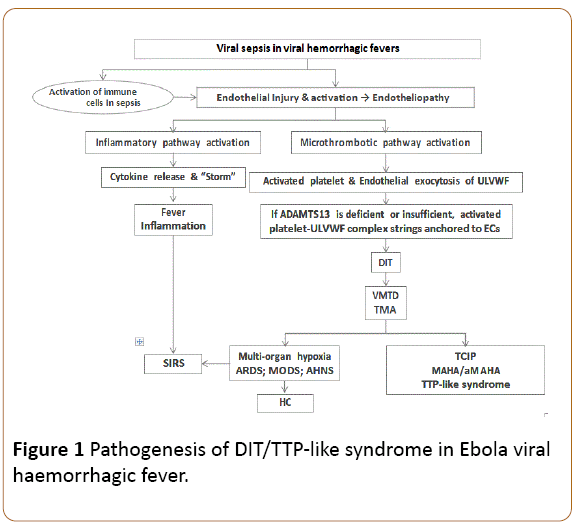
Figure 1: Pathogenesis of DIT/TTP-like syndrome in Ebola viral haemorrhagic fever.
Endotheliopathy-associated DIT is TTP-like syndrome
DIT is the underlying pathological condition leading to vascular microthrombootic disease (VMTD). Systemic VMTD includes two clinical disorders: thrombotic thrombocytopenic purpura (TTP) and TTP-like syndrome. In TTP, microthrombogenesis begins in circulation due to hyperactivity of ULVWF in both hereditary and antibody-associated type. On the other hand, in TTP-like syndrome developing in viral haemorrhagic fevers, it occurs at the intravascular surface of injured ECs. The pathogenesis and clinical characteristics of TTP and TTP-like syndrome are summarized in Table 1. DIT made of microthrombic that are consisting of platelet-ULVWF complexes and anchored to ECs can be called endotheliopathy-associated DIT/VMTD.
| |
Antibody-associated TTP |
Endotheliopathy-associated TTP-like syndrome |
| Etiology |
Anti-ADAMTS13 antibody |
ECs injury leading to dysfunction due to sepsis, surgery, trauma, Shiga toxin, preeclampsia, cancer, drugs, and others |
| Probable pathogenesis |
Increased destruction of ADAMTS13 |
Release of ULVWF and anchored to ECs |
| |
Hyperactive ULVWF® Aggregate with platelet |
Decorated ULVWF with platelets |
| |
Microthrombosis |
Microthrombosis |
| ADAMTS13 level |
Usually <5% |
Usually 20-70% |
| ADAMTS13 antibody |
Positive |
Negative |
| Intravascular ULVWF |
Increased |
Increased |
| Thrombocytopenia |
Present |
Present |
| Anemia |
MAHA |
MAHA/aMAHA |
| Schistocytes |
Always present at usually >2% |
May be present with fewer schistocytes |
| Hypoxic organ dysfunction |
Present |
Present |
| Typical example of examples of involving organs |
Brain (CNSD); Kidneys (ARF) |
Brain (CNSD); lungs (ARDS); liver (HELLP); kidneys (HUS); bowels (GIHS); liver (AHNS); adrenals (Waterhouse-Friderichsen syndrome) muscle (rhabdomyolysis); skin (purpura fulminans) |
| Advanced stage of organ involvement |
MODS |
SIRS; MODS |
| Coagulopathy |
Absent |
Absent unless hepatic coagulopathy occurs |
| Response to TPE |
Very good response |
Excellent response if treated early |
| Platelet transfusion |
Contraindicated |
Contraindicated |
Table 1: Acquired vascular microthrombotic disease (DIT/VMTD): Pathogenic and clinical characteristics of TTP and TTP-like syndrome; DIT: Disseminated Intravascular Thrombosis; VMTD: Vascular Microtrombotic Disease; TTP: Thrombotic Thrombocytopenic Purpura; SIRS: Systemic Inflammatory Response Syndrome; CNSD: Central Nervous System Dysfunction; ARDS: Acute Respiratory Distress Syndrome; GIHS: Gastrointestinal Hemorrhagic Syndrome; AHNS: Acute Fulminant Hepatitis/Acute Hepatic Necrosis Syndrome; ARF: Acute Renal Failure; HUS: Hemolytic Uremic Syndrome; HELLP: Hemolysis, Elevated Liver Enzyme, Low Platelet Syndrome; TPE: Therapeutic Plasma Exchange; ULVWF: Unusually Large von Willebrand Factor Multimers; MODS: Multi-Organ Dysfunction Syndrome; MAHA: Microangiopathic Hemolytic Anemia; aMAHA: Atypical Microangiopathic Hemolytic Anemia; ECs: Endothelial Cells.
In viral haemorrhagic fevers, endotheliopathy-associated DIT/ VMTD could trigger TTP-like syndrome [5,26-29], which is characterized by consumptive thrombocytopenia, microangiopathic hemolytic anemia (MAHA)/atypical MAHA (aMAHA) (if schistocytes are fewer in number) and hypoxic organ dysfunction syndromes. Unlike DIC, in which an abnormal hemostatic (coagulation) disorder occurs following tissue factor (TF) pathway activation, endotheliopathy-associated DIT/VMTD is a pathological microthrombotic disorder occurring as a result of microthrombogenesis. The TF pathway is not involved and coagulation factors are not depleted in the endotheliopathyassociated DIT/VMTD.
Is Ebola viral haemorrhagic fever “DIC”?
The simple answer is no. All the viral haemorrhagic fevers have been attributed to “DIC” [3,4,7]. In clinical medicine, “DIC” mainly has been diagnosed on clinical pretense and confirmed based on a scoring system of the International Society on Thrombosis and Haemostasis (ISTH). Because of misconception of “DIC”, DIT in the critically ill patient has been interpreted as the marker for a hemostatic (coagulation) disorder. This diagnosis hasn’t been based on more reliable coagulation factor assay of FVIII and FV, which are typically depleted in true DIC as seen in acute promyelocytic leukemia [30].
Donald McKay in early1950s coined the term “DIC” [31] for a coagulation disorder that is caused by abnormally activated intravascular thrombotic state. He and his followers believed intravascular microthrombi in the luminal arterioles and capillaries in the pathologic tissue examination were made of micro-clots of platelets, coagulation factors and fibrin. In coagulation profile, the supporting evidence was prolonged prothrombin and activated partial thromboplastin time, hypofibrinogenemia, and increased fibrin degradation products. In many patients with “DIC”, the coagulation profile was perfectly normal and haemorrhagic tendency did not occur. Puzzled but conveniently, the concept of “chronic/compensated/ covert” was introduced. This description, however, cannot explain inexplicably extensive microthrombi in the absence of depleted coagulation factors.
Clinically “DIC” and endotheliopathy-associated DIT/VMTD (i.e., TTP-like syndrome) are exactly the same in their underlying risk factors and presentation. Both almost always occur in critical illnesses (e.g. sepsis/septic shock, trauma, immunologic and collagen-vascular diseases, and complications of surgery, pregnancy and transplant) [32,33]. Pathologically both are characterized by arteriolar and capillary hyaline microthrombic with variable fibroblastic proliferation [34,35]. Hematologically they also present with TCIP and MAHA/aMAHA. Thus, “DIC” and DIT are exactly the same disorder.
Microthrombogenesis and activated TF coagulation pathway
According to the “two-activation theory of the endothelium” DIT induced by microthrombogenesis is completely different from true DIC occurring as a result of activated TF coagulation pathway. Their characteristic difference is shown in Table 2. The former is pathological microthrombotic disorder, but the latter is abnormal hemostatic (coagulation) disorder. Considering the difference in their pathogenic mechanisms, “DIC” must have been understood with a wrong pathogenesis. Hence, “DIC” is a misnomer. More than 60 years, this unfortunate misconception of “DIC” has created confusion in medical science and practice, including diagnostic dilemma and treatment failures until today.
| |
Endotheliopathy-associated DIT/VMTD |
True DIC |
| Examples |
TTP-like syndrome (endotheliopathy-associated) |
APL |
| Nature of the disorder |
Microthrombosis made of platelet-ULVWF complexes |
Coagulation activated by TF-FVIIa complexes |
| Mechanism of the genesis |
Intravascular microthrombogenesis |
Intravascular coagulation |
| Inciting events |
Sepsis; trauma; complications of surgery, pregnancy and transplant; and drugs/toxins leading to endotheliopathy |
Acute promyelocytic leukemia and drugs (?) leading to TF expression |
| Hematological manifestations |
TTP-like syndrome |
Hemorrhagic disease of APL |
| Pathogenesis |
| Mechanism |
Activation of microthrombotic pathway/microthrombogenesis |
Activation of TF-FVIIa pathway |
| Site of activation |
Endothelium |
Intravascular space |
| Pathology |
Endothelial activation/dysfunction® endotheliopathy |
TF expression ® coagulation and factor depletion |
| Result of pathogenesis |
Formation of platelet-ULVWF microthrombi |
Consumption of fibrinogen, FVIII, FV |
| Essence of pathology |
Arteriolar and capillary lumenal microthrombi |
Incoagulable blood/unstable blood clots |
| Effect on the involved organs |
Vascular microthrombosis leading to organ hypoxia |
Hemorrhage leading to organ damage |
| Coagulation tests |
| Fibrinogen; |
Normal |
Decreased |
| PT; aPTT; TT; |
Normal |
Prolonged |
| FDP |
Normal |
Increased |
| FVIII activity |
Normal or increased |
Markedly decreased |
| Thrombocytopenia |
Moderately severe |
Mild to very severe |
| Associated clinical syndromes |
TTP-like syndrome |
Hemorrhagic disorder |
| TMA |
| MODS |
| SIRS |
| Associate hematologic features |
| Schistocytes |
0 - +++ |
0 - + (?) |
| MAHA/aMAHA |
Often present |
Absent |
| Consumptive thrombocytopenia |
Always present |
Present (?) |
| Incidence in clinical practice |
Very common |
Extremely rare |
| Therapy |
| Platelet transfusion |
Contraindicated |
May be needed in leukemia |
| Treatment |
TPE; rADAMTS13 (expected to be very effective) |
Treat underlying pathology (e.g., ATRA in APL) |
Table 2: Hematological and clinical characteristics of endotheliopathy-associated DIT/VMTD and true DIC; APL: Acute Promyelocytic Leukemia; aPTT: Activated Partial Thromboplastin Time; aMAHA/MAHA: Atypical Microangiopathic Hemolytic Anemia/ Microangiopathic Hemolytic Anemia; ATRA: All-Trans Retinoic Acid; DIC: Disseminated Intravascular Coagulation; DIT: Disseminated Intravascular Thrombosis; FV: Factor V; FVIIa: Activated Factor VII; FVIII: Factor VIII; FDP: Fibrin Degradation Products; TMA: Microthrombotic Angiopathy; PT: Prothrombin Time; TF: Tissue Factor; TPE: Therapeutic Plasma Exchange; TT: Thrombin Time; MODS: Multi-Organ Dysfunction Syndrome; rADAMTS13: Recombinant ADAMTS13; SIRS: Systemic Inflammatory Response Syndrome; TTP: Thrombotic Thrombocytopenic Purpura; VMTD: Vascular Microthrombotic Disease.
If one understands and accepts the fact that “DIC” is a misnomer but its euonym is endotheliopathy-associated DIT, Ebola haemorrhagic fever can be explained perfectly well by the concept of DIT. The next question is how Ebola haemorrhagic fever gets haemorrhagic disorder. Another word, “What is the correct diagnosis for “DIC” that is associated with abnormal coagulation profile?” In Ebola, acute fulminating hepatitis/acute hepatic necrosis, especially multifocal necrosis type, occurs without a good explanation [5,36-40]. With “two-activation theory” this can be easily explained by endotheliopathyassociated DIT/VMTD causing hepatic microthrombosis and acute hepatic necrosis syndrome, leading to hepatic coagulopathy [5]. Indeed, hepatic coagulopathy shows exactly the same coagulation profile as seen in “acute DIC”.
True DIC is very rare but occurs perhaps in acute promyelocytic leukemia, presumably due to TF expression from leukemic cells [41]. As illustrated in Table 2, the predominant feature of true DIC is a haemorrhagic disorder without MAHA/ aMAHA or hypoxic organ dysfunction [30,41,42]. In differentiating true DIC from hepatic coagulopathy, the most important test is the assay of coagulation factors, especially FVIII and FV, which are depleted in DIC. More importantly, in hepatic coagulopathy, FVIII is normal or increased although it is markedly decreased in DIC [5,42-44]. Also, a markedly decreased liver dependent FVII occurs in hepatic coagulopathy. A suggested guideline for laboratory tests is presented in Table 3 to aid the differential diagnosis among complicated thrombopathies and coagulopathies [5].
| |
TTP and TTP-like syndrome (DIT) |
TTP-like syndrome (DIT) associated with HC (e.g. Ebola)=acute “DIC” |
DIC (e.g. acute promyelocytic leukemia) |
PF (e.g., amyloidosis) |
| Thrombocytopenia |
Always present |
Always present |
Always present |
Not present |
| MAHA/aMAHA |
Almost always present |
Usually present |
Very unlikely to be present |
Not present |
| Fibrinogen |
Normal |
Decreased |
Always decreased |
Always decreased |
| Factor VIII |
Normal |
Normal or increased |
Markedly decreased |
Decreased |
| Factor V |
Normal |
Decreased |
Decreased |
Decreased |
| Factor X |
Normal |
Decreased |
Usually normal |
Normal |
| Factor VII |
Normal |
Markedly decreased |
Normal |
Normal |
| Factor IX |
Normal |
Decreased |
Normal |
Normal |
| FDP |
Normal |
Positive |
Positive |
Strongly positive |
| Thrombin time |
Normal |
Prolonged |
Prolonged |
Prolonged |
| Thrombosis form |
Microthrombi |
Microthrombi |
Friable macrothrombi (?) or not formed |
Absent |
| Bleeding: Character |
Rare, mild petechiae |
May cause serious bleeding |
Common, serious bleeding |
Slow & persistent bleeding |
| Treatment |
Usually no need of treatment |
Controllable with FFP |
Abrogated with ATRA & chemotherapy |
Treatable with AFA |
| Platelet transfusion |
Contraindicated |
Contraindicated |
May be used with ATRA |
Not needed |
Table 3: Differential characteristic hematologic features among thrombopathies and coagulopathies (Adapted and modified from Chang [5] with permission); TTP: Thrombotic Thrombocytopenic Purpura; HC: Hepatic Coagulopathy; DIT: Disseminated Intravascular Thrombosis; DIC: Disseminated Intravascular Coagulation; PF: Primary Fibrinolysis; FDP: Fibrin Degradation Products; MAHA: Microangiopathic Hemolytic Anemia; aMAHA: Atypical MAHA; FFP: Fresh Frozen Plasma; AFA: Anti-Fibrinolytic Agents; ATRA: All- Trans Retinoic Acid.
In Ebola haemorrhagic fever, TCIP is the earliest indicator suggesting that microthrombogenesis is in progress. If a haemorrhagic disorder occurs, it is neither due to DIC nor due to thrombocytopenia alone, but most likely is due to hepatic coagulopathy occurring with endotheliopathy-associated DIT/ VMTD. In Ebola haemorrhagic fever, the “two activation theory” not only explains the concomitant inflammation, TCIP and progressive hypoxic organ dysfunction, but also would help to unmask unrecognized syndromes such as coming cytokine “storm”, TTP-like syndrome, MAHA/aMAHA, MODS and SIRS as illustrated in Figure 1.
Conclusion
Ebola haemorrhagic fever is due to endotheliopathyassociated DIT/VMTD, which hematologic manifestation is TTPlike syndrome. If the diagnosis is confirmed, in addition to the best supportive care, the treatment for Ebola haemorrhagic fever should include fresh frozen plasma for hepatic coagulopathy and therapeutic plasma exchange for TTP-like syndrome.
References
- About Ebola Virus Disease (2016) Centers for disease control and prevention
- Perng GC (2012) Role of bone marrow in pathogenesis of viral infections. J Bone Marrow Res 1.
- Sundberg E, Hultdin J, Nilsson S, Ahlm C (2011) Evidence of disseminated intravascular coagulation in a hemorrhagic fever with renal syndrome-scoring models and severe illness. PLoS ONE 6: e21134.
- Geisbert TW, Young HA, Jahrling PB, Davis KJ, Kagan E, et al. (2003) Mechanisms underlying coagulation abnormalities in Ebola hemorrhagic fever: Overexpression of tissue factor in primate monocytes/macrophages is a key event. J Infect Dis 188: 1618-1629.
- Chang JC (2016) A thought on possible pathogenesis of Ebola viral hemorrhagic disease and potential treatments: Could it be thrombotic thrombocytopenic purpura-like syndrome? J Ther Aph Dialysis 20: 93-98.
- Ebola virus disease (EVD) Information for clinicians in U.S. healthcare settings (2016) Centers for disease control and prevention.
- Zapata JC, Cox D, Salvato MS (2014) The role of platelets in the pathogenesis of viral hemorrhagic fevers. PLoS Negl Trop Dis 8: e2858.
- Sanchez A, Lukwiya M, Bausch D (2004) Analysis of human peripheral blood samples from fatal and nonfatal cases of Ebola (Sudan) hemorrhagic fever: Cellular responses, virus load and nitric oxide levels. J Virol 78:10370-10377.
- Williamson DR, Albert M, Heels-Ansdell D, PROTECT collaborators. Canadian critical care trials group; Australian and New Zealand intensive care society clinical trials group, Arnold DM, et al. (2013) Thrombocytopenia in critically ill patients receiving thromboprophylaxis: Frequency, risk factors and outcomes. Chest 144:1207-1215.
- Levi M (2016) Platelets in critical illness. Semin Thromb Hemost 42: 252-257.
- Venkata C, Kashyap R, Farmer JC, Afessa B (2013) Thrombocytopenia in adult patients with sepsis: Incidence, risk factors and its association with clinical outcome. J Intensive Care 1: 9.
- Ogura H, Gando S, Iba T, Eguchi Y; Japanese Association for Acute Medicine Disseminated Intravascular Coagulation Study Group (2007) SIRS-associated coagulopathy and organ dysfunction in critically ill patients with thrombocytopenia. Shock 28: 411-417.
- Stravitz RT, Ellerbe C, Durkalski V, Reuben A, Lisman T, et al. (2016) Thrombocytopenia is associated with multi-organ system failure in patients with acute liver failure. Clin Gastroenterol Hepatol 14: 613-620.
- Yang ZY, Duckers HJ, Sullivan NJ, Sanchez A, Nabel EG, et al. (2000) Identification of the Ebola virus glycoprotein as the main viral determinant of vascular cell cytotoxicity and injury. Nat Med 6: 886-889.
- Siragam V, Qiu X (2017) How can Ebola virus infection lead to endothelial dysfunction and coagulopathy? Future Virol 12: 89-92.
- Mackow ER, Gorbunova EE, Gavrilovskaya IN (2015) Endothelial cell dysfunction in viral hemorrhage and edema. Front Microbiol 5: 733.
- Peters CJ, Zaki SR (2002) Role of the endothelium in viral hemorrhagic fevers. Crit Care Med 30: S268-S273.
- Bodur H, Akinci E, Onguru P, Uyar Y, Basturk B, et al. (2010) Evidence of vascular endothelial damage in Crimean-Congo hemorrhagic fever. Int J Infect Dis 14: e704-707.
- Aird WC (2003) The role of the endothelium in severe sepsis and multiple organ dysfunction syndrome. Blood 101: 3765-3777.
- Xing K, Murthy S, Liles WC, Singh JM (2012) Clinical utility of biomarkers of endothelial activation in sepsis-A systematic review. Crit Care 16: R7.
- Janicek MJ, Van den Abbeele AD, Hollenberg NK, Kassis AI, Holman BL, et al. (1990) Platelet activation and aggregation after endothelial injury. Assessment with indium-111-labeled platelets and angiography. Invest Radiol 25: 988-993.
- Bockmeyer CL, Claus RA, Budde U (2008) Inflammation-associated ADAMTS13 deficiency promotes formation of ultra-large von Willebrand factor. Haematologica 93: 137-140.
- Valentijn KM, van Driel LF, Mourik MJ (2010) Multigranular exocytosis of Weibel-Palade bodies in vascular endothelial cells. Blood 116: 1807-1816.
- De Ceunynck K, De Meyer SF, Vanhoorelbeke K (2013) Unwinding the von Willebrand factor strings puzzle. Blood 121: 270-277.
- Padilla A, Moake JL, Bernardo A (2004) P-selectin anchors newly released ultralarge von Willebrand factor multimers to the endothelial cell surface. Blood 103: 2150-2156.
- Vaziri S, Navabi J, Afsharian M (2008) Crimean Congo hemorrhagic fever infection simulating thrombotic thrombocytopenic purpura. Indian J Hematol Blood Transfus 24: 35-38.
- Deepanjali S, Naik RR, Mailankody S, Kalaimani S, Kadhiravan T (2015) Dengue virus infection triggering thrombotic thrombocytopenic purpura in pregnancy. Am J Trop Med Hyg 93: 1028-1030.
- Ardalan MR, Tubbs RS, Chinikar S, Shoja MM (2006) Crimean-Congo haemorrhagic fever presenting as thrombotic microangiopathy and acute renal failure. Nephrol Dial Transplant 21: 2304-2307.
- Lopes da Silva R (2011) Viral-associated thrombotic microangiopathies. Hematol Oncol Stem Cell Ther 4: 51-59.
- Cooperberg AA (1967) Acute promyelocytic leukemia. Can Med Assoc J 97: 57-63.
- McKay DG, Margaretten W (1967) Disseminated intravascular coagulation in virus diseases. Arch Intern Med 120: 129-152.
- Franchini M, Lippi G, Manzato F (2006) Recent acquisitions in the pathophysiology, diagnosis and treatment of disseminated intravascular coagulation. Thromb J 4: 4.
- Nguyen TC, Kiss JE, Goldman JR, Carcillo JA (2012) The role of plasmapheresis in critical illness. Crit Care Clin 28: 453-468.
- Sueishi K, Takeuchi M (1993) Pathology of disseminated intravascular coagulation. Nihon Rinsho 51: 30-36.
- Tsai HM (2010) Pathophysiology of thrombotic thrombocytopenic purpura. Int J Hematol 91: 1-19.
- El Sayed SM, Abdelrahman AA, Ozbak HA, Hemeg HA, Kheyami AM, et al. (2016) Updates in diagnosis and management of Ebola hemorrhagic fever. J Res Med Sci 21: 84.
- Martines RB, Ng DL, Greer PW, Rollin PE, Zaki SR (2015) Tissue and cellular tropism, pathology and pathogenesis of Ebola and Marburg viruses. J Pathol 235: 153-174.
- Bradfute SB, Swanson PE, Smith MA, Watanabe E, McDunn JE, et al. (2010) Mechanisms and consequences of ebolavirus-induced lymphocyte apoptosis. J Immunol 184: 327-335.
- Talwani R, Gilliam BL, Howell C (2011) Infectious diseases and the liver. Clin Liver Dis 15: 111-130.
- Mehedi M, Groseth A, Feldmann H, Ebihara H (2011) Clinical aspects of Marburg hemorrhagic fever. Future Virol 6: 1091-1106.
- Tallman MS, Hakimian D, Kwaan HC, Rickles FR (1993) New insights into the pathogenesis of coagulation dysfunction in acute promyelocytic leukemia. Leuk Lymphoma 11: 27-36.
- Chang JC, Gross HM, Jang NS (1990) Disseminated intravascular coagulation due to intravenous administration of hetastarch. Am J Med Sci 300: 301-303.
- Senzolo M, Burra P, Cholongitas E, Burroughs AK (2006) New insights into the coagulopathy of liver disease and liver transplantation. World J Gastroenterol 12: 7725-7736.
- Castellone D (2010) Liver disease and coagulation outcomes. ANAIRA.