Mustafa Abd El Raouf1,2, Yufeng Zhang1, Jordi Caballé-Serrano3, Richard J. Miron4,5*
1The State Key Laboratory Breeding Base of Basic Science of Stomatology (Hubei-MOST) and Key Laboratory of Oral Biomedicine Ministry of Education, School and Hospital of Stomatology, Wuhan University, Wuhan, China
2Faculty of Veterinary Medicine, Zagazig University, Zagazig, Egypt
3Department of Oral and Maxillofacial Surgery, School of Dental Medicine, Universitat Internacional de Catalunya, Barcelona, Spain
4Department of Periodontology, Nova Southeastern University, Fort Lauderdale, Florida, USA
5Cell Therapy Institute, Center for Collaborative Research, Nova Southeastern University, Fort Lauderdale, Florida, USA
*Corresponding Author:
Richard Miron
Department of Periodontology
Nova Southeastern University
Fort Lauderdale, Florida, USA
Tel: 8005416682
E-mail: rmiron@nova.edu
Received Date: February 27, 2017 Accepted Date: March 13, 2017 Published Date: March 18, 2017
Citation: Miron RJ, Serrano JC, Zhang Y, et al. Novel Natural Bovine Bone Graft with Integrated Atelo-Collagen Type I: Atelo-collagen Bovine Bone Mineral (ABBM) Characterization in-vivo. Periodon Prosthodon 2017, 3:1.
Copyright: © 2017 Miron RJ, et al. This is an open-access article distributed under the terms of the Creative Commons Attribution License, which permits unrestricted use, distribution, and reproduction in any medium, provided the original author and source are credited.
Keywords
Bone graft; ABBM; Atelocollagen; Osteogenesis; Bone regeneration bone formation; Bone induction
Introduction
Bone grafting materials are commonly utilized to replace lost or missing bone [1,2]. Typically the osteopromotive potential of bone grafting materials are characterized based on three fundamental principles including osteogenesis, osteoinduction, and osteoconduction [3]. Osteogenesis refers to the grafts ability to contain living progenitor cells. Osteoinduction is the grafts ability to form ectopic bone formation and osteoconduction refers to the ability for the bone graft to support three-dimensional tissue ingrowth [3]. While it is known that autografts remain the gold standard for bone grafting procedures [4,5] a limited supply and additional donor site morbidity have necessitated alternatives.
Xenografts have been characterized as one of the most widely used bone grafting materials over the past 2 decades [6-8]. They are capable of supporting tissue ingrowth and favor long term stability of bone grafting materials. Interestingly, our group recently revealed how important the surface properties of xenografts are in order to facilitate growth factor adsorption [9]. It is well known that the role of bone grafting materials has gradually evolved from a passive structural-support replacement graft towards grafts that are better able to support tissue regeneration.
Recently, the engineering of a new class of xenograft has been developed by lyophilization of natural bovine bone with integrated atelo-collagen type I (termed ABBM). These processing techniques are more natural due to the use of atelopeptidation and lyophilization technologies that modify the immune-collagen components of the bone grafting material to non-immunogenic atelo-collagen. Processing of xenografts utilizing these technologies preserves the natural properties of collagen so that the end product contains roughly 30% collagen type I remaining incorporated within the xenograft in a natural way. The purpose of this in vivo study was first to investigate the surface characteristics of ABBM bone grafts and then to investigate its ability to form new bone formation in a rat femoral defect without eliciting an immune reaction to host tissues.
Materials and Methods
Bone grafting material
ABBM scaffolds containing atelo-collagen type I were processed utilizing atelopeptidation and lyophilization technologies modifying the collagen components of bone material within bone structure to non-immunogenic atelocollagen preserving the natural properties of collagen (ImploBone, BioImplon, Germany).
This processing technique does not use heat (thermal) processing when manufacturing which has been shown to negatively affect the natural crystalline micro-structure of hydroxyapatite, causing ceramization and destroying collagen components. The lyophilization technique, which involves the evaporation of water contained in a product by sublimation – a previously frozen material is placed in a vacuum, which turns the ice directly to vapor
This process preserves lyophilized collagen with lower humidity making the bone matrix hydrophilic. These bone grafts are found containing roughly 2% moisture, 65-75% hydroxyapatite, 25-35% atelo-collagen content and up to 0.1% non-collagenous proteins (proprietary information). The bone grafting material were visualized at various magnifications using scanning electron microscopy (SEM).
Animals and surgical protocols
Six female Wistar rats (mean body weight, 200 g) were used in this study. Animal handling and surgical protocols were conducted according to the guidelines for animal care and use committee of Wuhan University, People’s Republic of China, and approved by the Ethics Committee at the School of Dentistry. All animals were housed with constant temperature at 20–25° and had food and water ad libitum.
All operations were carried out under aseptic conditions with a gentle surgical technique. For surgery, the rats were generally anesthetized with intraperitoneal injections of chloral hydrate (10%, 4 ml/kg body weight). After disinfection of the skin, a 1 cm linear skin incision was made in the distal femoral epiphysis of hind limbs bilaterally, and blunt dissection of the muscles was performed to expose the femoral condyle.
A 3 mm diameter antero-posterior bicortical channel defect was created perpendicular to the shaft axis by removing cancellous trabecular bone using a dental trephine burr at a slow speed just above the growth plate and the contact surface between drill and bone tissues was kept wet and cool with a 0.9% sterile physiological saline solution to avoid thermal necrosis as previously described [10-13].
The drilled holes were rinsed with saline solution to remove bone fragments from the defect cavity completely. The defect holes were then gently filled with 10 mg of atelo-collagenized bovine bone mineral (ABBM, ImploBone, BioImplon, Germany).
A total of 6 surgeries were performed to investigate the immune response of the material. Postoperatively, a single intramuscular dose of penicillin (400,000 IU/ml, 0.1 ml/kg) was injected. After 4 weeks post-implantation, rats were sacrificed and samples were prepared for histological analysis.
Histological proceeding
The samples were fixed in 4% formaldehyde for 24 hours at room temperature, followed by decalcified in 10% ethylene diamine tetraacetic acid (EDTA) solution for 4 weeks. Then the femoral samples were dehydrated in a series of graded concentrations of ethanol from 70% to 95% and were then embedded in paraffin as previously described [14]. The samples were cut into 5 μm thick sections and stained with hematoxylin and eosin (H&E) (#S2255, Sigma) according to the manufacturer’s protocol.
Results
Surface properties of ABBM
In a first set of experiments to characterize the surface properties of ABBM, SEM was utilized at various magnifications. It was first shown that ABBM particles are found to be on average roughly 0.6-1 mm in size and show various surface topographies even at low magnification (Figure 1).
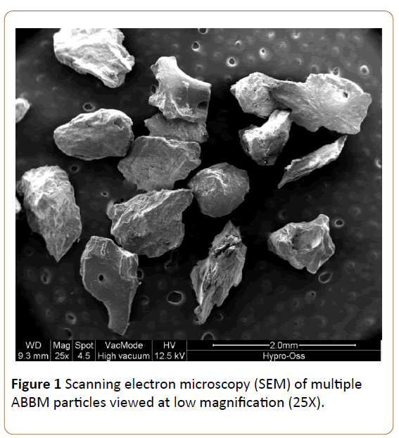
Figure 1: Scanning electron microscopy (SEM) of multiple ABBM particles viewed at low magnification (25X).
Figure 2 demonstrates an ABBM particle roughly 0.75 mm in width. Notice the number of roughened surface topographies that are observed at 100 times magnification.

Figure 2: Scanning electron microscopy (SEM) of an ABBM particle approximately 750 µm in size. Notice the number of macroscopic surface topography features found on the material surface.
When higher resolution SEM microscopy is utilized, the grafts clearly depict a highly collagenous surface texture with visible collagen fibrils found on the surfaces of ABBM particles at 400 and 1600 times magnifications (Figures 3 and 4).
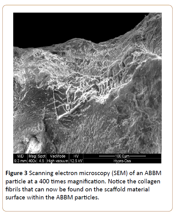
Figure 3: Scanning electron microscopy (SEM) of an ABBM particle at a 400 times magnification. Notice the collagen fibrils that can now be found on the scaffold material surface within the ABBM particles.

Figure 4: Scanning electron microscopy (SEM) of an ABBM particle at 1600 times magnification. ABBM particles appear with many roughened surface properties.
New bone formation/Immune response of ABBM in-vivo
Thereafter, the primary objective of this study was to determine whether these novel ABBM scaffolds can induce new bone formation without eliciting an immune response. All animals healed uneventfully with no signs of local inflammation. An inflammatory response was not observed in any of the defects and all bone grafts regenerated substantial new bone with ABBM and demonstrated a natural healing progression. Figure 5 demonstrates the ABBM graft surrounded by new bone formation.
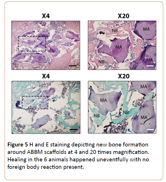
Figure 5: H and E staining depicting new bone formation around ABBM scaffolds at 4 and 20 times magnification. Healing in the 6 animals happened uneventfully with no foreign body reaction present.
It may be observed that the grafting material is found with large quantities of new bone formation observed at both low and high magnification. New blood vessel ingrowth and lack of multi-nucleated giant cells suggest that the material were well integrated into the host tissues without eliciting an immune response.
Discussion
Over the years, a large number of bone grafting materials are continuously brought to market with the aim of facilitating or improving new bone formation. Xenografts are one of the most commonly used bone grafting materials due to their low risk of disease transmission, available supply, and osteoconductive features. Their main disadvantage is that during the processing of typical bone grafting materials, xenografts are commonly devoid of proteins and growth factors therefore demonstrating very little osteopromotive ability when compared to other bone grafting materials [15]. Therefore, new types of xenografts are needed especially in countries that forbid the use of allografts.
The main purpose of this study was to investigate newly developed xenografts that contain integrated atelo-collagen naturally within their bone grafting particles. This relatively new technology makes use of processing techniques that utilize atelopeptidation and lyophilization thereby leaving roughly 25-35% atelo-collagen in a non-immunogenic fashion.
The processing technique does not require heat (thermal) processing when manufacturing which has been shown to negatively affect the natural crystalline properties of hydroxyapatite. Atelo-collagen type I is important for several reasons.
It is well known that collagen type I contains a variety of RGD peptides that allow for integrin bindings of various cell types that are later able to proliferate and differentiate on the material surface [16,17]. We have previously shown that ABBM scaffolds are able to facilitate an increase in cell numbers when compared to deproteinized bovine bone mineral that do not contain any collagen or growth factors [18]. Furthermore, collagen type 1 is known to inhibit bacterial pathogens that are often found in the oral cavity such as Staphylococcus aureus, Staphylococcus epidermidis, hemolytic Streptococcus, and Pseudomonas aeruginosa with some studies a decrease of bacterial activity by roughly 40% [19]. Parallel to this finding, atelo-collagen-based scaffolds have also been shown to provide protection against the early degradation of scaffolds by synovial fluids that contain various matrix metallopeptidases and plasmin proteins [20]. Therefore, the added features of these combined properties that are found when atelo-collagen are incorporated into such xenograft scaffolds are thought to markedly improve the bone regenerative potential of bone grafting materials.
In the present study, the bone grafting materials were first characterized via SEM to investigate the surface morphology of the bone grafting material. It was found that ABBM particles ideally demonstrated a roughened surface with visible collagen fibrils found on the scaffold material surface (Figures 3 and 4). It is well known that these visible collagen fibers are able to support mesenchymal cell adherence to biomaterials through a variety of RGD binding domains along with various fibronectin proteins [21]. Since ABBM contains visible collagen, it is hypothesized that these scaffolds will better support faster cell attachment and new bone formation at earlier time points [22,23].
Thereafter, the ABBM bone grafting materials were implanted into 3mm critical size bone defects and investigated for 2 important parameters: 1) for their ability to regenerate appreciable new bone formation and 2) to investigate the immune response of ABBM which incorporate a natural atelocollagen protein within its scaffold [24-26]. It was first observed that after a healing period of 4 weeks, appreciable new bone formation took place demonstrating the grafts ability to regenerate new bone in a short period of time [27]. Furthermore, no foreign body reaction was observed in any of the 6 animals and it may therefore be concluded that the material is well-suited to regenerate new bone with much further potential for future research and clinical applications. Future research is therefore eminently needed to compare the regenerative potential of ABBM versus some of the leaders in the bone grafting field [28-30].
Conclusion
The results from this study show for the first time the healing potential of ABBM scaffolds when implanted into an animal model for bone regeneration. It was found that ABBM particles demonstrated a roughened surface that was able to facilite appreciable new bone formation when implanted in the femurs of our 3 mm animal model. No foreign body reaction was observed in any of the animals and the atelocollagen was able to promote new bone formation in these defects. Future comparative and large animal models are now necessary to investigate the regenerative potential of ABBM in comparison to other leading bone grafting materials.
References
- Buser D, Chappuis V, Kuchler U, Bornstein MM, Wittneben JG, et al. (2013) Long-term stability of early implant placement with contour augmentation. Journal of dental research 92: 176s-182s.
- Jensen SS, Aaboe M, Janner SF, Saulacic N, Bornstein MM, et al. (2015) Influence of particle size of deproteinized bovine bone mineral on new bone formation and implant stability after simultaneous sinus floor elevation: A histomorphometric study in minipigs. Clinical implant dentistry and related research 17: 274-285.
- Miron RJ, Zhang YF. 2012. Osteoinduction: A review of old concepts with new standards. Journal of dental research 91: 736-744.
- Miron RJ, Gruber R, Hedbom E, Saulacic N, Zhang Y, et al. (2013) Impact of bone harvesting techniques on cell viability and the release of growth factors of autografts. Clinical implant dentistry and related research 15: 481-489.
- Miron RJ, Hedbom E, Saulacic N, Zhang Y, Sculean A, et al. (2011) Osteogenic potential of autogenous bone grafts harvested with four different surgical techniques. Journal of dental research 90: 1428-1433.
- Jensen SS, Bosshardt DD, Gruber R, Buser D. (2014) Long-term stability of contour augmentation in the esthetic zone: Histologic and histomorphometric evaluation of 12 human biopsies 14 to 80 months after augmentation. Journal of periodontology 85: 1549-1556.
- Fujioka-Kobayashi M, Sawada K, Kobayashi E, Schaller B, Zhang Y, Miron RJ (2016) Recombinant human bone morphogenetic protein 9 (rhbmp9) induced osteoblastic behaviour on a collagen membrane compared with rhbmp2. Journal of periodontology 1-14.
- Miron RJ, Wei L, Bosshardt DD, Buser D, Sculean A, Zhang Y. (2014) Effects of enamel matrix proteins in combination with a bovine-derived natural bone mineral for the repair of bone defects. Clinical oral investigations 18: 471-478.
- Fujioka-Kobayashi M, Schaller B, Saulacic N, Zhang Y, Miron RJ. (2016) Growth factor delivery of bmp9 utilizing a novel natural bovine bone graft with integrated atelo-collagen type i: Biosynthesis, characterization and cell behavior. Journal of biomedical materials research Part A.
- Yang S, Lan L, Miron RJ, Wei L, Zhang M, et al. (2015) Variability in particle degradation of four commonly employed dental bone grafts. Clinical implant dentistry and related research 17: 996-1003.
- Zhang Y, Cheng N, Miron R, Shi B, Cheng X. (2012) Delivery of pdgf-b and bmp-7 by mesoporous bioglass/silk fibrin scaffolds for the repair of osteoporotic defects. Biomaterials 33: 6698-6708.
- Zhang Y, Jing D, Buser D, Sculean A, Chandad F, et al. (2016). Bone grafting material in combination with osteogain for bone repair: A rat histomorphometric study. Clinical oral investigations 20: 589-595.
- Zhang Y, Wei L, Miron RJ, Shi B, Bian Z (2016) Bone scaffolds loaded with sirna-semaphorin4d for the treatment of osteoporosis related bone defects. Scientific reports 6: 26925.
- Wei L, Ke J, Prasadam I, Miron RJ, Lin S, et al. (2014) A comparative study of sr-incorporated mesoporous bioactive glass scaffolds for regeneration of osteopenic bone defects. Osteoporosis international : a journal established as result of cooperation between the European Foundation for Osteoporosis and the National Osteoporosis Foundation of the USA 25: 2089-2096.
- Miron RJ, Sculean A, Shuang Y, Bosshardt DD, Gruber R, et al. (2016) Osteoinductive potential of a novel biphasic calcium phosphate bone graft in comparison with autographs, xenografts, and dfdba. Clinical oral implants research 27: 668-675.
- Mizuno M, Fujisawa R, Kuboki Y. (2000) Type i collagen‚ÂÂinduced osteoblastic differentiation of bone‚ÂÂmarrow cells mediated by collagenÃÂα2β1 integrin interaction. Journal of cellular physiology 184: 207-213.
- Staatz W, Fok K, Zutter M, Adams S, Rodriguez B, et al. (1991) Identification of a tetrapeptide recognition sequence for the alpha 2 beta 1 integrin in collagen. Journal of Biological Chemistry 266: 7363-7367.
- Fujioka-Kobayashi M, Sawada K, Kobayashi E, Schaller B, Zhang Y, Miron RJ (2016) Osteogenic potential of rhbmp9 combined with a bovine-derived natural bone mineral scaffold compared to rhbmp2. Clinical oral implants research.
- Carlson GA, Dragoo JL, Samimi B, Bruckner DA, Bernard GW, Hedrick M, Benhaim P. (2004) Bacteriostatic properties of biomatrices against common orthopaedic pathogens. Biochemical and biophysical research communications 321: 472-478.
- Palmer M, Stanford E, Murray MM. (2011) The effect of synovial fluid enzymes on the biodegradability of collagen and fibrin clots. Materials 4: 1469-1482.
- Obara M, Kang MS, Yamada KM. (1988) Site-directed mutagenesis of the cell-binding domain of human fibronectin: Separable, synergistic sites mediate adhesive function. Cell 53: 649-657.
- Teitelbaum SL. (2010) Stem cells and osteoporosis therapy. Cell Stem Cell 7: 553-554.
- Tontonoz P, Pei LM. 2004. Fat's loss is bone's gain. J Clin Invest 113: 805-806.
- Garrett MP, Kakarla UK, Porter RW, Sonntag VK. (2010) Formation of painful seroma and edema after the use of recombinant human bone morphogenetic proteinà in posterolateral lumbar spine fusions. Neurosurgery 66: 1044-1049.
- Lamplot JD, Qin J, Nan G, Wang J, Liu X, et al. (2013) Bmp9 signaling in stem cell differentiation and osteogenesis. American journal of stem cells 2: 1-21.
- Munoz-Felix JM, Cuesta C, Perretta-Tejedor N, Subileau M, Lopez-Hernandez FJ, et al. (2016) Identification of bone morphogenetic protein 9 (bmp9) as a novel profibrotic factor in vitro. Cellular signalling 28: 1252-1261.
- Rodan GA, Martin TJ. 2000. Therapeutic approaches to bone diseases. Science 289(5484): 1508-1514.
- Shah MM, Smyth MD, Woo AS. (2008) Adverse facial edema associated with off-label use of recombinant human bone morphogenetic protein–2 in cranial reconstruction for craniosynostosis.
- Sreekumar V, Aspera-Werz RH, Tendulkar G, Reumann MK, Freude T, et al. (2016) Bmp9 a possible alternative drug for the recently withdrawn bmp7? New perspectives for (re-)implementation by personalized medicine. Archives of toxicology.
- Zhang Y, Yang S, Zhou W, Fu H, Qian L, et al. (2015) Addition of a synthetically fabricated osteoinductive biphasic calcium phosphate bone graft to bmp2 improves new bone formation. Clinical implant dentistry and related research.

