Keywords
Bovine cysticercosis; Economic importance; Zoonotic importance
Introduction
Cysticercosis is a muscular infection of cattle and is caused by larvae of the human intestinal cestod, Teania saginata. Its life cycle is entirely dependent on the link between man and cattle so that any break in this links can result in the total elimination of the parasite (Figure 1). Tapeworm infections have been recorded in history from 1500 B.C. and have been recognized as one of the earliest human parasite [1]. Cysts of Cysticercus bovis can be found anywhere in the carcass, meat and viscera [2]. The distribution of T. saginata is wider in developing countries where hygienic conditions are poor and where the inhabitants traditionally eat raw meat or insufficiently cooked meat [3,4]. Forty percent (40%) of the cases was reported to occur in Africa [3]. World Health Organization (WHO) [4] has reported that this disease is very common in developing countries like Ethiopia. It is associated with poor hygiene and local factors including cultural background (eating raw meat “Kurt”, Kitfo” semi cooked leblebtibis), economic condition and religious beliefs, close proximity of humans to cattle kept with little or no distinction between companion or utility functions [3]. In countries where T. saginata is common and cattle ingested tape worm ova frequently during grazing, an active immunity can develop and the incidence of C. bovis follows a definite age pattern [3]. Slaughtering is often carried out in open air in the absence of abattoirs [3]. Transmission of the parasite occurs most commonly in the environment characterized by poor sanitation, primitive livestock husbandry practice and inadequate meat inspection, management, control police [5]. Bovine cysticerosis is responsible for considerable amount of economic losses which can approach 30% when allowance is made for the loss in the carcass weight and the cost of freezing for the infected meat [6]. The health caused by the adult worms in human gives rise to high medical costs [7]. Generally, the loss is determined by disease prevalence, grade of the animals infected, potential markets, prices of cattle and treatment costs for detained carcass [8] and medical costs for infected human beings [7]. The average annual loss due to taenicidal drugs for treatment in Ethiopia was estimated to be 4,937,583.21 Ethiopian Birr [9-11]. Bovine cysticercosis is widely distributed in Ethiopia and a number of individuals reported the prevalence of bovine cysticercosis in different parts of the country. According to these reports, a prevalence of 9.7% in Gondar by Amsalu [12], 21% in Nekemte by Ahmed [9], 13.85% in Debre Zeit by Getachew [13], 19.5% in Bahir Dar by Mulugeta [14] and 3.2% in different agro climatic zones by Tembo [15] was recorded. The nation’s domestic meat consumption of about 45% comes from cattle, which generates export income mainly from the sale of live animals. In foreign trade, Ethiopia builds a big market in the Middle East. However, the country’s participation in exporting live animals is not yet flourished [16].
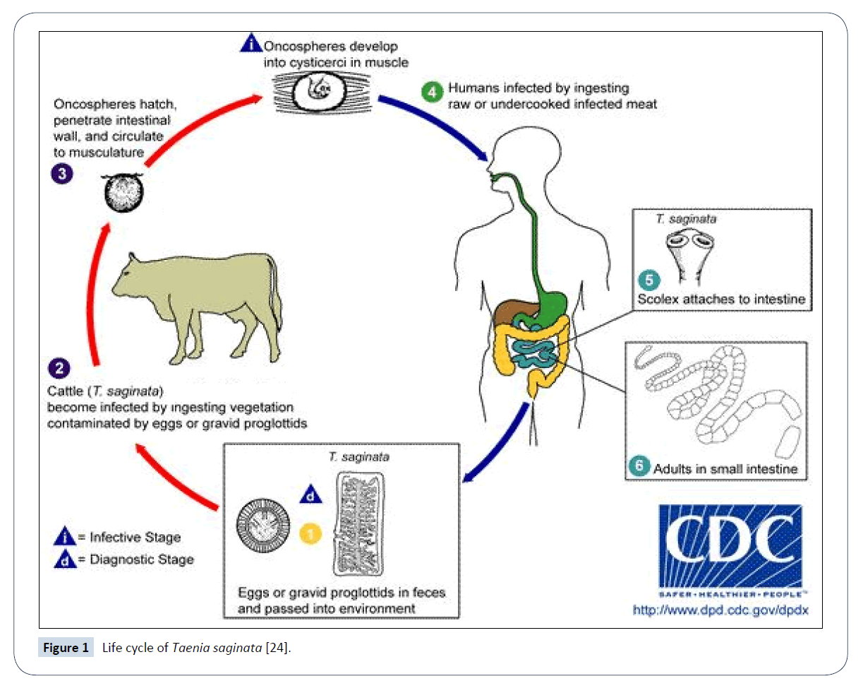
Figure 1: Life cycle of Taenia saginata [24].
On the other hand, the contributions of C. bovis to organ condemnation in slaughtered cattle at different abattoirs have been reported [17,18]. It is a great problem in developing countries like Ethiopia due to the cultural habit of eating raw meat in the form of “kurt” and “kitffo” as routine dish and during holidays has promoted the spread of human taeniasis throughout the country [19]. The above mentioned problems allow the parasite to continue its life cycle till to date and in the coming future [20]. Therefore, the objective of this Mini-Review is to compile some available information on bovine cysticercosis and to create awareness about the transmission and control strategies of the parasite.
Taxonomic Classification
The unarmed meat tape worm of human: Taeinea saginata, and its metacestode, Cysticercus bovis, belong to the class of cestoda, order Cyclophylidea, family Taeniidae and genus Taenia [1].
Adult worm
Taenia saginata is ribbon–shaped flattened, multi segmented and hermaphrodite. The body is divided in to three distinct parts of scolex (head), neck and strobilla. T. saginata measures 4-8 m in lengths and rarely measures up to 15 m. Anteriorly, the scolex has four muscular suckers of unarmed, no rostllum and number of these being relatively characteristics of a species [1].
Larvae
Over a period of three to four months the cysticerci are found after the egg is ingested and may remain in the intermediate host for up to 9 month or even up to the entire life of the host [21]. The larval stage or metacestodes are found in the masseter muscle, tongue, heart, diaphragm [22]. C. bovis is small (pea sized) in shape [23]. C. bovis is grayish white, about one cent meter in diameter and filled with fluid in which the scolex is often clearly visible. As in the adult tape worm, it neither has rostllum nor hooks. Although the cyst may occur anywhere in striated muscle, the predilection sites are the heart, the tongue, masseter and inter-costal muscles [1,24].
Epidemiology
Prevalence and distribution of Taeinea saginata
The prevalence and distribution of Taeniasis/cysticercosis (Taeinea saginata) could roughly be classified in to three groups: mainly those countries or regions with low infection rates less than 0.1%, moderate infection rate and highly endemic with prevalence in a population exceeding 10%. This includes central and east African countries like Ethiopia, Kenya and Zaire (Table 1). Those with moderate infection rate (endemic case) encompass areas like Caucasian and South central Asia Republics of USSR and in the Mediterranean (Syria, Lebanon and Yugoslavia) and countries with prevalence below 0.9% or even free from taeniosis include: North America, Australia and Newzland [1]. Out of 77 million bovine taeniasis carries of the world about 40% live in Africa. The highest being in Ethiopian, it has been reported by different travelers who has come to Ethiopian in ancient times which is documented in medical history of Ethiopia. A recent questionnaire survey in central and North West part of this country showed T. saginata to be in proportion of 84.9% and 67.7% among the population [15]. This situation of the disease in Africa is quite common, reaching 3.2.7.5% in Nigeria, 12.5% in Botswana, greater than 10% in Sudan, 30-36% in Kenya, 15% in Rwanda, 20% in Guinea and 31% in Burundi [21].
| Country |
Prevalence in % |
Source |
| Zambia |
6.1 |
Dorney, 2002 |
| Namibia |
6.2 in communal, 2.3 commercial |
Kumba, 2001 |
| Egypt |
0.23 in native cattle 7.2 in imported cattle |
Haridy, 1999 |
| Kenya |
33.02
14-18.2 |
Oyango –Abuje, 1996
Fulorovo, 1982 |
| Zaire |
22.3 |
Fulorovo, 1982 |
| Chad |
6.67 |
Fulorovo, 1982 |
| Nigeria |
10.2 |
Fulorovo, 1982 |
| Ethiopia |
2.2-3.2
3.2 |
Teka, 1997
Tembo, 2001 |
Table 1: Prevalence of Cysticercus bovis in some African countries [27].
Prevalence and distribution of Cysticercus bovis
The incidence of bovine cysticercosis varies depending on many factors, the major ones are, the degree of the contact between man and cattle and the livestock management system in livestock practice also some other factors. According to WHO [21], the incidence of C. bovis could be hyperendemic, endemic and epidemic cysticercosis. Hyper epidemic condition occurs in pastoral areas and it is also a common epidemiological picture in Africa. For instance; according some studies in Kenya, bovine cysticercosis is high in the market district where cattle graze along the side of stock men for most of the day and brought back to their houses in the evening. This practice ensures continuous man and cattle contact which makes the incidence of cysticercosis very high among their cattle [25]. Epidemics of cysticercosis is usually associated with feed lots is becoming serious problems, may arise either from direct contamination of feed lot with eggs or from the introduction of contaminated fodder silage contaminated with feces of laborers. Endemic cysticercosis or urban cysticercosis: this is associated concentration of localities with around urban long numeration, recreation areas, regions with developed agricultural industry as well as long rivers and main roads [21]. Contaminated with sewage workers of feed or effluent discharged in to rivers and to occasional flooding of pasture by infected individuals in contrast to the epidemiology in developing countries. Cattle of any age are susceptible to infected [1]. There are considerable variations in the prevalence of the disease in different areas that are not easily explained by existing information. However, it appears that taenia infections and cystic infections are usually associated with poor groups [26]. Among human or animal wastes, sewage sludge, manure and slurry used in agriculture are real health concern. Sewage sludge has been used for years as land fertilizer in many countries and this could serve as possible source of pathogens and important component [21].
Life Cycle
The life cycle of T. sginata is indirect where the definitive host is human and intermediate host is cattle [1]. Man is infected by the ingestion of raw uncooked parasitized meat [21]. Only fresh, viable C. bovis cysts are infective formally [27,28]. An infected human passes millions of eggs daily either free in the stool or as intact segment and each containing 250,000 eggs and these can survive on pasture for several month [1]. The oncosphere travels via blood to the striated muscle where it encysts and matures to be infective to man in about 12 weeks. Once in the animal body, the eggs reach and the larval work their way in to muscle tissue. Fortunately, cattle cannot transmit the disease among themselves [1]. However, they can ingest eggs from human sewage and excrement that have contaminated water and feed, or by liking soiled utensils. Eating cyst-laden and incompletely cooked meat, on the other hand may affect human. Humans cannot spread cysticercosis to their own species [21]. Finally, human being becomes infected by ingesting raw or inadequately cooked meat, which contains viable cysts [29].
Economic Importance
Economic losses resulting from food borne parasitic zoonoses are difficult to assess. Estimating the global economic impact, prevalence and public health importance of these parasitic zoonoses are handicapped by inadequate information [30]. Economic losses from cysticercosis are determined by disease prevalence, grade of animal’s infected, potential market price of cattle and treatment cost for detained carcasses [26]. For example, in Botswana and Kenya, the incidence of C. bovis at export abattoirs is about 8% and 12%, respectively. Annual losses in Botswana now approaches to 0.5 million pounds; while in Kenya it is about 1 million pounds. Similarly, the loss per animal slaughtered in Botswana is 2.25 million pounds; while in Kenya it is about 1 million pounds [21]. In fed lots situation, cysticercosis can cause a crippling economic below and may prevent the survival of the enterprise, particularly where it is not able to insure against such losses one such loss was estimated at 2,567,799 dollars in Yugoslavia [21]. The economic significance of adult parasite T. saginata and the proportion of carriers requiring hospital treatment was over 20% in Poland and 10% in France it is also assumed that each carrier misses one day work on average in France [21]. The economic impact of the disease in the cost implication can be broken down in to those involved in treating human taeniosis and cattle carcass (cost of freezing) Or condemned as well costs involved in the inspection procedures amount to a million of dollars. Lightly infected carcasses can be easily missed and passed for human consumption; thus infection transmission is maintained between human and cattle taeniosis (Cysticercosis) remaining a wide spread zoonosis that affects human health and economy through condemnation, quality degradation of frozen meat, cost of refrigeration, cost of human therapy efficiency by creating un easiness (Figure 1) [27]. In Ethiopia, there is a wide usage of both traditional and modern taenicidal drugs (Table 2) [26], which is an indication of the economic importance of the drug in each house hold. The total dose of Niclosamide and Diclorophen production in the drug factories in this country between 1996 and 2000 was 31,814,833. Generally, condemnation of meat and organs from infected animals are the causes of reduction of meat production and restriction on import export trade [27].
| Local Name |
Scientific Name |
Parts Used |
| Bisana |
Crrotonmacro satchuys |
Bark |
| Bisana |
Crotonmacro satchuys |
Seed |
| Enkoko |
Embelia schimperi |
Fruits |
| Dubafire |
Cucurbita pepo |
Seed |
| Kosso |
Hagenica abyssinia |
Flowers |
Table 2: Major taenicidal herbs used in Ethiopia arranged in decreasing order of potency [26].
Zoonotic Importance
Man is the only final host where the adult Taenia saginata resides in the small intestine. The size reached by the adult worm is related to the number of worms present [31]. In a single worm infection, a worm can develop longer and produce large number of proglottids [24]. Multiple infections up to 20 tapeworms in one host are often occurring in developing countries [5]. Tapeworms can also cause intestinal obstruction [32]. Taenia saginata in the small intestine of man absorbs digested food from the day the cysticercus is ingested it may take 2-3 months for the parasite to produce ripe segments. As long as the scolices are attached to the intestinal mucosa of the victim new segments will continually grow to replace those, which are being detached from the worm [33]. Transmission between animals and man depend on etiological factors such as human habits, behavior, religion and beliefs. They influence the type of food man consumers and the manner of preparation. Some of these practices are based on the hundreds of years of traditional Example of this could be eating of raw meat in the form of ‘kitfo’ ‘Lebleb’, and kurt in Ethiopia. In Egypt, Turkey and Middle East a beef dish known as caunters “basterma” or kebaba-like fishes “basterma “or semi-raw meat used as staffing for regional: dishes are responsible for transmission teniasis [21].
Diagnosis
In human
Adult cestodes can be expelled from human using Anthelmentics followed by a saline purgative and identified based on the scolex and proglottid morphology. In human beings, the diagnosis is established by examination of the eggs in the stools or gross examination of the proglosttids or segments passed in the stool. Diagnosis is based on symptoms, fecal examination and rectal swabs, although it is difficult to discover the disease during the first three months. A person should not be considered uninfected before having three negative test completed over a 2-3 days interval. T. saginata egg can be distinguished from T. solium or other tape worms such as echinococcus by their morphology. However ELISA and PCR can differentiate the eggs of T. solium from T. saginata and morphology can be used to distinguish these proglottides [34].
In animals
It is difficult in live animals to diagnoses the presence of C. bovis in the muscles. Thus diagnosis can be performed only at post mortem examinations by direct observation of C. bovis in the muscles [28]. Live cattle show no symptoms, so that it becomes extremely important to identify the cysts during met inspection. A previous history of infestation on the animal premises also acts as available diagnostic tool. Serological tests are also available to detect the disease in live animals. The IHAT with 100% sensitivity and 91-100% specificity can be used as a diagnostic test for epidemiological survey, to map infected and disease free areas and to estimate the natural prevalence of the disease [27].
Risk Factors for Taenia saginata and Cysticercus bovis
Environmental factors
Emphasis has been placed on the environmental factors that affect the free living egg. The infective pattern of taeniids in their intermediate host is determined by complex interaction of parasite and host related factors. Many of these have been examined but particularly emphasis placed on environmental factors that affect determining the infective pattern. The dynamic of this aspect of transmission has been previously neglected [35].
Egg output and dispersal in the system
The biotic potential of micro climatic effects of the environment, the egg output of T. saginata worms have been earlier described in this paper. The daily output runs in to several hundred thousand eggs. The important epidemiological aspects is the ways in which the enormous number of eggs dispersed following expulsion in proglottids and feces so that they become readily available to the intermediate hosts [36]. The primary sites of egg deposition determine the movement and defecation habit of the definitive host. How every, evidence is accumulating the considerable dispersion occurs almost immediately after defection. Intermediate host, cattle generally avoid grazing areas contaminated with feces. Thus, dispersal increases the chance the being ingested. The combination of high biotic potential and long range dispersal means that individual infective host over a very wide area [37]. As a risk for the occurrence of the disease recently such idea has been reported by Cabart et al. [37]: emphasizing the need of surveillance on the use of urbane sewage on pastures because of the cysticercosis threat. Birds like sea gulls and other scavenging birds have been implicated in the transfer of egg of T. saginata from sewage to pasture [24]. The possible role of the soil fauna in the epidemiology of cysticercosis especially worth worms could be a possible candidate for dispersion of the eggs. Furthermore many arthropods have been implicated in egg dispersal, including flies, beetles, ants, fleas, and cockroaches. The blow flies and dung beetles appear to be the most likely candidate due to their ecological association with fecal material [38].
Egg longevity and duration of infectivity
It will be very important to define factors that are lethal to the eggs because it will give us an over view of the epidemiological significance of the ageing process and some other factors on the duration of infectivity. In damp temperate climates, the oncosphere of the beef tape worm can with stand desiccation in the environment for about 10 months are viable for 130 days in pure water, liver for up to 70 days at temperature varying from +4 to -38°C and do not die in winter. In summer, solar radiation destroys the oncospheres on soil surface with in two days. How every, under the protection of a plant cover they can survive for up to 40 days. In the micro climate of cattle the egg can stay up to 18 months [35].
Temperature
Under laboratory conditions the viability of isolated eggs of T. saginata is much higher when compared with those inside a prognostic. At temperatures 19-37°C longevity varies from 27-29 h to 2-3 days, and at 40°C it is about 62-64 days. With regards to humidity, the eggs of T. saginata do not survive in vitro in the absence of surface moisture [21].
Intrinsic factors (Host factors)
The survival of the parasite population is also dependent up on the intrinsic regulatory mechanisms of the intermediate host population. In addition, immunity to super infection is acquitted and larva survives in only proportion of intermediate population. Only that proportion will play any active part in the transmission of cysticercoids, before the onset of acquired resistance in the intermediate hosts. The infective pattern of larva cestodes will be modified by the infection pressure, the age of the host at the time of the first infection, feeding habit, livestock movement, egg dispersed and the invasiveness of the embryo. Workers in Kenya have shown that most cases of bovine cysticercosis are acquired in early calf hood, calves from birth to 28 days being highly susceptible to artificial infection [26]. However, all age groups of cattle are susceptible to infection young animal being more susceptible than older ones which tend to be more resistant [29]. In heavily endemic areas it is likely that calves encounter infection in the first few days of life after which they become progressively more resistant. Both sexes are susceptible. One of the important factors that determine the infective pattern is the time interval after ingestion of the first egg by immunological competent animals where thy remain susceptible to super infection cattle produce immunity within 12-16 days after infection, the level of titers stabilized in the haemagglutination reaction which remain positive throughout the time of serological study (250 days post infection). In calves have demonstrated that cysticerci perish and become calcified after 1-12 months not only in strongly infected animals, but even when infestations were relatively low. On the contrary; researchers in Kenya have indicated that calves infected with beef tape worm onchospher in the first days after birth carry viable cysts during their entire life, developing persistent immunity to reinfection even if the rate of infestation is very low [3].
Treatment
For the treatment of the taeniosis there are a number of taenicidal drugs available in the market. However, the drugs of choice in treating taeniosis are Niclosamide. Adult dose rate of 200 mg is effective in damaging the worm. Albendazole is a broad spectrum anthelmentic of the Benzimadazole Carbanate class, which is effective against larva and adult stage of cestodes and treamtode [21]. In addition, Praziquentel, 5-15 mg/kg, is effective. Alternatively, a single 2 g dose of Niclosamice is given as 4 tablets (500 mg each that are chewed one at a time and swallowed with a small amount of water (0.5 g is the dose of children 2-5 years old, 1 g for old children) Both drugs have cure rates of about 90% Treatment can be considered successful when no proglottides are passed again within 4 months [39]. In Ethiopia, the majority of the rural inhabitants use traditional herbal drugs in routine self-deworming practices as a taenicidal herb, which has been a major concern for researchers [26].
Control and Prevention
Prevention of infection in cattle
Health education should be regarded as the key factor in obtaining the commitment for development of, and continuing involvement in a control program. Proper health education should principally be oriented to diminish the number of tape worm carriers, thus lowering the egg output, change in attitudes, traditional, socio-cultural and behavioral factors that favor a high infection pressure from carriers, and educate people on the prevention and control [21]. In the prevention of infection from man to animals; special attention should be paid to persons likely to infect food animals (cattle), such as farmers, agricultural workers, hunters and tourists. Farmers should be informed the risk of association with the life cycle of the parasite. They should be encouraged to report the presence of infection, use toilets and avoid contamination of the environment [19]. Immunity to larval tapeworm infection plays an important role in the dynamic transmission of the parasite. In general, extensive studies have been under taken to characterize the immune response to cysticercosis in cattle [40].
Prevention of human infection
The traditional methods of slaughter, dressing and marketing create difficulties for inspection. Time is lost during manual killing and dressing of caresses and butchers may also oppose inspection due to fears loss of earnings [31]. Health education of the general public is a key after in the prevention of taeniosis/cysticercosis: however, the follow methods of control are important. Meat inspection in abattoir is the major way of ensuring that consumers are supplied with ‘measly’ free beef as long as the law enforcement concerning a carcasses infection with Cysticercus bovis are strictly followed. It is suggested that effectively improve meat inspection based on the type of the animal, husbandry history and the target of human population of consumption [41]. In addition, other control approaches such as immune diagnosis should be developed and implemented to complete meat inspection procedures. When infection is detected in a carcass the judgment varies from country to country but it has been recommended accordingly [21]. Less contaminated meat should be trimmed and slightly or moderately infected carcass should be thoroughly looked and frozen. From the control of T. Saginata freezing the cyst is the most ideal method used in abattoirs of international standards. In general, freezing for 48 h at a temperature of -10°C for ten days is recommended [39].
Conclusion
The wide distribution of Taenia saginata (Cysticercus bovis) is associated with several factors including: Raw and under cooked beef consumption, bush defecation and poor waste disposal, poor sludge and sewage treatment system, low level of public awareness, and presence of backyard (village) slaughtering practices. Therefore, Veterinarians and Medical professions need to work in collaboration for the control of the disease. In addition, public health awareness should be created through public media on improving personal and environmental hygiene for breaking the life cycle of the disease.
References
- Urquhart GM, Armour J, Duncan JL, Dunn AM, Jennings FW (1996) Veterinary Parasitology (2nd edn.). Black Well Science, London pp: 120-137.
- Ginsberg A (1960) The Detection of Cysticercus bovis in the abattoir. Vet Res 72: 310-311.
- Fralova A (1985) Taeniosis. In: Aed L (ed.) Zoonotic control. UNEP publication, Moscow pp: 192-235.
- World Health Organization (WHO) (1996) Investigating in health research and development. Report of the committee on Health research relating to future intervention options. WHO, Geneva, Switzerland p: 278.
- Mann I (1984) Environmental Hygiene and sanitation based on the concept of primary health as a tool for surveillances, prevention and control of taeniosis/Cysticercosis. Current Public Health Research in Tropics pp: 127-140.
- Pawlowski ZS, Schuitz MG (1972) Taeniasis and cysticercosis (Taenia saginata). Adv Parasitol 10: 269-343.
- Fan PC (1997) Annual economic losses caused by Taenia saginata taeniasis in East Asia. Parsitol Today 13: 194-196.
- Gruindle RG (1978) Economic loss resulting from bovine cysticercosis with special reference to Botswana and Kenya. Tropical Animal Health and Production 10: 127-140.
- Ahmed I (1990) Bovis cysticercosis in animals slaughtered at Nekemte abattoir, Ethiopia. DVM thesis, Addis Ababa University, Faculty of Veterinary Medicine, Debre Zeit, Ethiopia.
- Dawit S (2004) Epidemiology of Tenia saginata taeniosis and cysticecosis in North Gondar Zone, DVM thesis, Addis Ababa University, Faculty of Veterinary Medicine, Debre Zeite, Ethiopia.
- Fufa A (2006) Study on the prevalence of Bovine cysticercosis in Hawassa Municipal Abattior and Taenia saginata in Hawassa town and its surrounding; South Ethiopia, Msc thesis, FVM, AAU, Debre Zeit, Ethiopia.
- Amsalu D (1989) Prevalence and significance of Cystcercus bovis among slaughtered cattle at Debre Zeit abattoir; DVM thesis, Debre Zeit, Ethiopia.
- Getachew B (1990) Prevalence and significance of Cystcercus bovis among cattle slaughter at Debre Zeit Abattior, DVM thesis, FVM, AAU, Debre Zeit, Ethiopia.
- Mulugeta A (1997) Bovine cysticercosis: prevalence, economic and public health importance at Bahir Dar municipal abattoir. DVM thesis. Addis Ababa University, Faculty of Veterinary Medicine, Debre Zeit, Ethiopia.
- Tembo A (2001) Epidemiology of Teania saginata Taeniosis/Cysticercosis in three selected Agro climatic Zones in centeral, Ethiopia, MSc thesis, FVM, AAU, Debre Zeit, Ethiopia.
- EARO (Ethiopian Agricultural Research Organization) (2000) Beef Research Strategy. Animal Science Directorate Adiss Abeba, Ethiopia pp: 241-243.
- Fekadu D (2003) A study on cestodes and metacestod of sheep in Sheno agricultural Research (SHARC), North Shoa. DVM thesis, Faculty of Vetrenary Medicine, Addis Ababa University, Debrezite. Ethiopia.
- Mezgebu Y (2003) Major causes of organ condemnation in ruminant slaughtered at Gondar abattoir. DVM thesis, Faculty of Veterinary Medicine, Addis Abeba University, Debrezite, Ethiopia.
- Gebreemannel T (1997) Food hygiene principles and methods of food borne disease control with special reference to Ethiopia, Addis Ababa University, Faculty of veterinary medicine Department of community health, Debre Zeit, Ethiopia pp: 104-115.
- Eckert J (1996) Food safety, meat and fish born zoonoses. Vet Parasitol 64: 143-147.
- World Health Organization (WHO) (1983) Guidelines for Surveillance, Prevention and Control of Taeniasis/Cysticercosis. In: Gemmell M, Matyas Z, Pawlowski Z, Soulsby EJL (eds.) VPH 83: 49-207.
- Food and Agriculture Organization (FAO) (2007) Animal production and health division relating to animal health disease cards of cysticercosis (bovine).
- The World Organisation for Animal Health (OIE) (2004) Manual of Diagnosis Tests and vaccines for terrestrial Animals (mammals, birds and bees) (5th edn.). Paris pp: 997-1005.
- Smyth JD (1994) Introduction to Animal Parasitology (3rd edn.). Hodder and Stoughton: London pp: 259-273.
- Onyango AJA, Hughes G, Opicha M, Ninyi KM, Rugutt MK, et al. (1996) Diagnosis of Teania saginata Cysticercosis in Kenya cattle by antibodies and antigen ELISA. J Vet Parasitol 66: 221-230.
- Feseha G (1995) Zoonotic disease in Ethiopia. Ethiopia Vet Assoc Proc pp: 22-38.
- Nigatu K (2004) Cystcercus bovis: Development and Evaluation of serological Tests and prevalence at Addis Ababa Abattoir, Ethiopia.
- Gracey FJ, Collins SD, Hucy RJ (1999) Meat hygiene (10th edn.). Bailliere Tindall, Oval roads, London pp: 413-420.
- Dunn AM (1978) Veterinary Helminthology (2nd edn.) London: Balter and Tanner Ltd 118: 276-278.
- Murell B (1991) Economic losses resulting from food born parasitic zoonoses. In: Hailu D (ed.) Prevalence and risk factor for Teania saginata cystycercosis in three selected areas of Eastern Shoa. South Asian Journal of Tropical Medicine 22: 268-270.
- Maedia GE, Kyvsgaard NP, Nansen C, Bogh HO (1996) Distribution of Teania saginata cyst by cattle group on naturally infected cattle in Tanzania. Prev Vet Med 28: 81-89.
- Wanzala W, onyango-Abuje JA, kang’ethe Ek, Zessin, kyule NM, et al. (2003) Analysis of post mortem diagnosis of bovine cysticercosis in Kenya cattle. Onl J Vet Res pp: 1-9.
- Teka G (1997) Food Hygiene Principles and Food Born Disease Control with special Reference to Ethiopia. Addis Ababa University, FVM, Department of Community Health, Addis Ababa, Ethiopia.
- The World Organisation for Animal Health (OIE) (2005) Manual of Diagnosis Tests and vaccines for Terrestrial Animals (mammals, birds and bees) (5th edn.). Paris pp: 997-1005.
- Ferting DL, Dom CR (1985) Teania saginata cysticercosis in Ohio cattle feeding operation. Journal of Am Vet Med Assoc pp: 1281-1286.
- The World Organisation for Animal Health (OIE), World Health Organization (WHO) (2005) Cestodes.
- Carbaret J, Geerts S, Madeline M, Ballandone C, Barbir D (2002) The use of urban sewage sludge on pastures: the cysticercosis threat. Vet Res London pp: 575-597
- Abusers T, Epe G, Schnier, Klein M (2006) Visual diagnosis of Taenia saginata cysticercosis during meat inspection: is it unequivocal. Parasitol Res 99: 405-409.
- CTA (1989) Mannual of Tropical Veterinary Parasitology (2nd edn.). Harcount Brace & Company. Asia: PTE Ltd pp: 387-443.
- Soulsby EJL (1982) Helminthes, Arthropods and protozoa of domesticated animals (7th edn.). London Lead and Febiger. Philadelhia: Bailliere pp: 107-111.
- Gordon N, Geelong G (2006) Beef measles.