María Vallet-Regí1,2*, Juan L Paris1,2, Paz de la Torre3, Victoria Cabañas M1, Miguel Manzano1,2,and Ana I Flores3
1Faculty of Pharmacy, Department of Chemistry in Pharmaceutical Sciences, Hospital Health Research Institute, Spain
2Biomedical Research Center in Bioengineering, Biomaterials and Nanomedicine Network (CIBER-BBN), Spain
3Group of Regenerative Medicine, Hospital Research Institute, Spain
*Corresponding Author:
María Vallet-Regí
Faculty of Pharmacy
Department of Chemistry in Pharmaceutical Sciences
Hospital Health Research Institute
Spain
Tel: +34 913941843
E-mail: vallet@ucm.es
Received date: December 21, 2017; Accepted date: January 10, 2018; Published date: January 12, 2018
Citation: Vallet-Regí M, Paris JL, Torre PDL, Cabañas MV, Manzano M, et al. (2018) Mesenchymal Stem Cells from Human Placenta as Nanoparticle Delivery Vectors. Insights Stem Cells Vol.4: No.1: 1.
Commentary
The use of cells with migratory properties towards different pathological sites holds great promise in the development of future therapeutics [1,2]. Some of these cell types can exert some positive effect on the development of a variety of diseases [3]. Furthermore, these effects could be improved by also transporting some drug of interest within the migrating cells [4]. However, the number of different drugs that can be introduced within the vehicle cells without compromising their viability or migratory behavior is very low. For this reason, the introduction of drug-loaded nanoparticles appears as an interesting strategy to increase the amount of drug that the cells can carry, allowing us to also ensure the retention of the active molecule inside the cell during its journey in the bloodstream [5].
5]. Among the proposed cell types for this application it is worth mentioning macrophages, lymphocytes, different bacteria and mesenchymal stem cells (MSCs) [3,6-9]. MSCs are multipotent stem cells which present several advantages compared to other migratory cells, such as their immunomodulatory properties, which would allow their therapeutic use in allogeneic settings [10,11]. MSCs can also secrete different growth factors, hormones and other signaling molecules, and some MSC types have been shown to hinder tumor development. Several signaling molecules are known to be involved in MSC migration towards tumors, in a concerted manner in which all the signaling factors cooperate to drive tumor homing [12,13]. Some authors have reported that, under certain conditions, some MSC types can also favor tumor growth and progression, so a deeper understanding of the interactions between MSCs and solid tumors is vital to enable their use in the clinical setting [12].
MSCs obtained from the decidua of human placenta (DMSCs) are especially promising (for cancer therapy and other applications), since they have been shown to migrate towards tumors in vitro and in vivo, and they can slow down the growth of primary mammary tumors in immunocompetent rats, as well as they can reduce the appearance of secondary tumors [10,14].
Different types of nanoparticles (made of organic, inorganic or hybrid materials) could be included inside these DMSCs in order to be transported towards the diseased site [15]. Among all of these nanoparticle types, mesoporous silica nanoparticles (MSNs) are attractive candidates as drug carriers due to their high physicochemical stability and their textural properties, such as a high surface area, which provide a high drug loading capacity [16-18]. For all the reasons exposed above, we proposed the use of DMSCs as vehicles to carry MSNs towards diseased sites, especially in the context of cancer (Figure 1a) [19]. In that first work, we employed positively-charged nanoparticles to enhance particle uptake by DMSCs. Other research groups have developed nanoparticles modified with ligands capable of interacting specifically with the cell membrane of MSCs to increase their association with the cells. A couple of examples of these strategies consist in decorating MSN surface with anti-CD90 antibodies [20], or with hyaluronic acid, which interacts with CD44 [21], being both of these markers expressed on the MSC membrane.
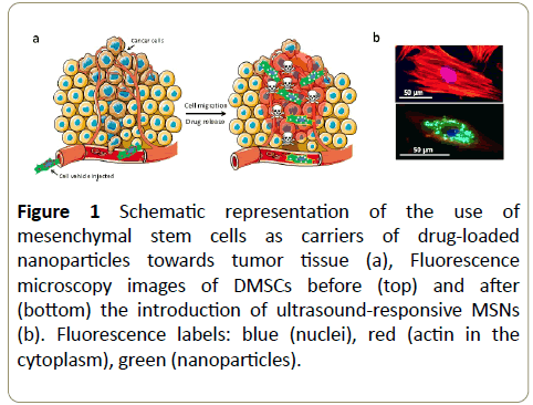
Figure 1: Schematic representation of the use of mesenchymal stem cells as carriers of drug-loaded nanoparticles towards tumor tissue (a), Fluorescence microscopy images of DMSCs before (top) and after (bottom) the introduction of ultrasound-responsive MSNs (b). Fluorescence labels: blue (nuclei), red (actin in the cytoplasm), green (nanoparticles).
Other authors have also proposed similar cell-transport strategies employing different cell vehicles with MSNs or other types of nanoparticles [20-23]. However, the high toxicity of anticancer drugs might compromise the viability and migration of the cell vehicles in all of those systems. For that reason, we introduced inside DMSCs ultrasound-responsive MSNs which could retain the toxic cargo within them until exposed to an external stimulus (Figure 1b) [24]. Recently, other researchers have developed similar approaches, employing nanoparticles sensitive to other external stimuli, such as Near Infrared (NIR) light [25,26]. Even though the penetration capacity of NIR light is enhanced compared to visible or ultraviolet light, ultrasound can still penetrate much deeper in the body in a non-invasive manner. This provides a significant advantage over the cited works and it could also ease the future clinical translation of our platform.
References
- Stuckey DW, Shah K (2014) Stem cell-based therapies for cancer treatment: Separating hope from hype. Nat Rev Cancer 14: 683-691.
- Harrington K, Alvarez VL, Crittenden M (2002) Cells as vehicles for cancer gene therapy: The missing link between targeted vectors and systemic delivery? Hum Gene Ther 13: 1263-1280.
- Karp JM, Leng, Teo GS (2009) Mesenchymal stem cell homing: The devil is in the details. Cell Stem Cell 4: 206-216.
- Shah K (2012) Mesenchymal stem cells engineered for cancer therapy. Adv Drug Deliv Rev 64: 739-748.
- Gao Z, Zhang L, Hu J, Sun Y (2013) Mesenchymal stem cells: A potential targeted-delivery vehicle for anti-cancer drug loaded nanoparticles. Nanomedicine Nanotechnology. Biol Med 9: 174-184.
- Roger M, Clavreul A, Venier-Julienne MC (2010) Mesenchymal stem cells as cellular vehicles for delivery of nanoparticles to brain tumors. Biomaterials 31: 8393-8401.
- Kidd S, Spaeth E, Dembinski JL (2009) Direct evidence of mesenchymal stem cell tropism for tumor and wounding microenvironments using in vivo bioluminescent imaging. Stem Cells 27: 2614-2623.
- Hu YL, Fu YH, Tabata Y, Gao JQ (2010) Mesenchymal stem cells: A promising targeted-delivery vehicle in cancer gene therapy. J Control Release 147: 154-162.
- Pawelek JM, Low KB, Bermudes D (2003) Bacteria as tumour-targeting vectors. Lancet Oncol 4: 548-556.
- Vegh I, Grau M, Gracia M (2013) Decidua mesenchymal stem cells migrated toward mammary tumors in vitro and in vivo affecting tumor growth and tumor development. Cancer Gene Ther 20: 8-16.
- Jossen V, Pörtner R, Kaiser SC (2014) Mass production of mesenchymal stem cells- impact of bioreactor design and flow conditions on proliferation and differentiation. Cells Biomater Regen Med.
- Cuiffo BG, Karnoub AE (2012) Mesenchymal stem cells in tumor development. Cell Adh Migr 6: 220-230.
- Caplan AI, Dennis JE (2006) Mesenchymal stem cells as trophic mediators. J Cell Biochem 98: 1076-1084.
- Macias MI, Grande J, Moreno A (2010) Isolation and characterization of true mesenchymal stem cells derived from human term decidua capable of multilineage differentiation into all 3 embryonic layers. Am J Obstet Gynecol 203: 495.e9-495.e23.
- Chen G, Roy I, Yang C, Prasad PN (2016) Nanochemistry and nanomedicine for nanoparticle-based diagnostics and therapy. Chem Rev 116: 2826-2885.
- Li Z, Barnes JC, Bosoya (2012) Mesoporous silica nanoparticles in biomedical applications. Chem Soc Rev 41: 2590-2605.
- Vallet-Regí M, Rámila A, Del Real RP, Pérez-Pariente J (2001) A new property of MCM-41: Drug delivery system. Chem Mater 13: 308-311.
- Martínez CM, Colilla M, Vallet RM (2015) Smart mesoporous nanomaterials for antitumor therapy. Nanomaterials 5: 1906-1937.
- Paris JL, de la Torre P, Manzano M (2016) Decidua-derived mesenchymal stem cells as carriers of mesoporous silica nanoparticles. In vitro and in vivo evaluation on mammary tumors. Acta Biomater 33: 275-82.
- Li L, Guan Y, Liu H (2011) Silica nanorattle-doxorubicin-anchored mesenchymal stem cells for tumor-tropic therapy. ACS Nano 5: 7462-70.
- Huang X, Zhang F, Wang H (2013) Mesenchymal stem cell-based cell engineering with multifunctional mesoporous silica nanoparticles for tumor delivery. Biomaterials 34: 1772-1780.
- El-Sadik A (2010) Nanoparticle-labeled stem cells: A novel therapeutic vehicle. Clin Pharmacol Adv Appl 9.
- Tang C, Russell PJ, Martiniello WR (2010) Concise review: Nanoparticles and cellular carriers-allies in cancer imaging and cellular gene therapy? Stem Cells 28: 1686-1702.
- Paris JL, de la Torre P, Cabañas MV (2017) Vectorization of ultrasound-responsive nanoparticles in placental mesenchymal stem cells for cancer therapy. Nanoscale 9: 5528-5537.
- Shammas RL, Fales AM, Crawford BM (2017) Human adipose-derived stem cells labeled with plasmonic gold nanostars for cellular tracking and photothermal cancer cell ablation. Plast Reconstr Surg 139: 900e-910e.
- Kang S, Lee J, Ryu S (2017) Gold nanoparticle/graphene oxide hybrid sheets attached on mesenchymal stem cells for effective photothermal cancer therapy. Chem Mater 29: 3461-3476.