Keywords
Toxicity Properties, ZnO Nps, Liver Enzymes, Male Rat
Introduction
Nanotoxicology is an emerging field, with a small number of peer-reviewed studies published to date. It is often
suggested by nano proponents that we do not yet know enough about the behavior of nanoparticles to determine
whether they pose enhanced risks to human health [1-3]. However, researchers suggest clearly that nanoparticles
have a greater risk of toxicity than larger particles. This body of evidence has been sufficient for the world’s oldest
scientific organization to warn that we should not continue to release products containing nanomaterials until we
have vastly improved requirements for safety testing [4,5]. The study of the toxicity of nanomaterials toxicity on
living cells and within the context of environmental air pollution is a very large research field [6,7]. The same
properties that make nanoparticles useful in a variety of applications can potentially make them toxic and harmful to
the environment. In general, the toxicological data specific to nanoparticles remains insufficient due to the small
number of studies, the short exposure period, the different composition of the nanoparticles tested (diameter, length
and agglomeration), and the often-unusual exposure route in the work environment, among other factors; in fact
Nanotoxicology is a field of study centered on trying to understand how nanomaterials may affect cellular function
and their degree of toxicity. Because of their small size (1-100 nanometers), nanomaterials oftentimes exhibit
unusual physical, chemical, and biological properties [8]. Although not well understood, it is thought that these properties may be related to the increased surface area to volume, chemical composition, shape, surface structure,
surface charge, aggregation and solubility. Additional studies (absorption, translocation to other tissues or organs,
biopersistence, carcinogenicity, etc.) are necessary to assess the risk associated with inhalation exposure and
percutaneous exposure of workers. Four separate liver enzymes are included on most routine laboratory tests [9].
They are- aspartate aminotransferase (AST or SGOT) and alanine aminotransferase (ALT or SGPT), which are
known together as transaminases; and alkaline phosphatase (ALP) and gamma-glutamyl transferase (GGT), which
are known together as cholestatic liver enzymes. Elevations of these enzymes can indicate the presence of liver
disease. Aspartate aminotransferase (AST or SGOT) is an exception to the rule that aminotransferases transfer
amino groups to α-Ketoglutarate to form glutamate [10,11]. Since during amino acid catabolism, aspartate
aminotransferase transfers amino groups from glutamate to oxaloacetate, to form α-Ketoglutarate and aspartate
respectively, aspartate is then used as a source of nitrogen in the urea cycle [12]. Alanine aminotransferase (ALT or
SGPT), catalyzes the transfer of the amino group of alanine to α-ketoglutarate, resulting in the formation of pyruvate
and glutamate [13,14]. Here we investigated toxicity effect of zinc oxide nanoparticles on ALT, ALP and AST
enzymes in male Rat and seem this results can be used for increase health of human against toxicity effect of
nanoparticles.
Materials and Methods
Reagents
The biologic material used for the experiment consists in whole Rat blood freshly withdrawn in the presence of
heparin. The blood contained serum for Enzymology measurements. All other chemicals used were of reagent grade
and were from standard commercial sources such as Merck and sigma.
Preparation of Zno nanoparticles
To prepare of ZnO NPs, in a typical experiment, a 0.45M aqueous solution of zinc nitrate (Zn (NO3)2·4H2O) and
0.9M aqueous solution of sodium hydroxide (NaOH) were prepared in distilled water. Then, the beaker containing
NaOH solution was heated at the temperature of about 55.C. The Zn (NO3)2 solutions were added drop wise (slowly
for 1 h) to the above-heated solution under high-speed stirring. The beaker was sealed at this condition for 2 hour.
The precipitated ZnO NPs were cleaned with deionized water and ethanol then dried in air atmosphere at about
60.C.
Investigation study methods of zinc oxide nanoparticles
The phase characterization of nanoparticles was performed by means of X-ray diffraction (XRD) using a D/Max-RA
diffractometer with CuKa radiation. The morphologies and particle sizes of the samples were characterized by JEM-
200CX transmission electron microscopy (TEM) working at 200 kV and Scanning electron microscopy (SEM)
images were obtained with a ZIESS EM 902A scanning electron microscope. Samples were measured and recorded
using a TU-1901 double-beam UV–visible spectrophotometer.
Investigation of Rats and Enzymology method
These experimental studies were performed on 40 male rats that used for treatment with ZnO Nps. The animals were
purchased from Pasteur Institute of Tehran; and to prepare condition, they were kept for a month in the animal’s
room. Animals were kept in proper laboratory and temperature conditions in enough room light (12 h light and 12 h
dark). The average weight of animals were (250±15 g) that divided into ten octet groups. These groups included a
control group that received 1 ml of rats physiological saline, until the shock effect of injection in treatment and
control groups been equal; The second group was received 1 ml of Zinc oxide nanoparticles with 25ppm
concentration; The third group was received 1 ml of Zinc oxide nanoparticles with 50ppm concentration; The fourth
group was received 1 ml of Zinc oxide nanoparticles with 100ppm concentration and the fifth group was received 1
ml of Zinc oxide nanoparticles with 200ppm concentration; These injections were continued for a week. The method
of injection was Intra peritoneal in all groups. After mentioned treatment, the blood sampling was done of rats. The
blood sampling was done from the corner of the eye lids of animals by using of Capillary tube. For measurement of
ALT, ALP and AST enzymes, some of taken blood for 15 minutes Centrifuged With 3000 rpm to separate serum
from clot. After separation the serum from clot by using of sampler, Samples until the enzymatic measurements
were frozen and kept at -20 oc. then by using of enzymatic kits from biochemistry CO and by suggested method of
International Federation of Clinical Chemistry (IFCC), Enzymatic assays were performed. In the measurement of
activity of AST and ALT, activity of both enzymes indicates reduction of Nicotine amide adenine dinucleotide
(NADH) in equations 1&2:
L-Aspartate (L-Alanine) + α-ketoglutarate AST (ALT) ↔ Oxalactate (pyruvate) + L-Glutamate (1)
Oxalactate (pyruvate) + NADH+ H+ MDH (LDH) ↔ L-malate (lactate) + NAD+ (2)
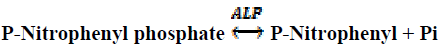
Activity of alkaline phosphatase (ALP) with standard method of IFCC is a reflex of conversion of P-Nitrophenyl
phosphate to P-Nitrophenyl that shown in equation 3:
 (3)
(3)
After data collection, statistical analysis was done with using of SAS software and also Tukey Dunnett and T tests
were done. The p˂0/05 was considered as a significant Index and results display as Mean±SD.
Results
X-Ray diffraction of Zno nanoparticles
The x-ray diffraction data were recorded by using Cu Kα radiation (1.5406 A°). The intensity data were collected
over a 2θ range of 20-80°. The average grain size of the samples was estimated with the help of Scherrer equation
using the diffraction intensity of (101) peak. x-ray diffraction studies confirmed that the synthesized materials were
ZnO with wurtzite phase and all the diffraction peaks agreed with the reported JCPDS data and no characteristic
peaks were observed other than ZnO. The mean grain size (D) of the particles was determined from the XRD line
broadening measurement using Scherer equation[15]:
D=0.89λ / (βCosθ) (1)
Where λ is the wavelength (Cu Kα), β is the full width at the half- maximum (FWHM) of the ZnO (101) line and θ
is the diffraction angle. A definite line broadening of the diffraction peaks is an indication that the synthesized
materials are in nanometer range. The lattice parameters calculated were also in agreement with the reported values.
The reaction temperature greatly influences the particle morphology of as-prepared ZnO powders. Figure 1 (a &b) shows the XRD patterns of ZnO nanoparticles.
Figure 1: XRD patterns of ZnO nanoparticles. (a) Indicate standard XRD pattern and (b) indicate sample XRD pattern
UV–visible absorption spectra for ZnO nanoparticles
The UV–visible absorption spectra of ZnO nanoparticles are shown in Figure 2 although the wavelength of our
spectrometer is limited by the light source, the absorption band of the ZnO nanoparticles have been shows a blue
shift due to the quantum confinement of the exciting present in the sample compare with bulk ZnO particles. This
optical phenomenon indicates that these nanoparticles show the quantum size effect [16].
Figure 2: UV-Vis absorption spectra for ZnO nanoparticles
Electron microscopic investigation of Zno nanoparticles
Morphology of the sample was investigated using SEM and TEM. Parts (a) and (b) of Figure 3 show the typical
SEM and TEM images of the sample respectively. The SEM image was captured in 10-nanometer scale bar size of
Zno nanoparticles and the TEM image was captured in 20 nanometer scale bar sizes of Zno nanoparticles.
Figure 3: (a) SEM image and (b) TEM image, of ZnO NPs
Toxicological Results
The results showed that activity of ALT enzyme Increased in all groups (Figure 4). This increase compare to the
control group in the second, third and fourth groups that received 50ppm, 100ppm and 200ppm nanoparticles
respectively is significant from the statistical point (p˂0/05).
Figure 4: Effect of different concentrations of ZnO nanoparticles on ALT enzyme
The results showed that activity of AST enzyme Increased in all groups (Figure 5). This increase compare to the
control group in the fourth group that received 200ppm nanoparticles is significant from the statistical point
(p˂0/05).
Figure 5: Effect of different concentrations of ZnO nanoparticles on AST enzyme
The results showed that activity of ALP enzyme Increased in all groups (Figure 6). This increase compare to the
control group in the third and fourth groups that received 100ppm and 200ppm nanoparticles respectively is
significant from the statistical point (p˂0/05).
Figure 6: Effect of different concentrations of ZnO nanoparticles on ALP enzyme
Discussion
Very small particles, so-called nanoparticles, have the ability to enter, translocate within, and damage living
organisms [17,18]. This ability, results primarily from their small size, which allows them to penetrate physiological
barriers, and travel within the circulatory systems of a host [19]. While natural processes have produced
nanoparticles for eons, modern science has recently learned how to synthesize a bewildering array of artificial
materials with structure that is engineered at the atomic scale [20,21]. The smallest particles contain tens or
hundreds of atoms, with dimensions at the scale of nanometers - hence nanoparticles. They are comparable in size to
viruses, where the smallest have dimensions of tens of nanometers (for example, a human immunodeficiency virus,
or HIV, particle is 100 nm in diameter), and which in the emerging science of nanotechnology might be called
‘Nanoorganisms’ [22]. Like viruses, some nanoparticles can penetrate lung or dermal (skin) barriers and enter the
circulatory and lymphatic systems of humans and animals, reaching most bodily tissues and organs, and potentially
disrupting cellular processes and causing disease [23-25]. The toxicity of each of these materials depends greatly,
however, upon the particular arrangement of its many atoms. Due to their small size, nanoparticles can translocate
from these entry portals into the circulatory and lymphatic systems, and ultimately to body tissues and organs [26].
Some nanoparticles, depending on their composition and size, can produce irreversible damage to cells by oxidative
stress or/and organelle injury [27,28]. Here we investigated toxicity effect of Zinc oxide nanoparticles on ALT, ALP
and AST enzymes in male Rat. Understanding the specific mechanisms of nanoparticles and its interaction Require
very extensive research in this field. When the nanoparticles are accumulated in a tissue, may be absorbed into the
cells or not to be absorbed. If these particles are absorbed, the finally replacement in cell lysosomes or cell
cytoplasm will depend on the characteristics of nanoparticles. If the nanoparticles are located in the cytoplasm, the
presence of some coarse grain material can cause direct damage or cell death is caused by this interactions. In this
study, to evaluate the toxicity effect of nanomaterials on the rat’s liver, the ALT, ALP and AST were measured.
That with increasing concentration of nanoparticles also increased levels of these three enzymes. And found linear
equation between concentration of nanoparticles and levels of these three enzymes. ALT and AST were located in
cell and ALP was located in cell membrane. In effect the loss of liver cells, these enzymes are released in the blood.
Therefore, increases of these enzymes are a sign of liver cells damage. ALT and AST indicate Status of liver cells.
ALP further demonstrates the performance and biliary Hungarian injuries, especially Hungarian extra hepatic. We
conclude that the development of nanotechnology and the study of nanotoxicology have increased our awareness of
environmental particulate pollution generated from natural and anthropogenic sources, and hope that this new
awareness will lead to significant reductions in human exposure to these potentially toxic materials. With increased
knowledge, and ongoing study, we are more likely to find cures for diseases associated with nanoparticle exposure,
as we will understand their causes and mechanisms. We foresee a future with better-informed, and hopefully more cautious manipulation of engineered nanomaterials, as well as the development of laws and policies for safely
managing all aspects of nanomaterial manufacturing, industrial and commercial use, and recycling.
Conclusion
The current knowledge of the toxic effects of nanoparticles is relatively limited. Nonetheless, the available data
indicate that some insoluble nanoparticles can pass through the different protective barriers, be distributed in the
body and accumulate in several organs. In this study results I saw that zinc oxide nanoparticles had toxicity on liver
enzymes and will lead to harmful effects on body metabolism, according to my results, I recommend to all
researchers that used of nanomaterials in their researches, have notice to these toxicity effects of nanoparticles.
References
- M. J. Sailor and J. R. Link, Chemical Communications, 2005, 11, 1375–1383.
- V. Shah and I. Belozerova, Water, Air, and Soil Pollution, 2009,197, 143–148.
- W.-X. Zhang, Journal of Nanoparticle Research, 2003, 5, 323–332.
- D. W. Galbraith, Nature Nanotechnology, 2007, 2, 272–273.
- H.-J. Park, S. H. Kim, H. J. Kim, and S.-H. Choi, Plant Pathology, 2007, 22, 295–302.
- L. Yang and D. J. Watts, Toxicology Letters, 2005, 158, 122–132.
- Reinhardt K; Winkler H, Weinheim, Germany: Wiley-VCH, 2002, 7, 285–300.
- Laberty-Robert, C., J. W. Long, E. M. Lucas, K. A. Pettigrew, R. M. Stroud, M. S. Doescher, and D. R. Rolison. Chem. Mater. 2006, 18, 50–58.
- Laberty-Robert, C., J. W. Long, K. A. Pettigrew, R. M. Stroud, and D. R. Rolison. Adv. Mater. 2007, 19, 1734–1739.
- Gu, H., and M. D. Soucek. Chem. Mater., 2007.19, 1103–1110.
- M. Arruebo et al. Small, 2008, 4, 2025-2034.
- Moore, M. N. Environment International. 2006, 32, 967-976.
- Shvedova AA, Castranova V, Kisin E, Schwegler-Berry D, Murray AR, Gandelsman VZ, Maynard A, Baron P, J Toxicol Environ Healthm, 2003, 66 , 1909-1926.
- Moore, M. N. Environment International., 2006, 32, 967-976.
- Neumann HG. Chemosphere., 2001, 42, 473–479.
- Carithers, RL. Alcoholic Hepatitis and Cirrhosis in Liver and Biliary Diseases ed. By Kaplowitz N, Williams & Wilkins, Baltimore, MD, 1992, 334-346.
- www.healthoracle.org.
- Hafkenscheid, J.C.M.; Dijt, C.C.M. Clin. Chem., 1979, 25, 55-59.
- Sampson, E.J.; Whitner, V.S.; Burtis, C.A.; McKneaily, S.S.; Fast, D.M.; Bayse, D.D. Clin. Chem., 1980, 26, 1156-1164.
- Jacobs, D.S. Laboratory Test Handbook. 4th ed. Cleveland, OH: Lexi-Comp Inc. 1996.
- Purcell,G.V.; Behenna, D.B.; Walsh, P.R., Clin.Chem., 1979, 25, 780-782.
- https://www.medfriendly.com.
- Jani P, Halbert G W, Langridge J, Florence A T, J. Pharm. Pharmacol., 1990, 42, 821-826.
- Maynard A D, Kuempel E D, J. Nanopart. Res. 2005, 7, 587-614.
- Nel A, Xia T, Madler L, Li N, Toxic potential of materials at the nanolevel Science, 2006, 311, 622-627.
- Donaldson K, Tran L, Jimenez L A, Duffin R, Newby D E, Mills N, MacNee W, Stone V, Fibre Toxicol. 2005, 2, 10.
- Powers K W, Brown S C, Krishna V B, Wasdo S C, Moudgil B M, Roberts S M, Part VI. Characterization of nanoscale particles for toxicological evaluation Toxicol. Sci. 2006, 90, 296-303.
- Colvin V L , Nature Biotechnol. 2003, 21, 1166-1170.


