Keywords
Bioassay; Statistical correlation; Toxicity; Aquaculture
Introduction
Toxicity expresses the degree to which a substance is poisonous, destructive or generally harmful to life [1]. Poisons or toxicants are chemicals which have harmful or adverse effects on living organisms and the study of toxic substances is known as toxicology [2]. In a broader sense, toxicity is concerned with the chemical nature, interaction with biological systems and safety evaluation of potentially poisonous materials [2,3]. Toxicity could be acute or chronic, depending on the dosage, nature and duration of effects of the toxin [2,4]. It is acute when the effects are rapidly occurring resulting from exposure to a relatively large quantity of the poison administered often in a single dose [2]. Chronic toxicity occurs when an organism is frequently or infrequently exposed to low levels of poison over a long period of time often years. It is characterized by long term effects which could be lethal or sub-lethal [2].
Senna siamea is an angiosperm, native of Southeast Asia and widely distributed in Africa, Latin America and in Oceania [5]. The plant is of the genus, Senna, specie, siamea and family, initially was Caesalpiniceae, later reclassified in Leguminoseae and now in Fabaceae [6]. S. siamea is a tree plant, 10-12 m tall (occasionally reaching 20 m). It has a short bole, the crown is dense and rounded at first and later become irregular and spreading. The young bark is grey and smooth and later develops longitudinal fissures. The leaves are alternate, 15-30 cm long, with 6-14 leaflets, each ending in a tiny bristle. The flowers are bright yellow and projects up to 60 cm long upright, with pyramid-shaped pinnacles. The fruits are flat with indehiscent pad 5-30 cm long, constricted between the seeds and containing about 20 seeds per pod. The seeds are bean-shaped, greenish-brown and 8-15 mm long [7]. In Nigeria, the plant is known as Bikini raskata, odan in different localities [7].
C. gariepinus (African Sharp tooth catfish) indigenous from Africa is classified into Kingdom: Animalia, Phylum: Chordata, Class: Actinopterygii, Order: Siluriformes, Family: Clariidae, Genus: Clarias and species: Clarias gariepinus [8]. The African Sharp tooth catfish is a large, eel-like fish, usually of dark gray or black colouration on the back, fading to a white belly. It has an adult length of 1-1.5 m and reaches a maximum total length of 1.7 m and can weigh up to 60 kg. C. gariepinus is one of the most important tropical catfish species for aquaculture in spite its commanding presence in the wild [8]. In Nigeria, it is widely cultured in ponds and occurs freely in natural freshwater. The fish has hardiness with high resistance to handling and stress [9,10]. C. gariepinus has high adaptation for low dissolved oxygen in water especially by fishes above 14 days old with functionally developed accessory respiratory organs [11,12]. It has long tolerance for drought but cannot survive long in water temperature below 9-10°C. These qualities account for its wide application in aquaculture and increased importance in ecotoxicological studies, hence its choice as test organism for the present study [8].
Materials and Methods
Location and materials for the experiment
The study was carried out in the Department of Fisheries and Aquatic Environmental Management, University of Uyo, (latitude 50 026´N and Longitude 70 55´E), Uyo, Nigeria (Figure 1).

Figure 1: Map of University of Uyo showing study location.
Sources of experimental materials
Plant materials: The plant materials (fresh leaves of S. siamea) were obtained from the arboretum of the Department of Forestry and Natural Environmental Management, University of Uyo, Nigeria. The plant was identified according to Mamadou et al. [7]. The extraction and identification of phytochemicals were carried out in the laboratory of the Department of Pharmacognosy of the University of Uyo.
Test organism: The test organisms, 400 juveniles of C. gariepinus were purchased from Akanse’s Farms Ltd. Abak. The fish were of age ten (10) weeks, weight (33.8 ± 1.6 g) and total length (19.70 ± 0.76 cm) (total length and weight are presented in Mean ± Standard Deviation). The organisms were transported in oxygenated bags to the Hatchery/production unit of the Department of Fisheries and Aquatic Environmental Management, University of Uyo, Nigeria, where the study was conducted.
Fish feed: The feed was 2.5 mm skretting commercial fish feed supplied by De Dino Resources, Ltd. Uyo, Nigeria. Nutritional composition of the feed is presented in Table. The feed was stored under standard conditions as prescribed by the manufacturer (Table 1).
| Macronutrient |
% Composition |
Micronutrient |
Composition(mg/Kg) |
| Crude protein |
43 |
Iron |
58 |
| Crude fat |
12.5 |
Iodine |
2.9 |
| Crude fiber |
2.5 |
Copper |
7 |
| Ash |
7 |
Manganese |
22 |
| Calcium |
1.5 |
Zinc |
130 |
| Sodium |
0.3 |
Antioxidant |
150 |
| Phosphorus |
0.9 |
- |
- |
Table 1 Nutritional composition of feed.
Methods
Fish feed: Extraction was done by solvent extraction [13]. Isolation and quantification of active compounds from the extract were done by chromatography [14,15]. Collected plant specimen was cleaned, cut into smaller pieces, shade-dried to constant weight to obtain the dry matter and then pulverized to finer particles. Known weight of the pulverized dry matter was macerated in 10.0 L of 96% ethanol with intermittent agitation for 72 h to extract the active ingredients. The macerated dry matter was filtered to obtain the extract. Extract obtained was concentrated to constant weight using model HH-S stainless steel thermostatic water bath at 40 ºC. Concentrated extract obtained was covered with foil paper and used as toxicant for the study.
Tannins: The sample, 500 mg of extract was dissolved in 50 ml of distilled water, homogenized in a mechanical shaker for 1 h and filtered into 50 ml volumetric flask. 5 ml of the filtrate was pipetted into a test tube and mixed with 2 ml of 0.1M FeCl3 in 0.1N HCL and 0.008M Potassium Ferro cyanide. The absorbance of the resulting solution was measured at 120 nm using UV-2700 UV-VIS Spectrophotometer within 10 min to be the total tannins composition of the extract [15].
Saponins: The sample, 20 g of pulverized dry matter was macerated in 100 cm3 of 20% aqueous ethanol and heated for 4 h at 55ºC (HH-S stainless steel thermostatic water bath) with continuous stirring. The mixture was filtered and the residue re-extracted with another 200 ml 20% ethanol. The combined extracts were concentrated to 40 ml at 90 ºC (HH-S stainless steel thermostatic water bath). The concentrated extract was transferred into a 250 ml separating funnel and 20 ml of diethyl ether was added and shaken vigorously. The aqueous layer was recovered and the ether layer discarded. The purification process was repeated and 60 ml of n-butanol was added. The combined n-butanol extract were washed twice with 10 ml of 5% aqueous sodium chloride. The remaining solution was evaporated (HH-S stainless steel thermostatic water bath) and the resulting residue oven dried at 60 ºC using Grab CAD Oven to constant weight. The saponin content was then calculated from the resulting residue [16].
Flavonoids: The method is based on the formation of flavonoidaluminuim complex which has an absorptivity maximum at 415 nm [17]. 100 ml of the plant extract in methanol (10 mg/ ml) was mixed with 100 ml of 20% AlCl3 in methanol and a drop of acetic acid and then diluted with methanol to 500 ml. The absorption at 450 nm was read after 40 min using UV-2700 UVVIS Spectrophotometer. Blank sample were from 100 ml of plant extracts and a drop of acetic acid and then diluted to 500 ml with methanol. The absorption of standard rutin solution (0.5 mg/ ml) in methanol was measured under the same condition. All determinations were carried out in triplicate [18].
Alkaloids: The sample, 5 g of pulverized dry matter was macerated in 200 ml of 10% acetic acid in ethanol covered for 4 h. The mixture was filtered and concentrated (water bath) to one quarter of its original volume. Concentrated NH4OH was added drop wise to the extract until the precipitation was complete. The whole solution was allowed to settle and the precipitate was collected and washed with dilute NH4OH. The residue was dried and weighed as the total alkaloid content of the extract [14,15].
Steroids: The sample, 1 g of pulverized dry matter was extracted by heating under reflux for 15 min with 10 ml Dichlomethane (DCM). Filtrate was evaporated to dryness (HH-S stainless steel thermostatic water bath) and residue was dissolved in 1 ml toluene. 50 μl of the toluene solution was applied to the chromatogram and was put in a pre-saturated tank containing toluene ethyl acetate and left in air-conditioned room at 18 ºC for the solvent to rise to a distance of 15 cm. Reading was taken at 254 nm using UV-2700 UV-VIS Spectrophotometer, after treating the chromatogram with komarowsky reagent [14].
Glycosides: The sample, 1 g of pulverized dry matter was mixed with 5 ml of 50% methanol and 10 ml of 10% Lead (II) acetate solution, then heated (HH-S stainless steel thermostatic water bath) for 10 min and allowed to cool. Cooled filtrate was extracted with two separate 10 ml quantities of dichloromethane (DCM) and the extracts combined and completely evaporated (HH-S stainless steel thermostatic water bath). The residue was dissolved in DCM-methanol (1:1) and 100 μl of the solution was applied to the chromatogram and was put in pre-saturated tank containing ethyl acetate methanol-water (100:13.5:10) and was cooled in air conditioned room (18 ºC) for the solvent to rise to a distance of 15 cm. Reading was taken at 365 nm using UV-2700 UV-VIS Spectrophotometer, after treating with Kedde reagent [19,14].
Preparation of test solutions: Test solutions were prepared by simple dilution principle and concentrations presented in milligrams per litre (mg/L). Desired weight of toxicant was accurately measured using electronic balance, spatula and 100 ml beaker of known weight. The desired weight of toxicant was dissolved in a known volume of water and the solution made up to required volume with water [6].
Acclimatization of test organisms: The juveniles of C. gariepinus used for this study were acclimatized for 14 days in the Hatchery/ Production unit of the Department of Fisheries and Aquatic Environmental Management of the University of Uyo. During this period, the fish were fed twice daily with 2.5 mm skretting commercial fish feed supplied by De Dino Resources, Uyo, Nigeria and water was changed after every 48 h. Feeding of organisms was ended 48 h prior to resumption of acute test and throughout the period of test to avoid interferences with stomach content [20].
Water quality tests: Water quality tests were carried out to ensure that optimum water conditions were maintained during the experiment, and that the results obtained were solely due to the effects of the toxicants on the fish samples. The physicochemical parameters of the test water with respect to temperature, dissolved oxygen (DO), pH, Total Dissolved Solids (TDS) and conductivity were measured using suitable water quality measurement instruments. Temperature was measured with HM Digital temperature thermometer, pH with ORP Pocket pH meter, TDS with ORP Pocket TDS tester, conductivity with ORP Combo-multimeter and DO with ORP Combo-multimeter. The measurements were taken before and during acclimation of the organisms, twice daily for the 96 h period of exposure of the organisms for the acute test and before and after every renewal of treatments for the chronic test.
Acute toxicity bioassay
Range finding test: Range finding test was conducted following standard procedure, to determine range of concentration for the definitive test [20]. The test was conducted in white translucent plastic aquaria (30 litres capacity) of dimension 52 22 cm each containing 20.0 L of standard solution of toxicant (treatment). Five (5) experimental set up of graded concentrations (0.1 mg/L, 1.0 mg/L, 10.0 mg/L, 100.0 mg/L, 1000 mg/L) of toxicant and a control, each with one replicate was considered for the test [20]. Aquaria were cleaned and sterilized using suitable reagents, sundried two days before commencement of test. Randomly selected ten (10) organisms were exposed to each aquarium containing 20.0 L of treatment. The experimental set up was covered with netting material to prevent the fish from jumping out and also prevent infestation by insects. Mortality in organisms was indicated by inability of fish to respond to external stimuli even on gentle prodding with a glass rod and no visible opecula movement [20-22]. Experimental set-up was monitored hourly in the first 4 h, four (4) hourly in the next 24 h and daily for the remaining 96 h and records of mortality and behavioural responses of organisms for the respective test solutions were recorded [19]. LC50 for the extract was established from mortality data generated using logit method [21,22].
Definitive test: Definitive test was conducted based on the result of the range finding test to establish LC50 for the extract. The test considered concentrations range of 10.0 mg/L to 100 mg/L for the extract, each with a replicate and using a spacing factor of 1.5. 10 healthy juveniles of C. gariepinus were randomly selected and exposed to 20 L of each treatment. The set up was observed hourly for the first four (4) h, four (4) hourly for the remaining twenty four (24) and daily for the remaining 96 h [20]. Water quality parameters were tested throughout the experiment and the Fish mortality data was generated. Inability of fish to respond to external stimuli even on gentle prodding with a glass rod and no visible opecula movement were used as indices of mortality in fish [20-22]. Confirmed death fishes were removed from the test media.
Maximum allowable toxicant concentration (MATC): MATC for the extract was determined using Boyd application factor equation, presented as eqn. (1).
AF=MATC/LC50
AF is application factor, LC50 is 96 h LC50. AF of 0.05 is suitable for most natural toxins, while 0.01 should be used for industrial chemicals or pesticides [23].
Chronic toxicity bioassay: Chronic toxicity bioassay was conducted to determine the toxicity of the extract on the blood parameters and tissue of sensitive organs of the exposed fish. The bioassay was designed to be intermediate termed chronic test with 21 days exposure using sub-lethal concentrations of the treatment (0.0 mg/L, 2.5 mg/L, 5.0 mg/L, 7.5 mg/L, 10.0 mg/L and 12.5 mg/L. 10 randomly selected juveniles of C. gariepinus were exposed to respective treatments for 21 days [20,22,24]. The treatments were renewed every 24 h and physicochemical parameters of the test media measured before and after every renewal [19]. At the end of the 21 days exposure, blood and tissue samples were collected from randomly selected fish from each treatment for further analysis.
Collection of blood and tissue samples: Blood samples were collected from randomly selected fishes from each treatment. The sample was collected from the caudal fin of the fish using hypodermic syringe and needle and preserved in EDTA treated bottles for haematological analysis. Fish tissues (gills, liver and guts) were obtained by dissecting open the lower part of the operculum to access the internal organs of fish. The organs of interest were carefully removed using suitable equipment’s. Collected samples were preserved immediately in sample bottles containing 10% formalin and properly labelled against the respective organs and treatments. The samples were subsequently taken to anatomy laboratory for histological analysis.
Blood and tissue analysis: Collected blood samples were subjected to auto-haem analysis using BC 2800 auto-haem analyser in the haematology laboratory of the Department of Physiology, University of Uyo, Nigeria. Blood parameters of interest (Hb, PCV, RBC, WBC, PLT, MCV and MCH) were analyzed. Histological analysis was carried out on the tissue samples using Haematoxylin and Eosin Method [7]. The analysis involved the following histological operations/laboratory procedures, tissues were De-waxed and hydrated, stained in Ehrlich haematoxyline solution for 20 min, washed thoroughly in running tap water, differentiated in 1% acid and alcohol until only the cell nuclei retain the stain, blued in running tap water for 5 min, tissues were counter stained in 1% Eosin for 3 min, washed in water to remove excess Eosin, dehydrated in absolute alcohol, cleared in xylene, mounted in Polysterene Dibutylephthalate xylene (DPX), examined under x 10 objective for deformities using Olympus B x 51 Microscope.
Statistical analysis: Data collected were analyzed using descriptive statistics (mean, standard deviation, frequencies and percentage). Comparison of data of physicochemical parameters and Blood parameters between the control and other treatments were carried out using Analysis of Variance (ANOVA). Duncan’s statistical test was used to further analyze for significance in changes in physicochemical parameters of test media and changes in blood parameters as the concentration of treatments increased. Statistical correlation of changes in physicochemical parameters with changes in blood parameters was tested using Pearson correlation matrix.
Ethical issues
The ethical issues of conducting toxicity test were all observed in every aspect of this research work. All physical and emotional harm that would have resulted from the conduct of this research were carefully avoided at every stage of the work. The test organisms were treated in line with professional recommendations throughout the period of the study. Experimental wastes generated in the course of the study were properly disposed in an environmentally friendly manner. Plagiarism policies were strictly adhered to in developing the research report. Data generated from the experimental work were reported exactly as observed, devoid of data doctoring or any form distortion of experimental outcome [25]. Every illegitimate use of computer and internet were carefully avoided in the conduct of this research work. The printing materials used in printing this report were those manufactured from sustainable and environmentally friendly processes.
Results and Discussion
Results
Extraction and phytochemical screening: Extraction and concentration of the plant extract yielded 117 g and its phytochemical analysis yielded as presented in Table 2. The results show that S. siamea contains plant secondary metabolites. These metabolites have been reported to be toxic bioactive substances [22,26] (Table 2).
| Phytochemical |
Extract |
| Alkaloids |
√√ |
| Steroids |
√√ |
| Saponins |
√√ |
| Glycocides |
√√ |
| Tannins |
√√ |
| Flavonoids |
√√ |
√√: High volume >1.0 mg
Table 2 Phytochemical contents of the extracts.
Acclimatization: Test organisms were successfully acclimatized for 14 days with less than 2.0% total mortality (3 organisms of 400 died, 0.75% mortality).
Physicochemical parameters of water of acclimatization: Changes in physicochemical parameters of water of acclimatization were observed to be within acceptable limit for survival and growth of C. gariepinus [26].
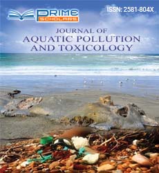
Range finding test: LC10 and LC50 of the extract were established as shown in Figure 2. Other behavioral responses to toxicity (restlessness, erratic movement, repeated attempts to jump out of culture medium, frequent opecula movements, occasional state of motionlessness, excessive mucus secretion, adoption of different postures, swirling movements, loss of balance and loss of reflex) were also observed in exposed fish (Figure 2).
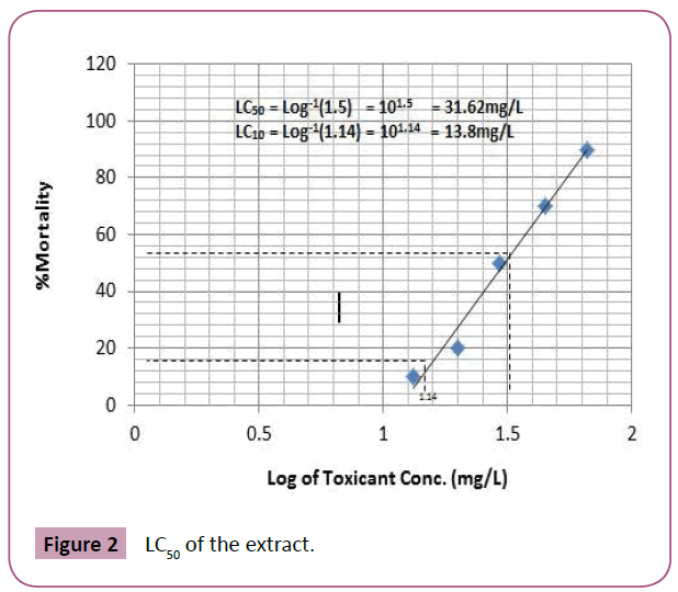
Figure 2: LC50 of the extract.
Definitive test: Results of definitive test are presented in Figures 3 and 4. Raw data generated from the definitive test are summarized in Tables 3 and 4.
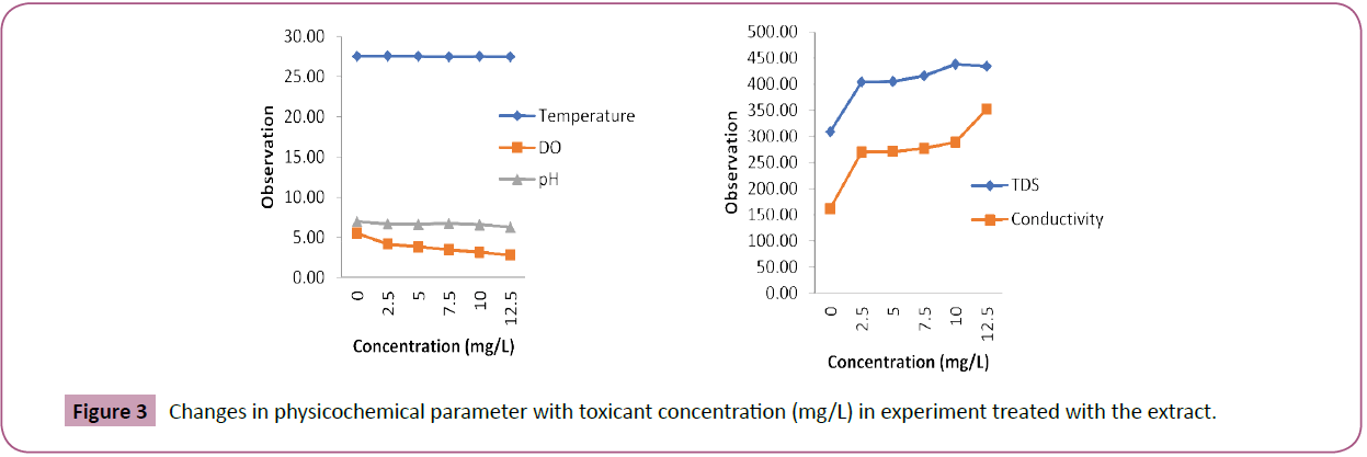
Figure 3: Changes in physicochemical parameter with toxicant concentration (mg/L) in experiment treated with the extract.
Figure 4: Changes in blood parameters with toxicant concentration in fish exposed to various concentrations of the extract.
| |
0.0 mg/l |
2.5 mg/l |
5.0 mg/l |
7.5 mg/l |
10.0 mg/l |
12.5 mg/l |
Total |
| Temperature |
27.50 ± 0.50a |
27.55 ± 0.45a |
27.50 ± 0.50a |
27.45 ± 0.55a |
27.50 ± 0.50a |
27.45 ± 0.45a |
27.49 ± 0.15 |
| DO |
5.45 ± 0.12a |
4.15 ± 0.68b |
3.81 ± 0.51bc |
3.44 ± 0.25bc |
3.13 ± 0.04bc |
2.77 ± 0.16c |
3.79 ± 0.28 |
| pH |
6.94 ± 0.01a |
6.65 ± 0.05ab |
6.62 ± 0.02ab |
6.70 ± 0.20ab |
6.55 ± 0.05bc |
6.25 ± 0.05c |
6.62 ± 0.07 |
| TDS |
310 ± 0.00a |
405 ± 11.00b |
406 ± 10.00b |
417 ± 19.00b |
439 ± 24.00b |
435 ± 16.00 b |
402 ± 13.81 |
| Conductivity |
163 ± 0.00a |
271 ± 46.00ab |
272 ± 35.00ab |
278 ± 32.00ab |
290 ± 20.00b |
353 ± 47.00b |
271.17 ± 19.76 |
Means with different superscripts along the same row are significantly different (Duncan’s test) p<0.05
Table 3 Physicochemical parameters with toxicant concentration in experiment treated with the extract.
| |
0.0 mg/l |
2.5 mg/l |
5.0 mg/l |
7.5 mg/l |
10.0 mg/l |
12.5 mg/l |
Total |
| Hb (g/L) |
95 ± 0.00a |
88 ± 1.00ab |
79.5 ± 6.50bc |
72.5 ± 11.50bc |
58.5 ± 5.50cd |
46.5 ± 3.50d |
73.33 ± 5.34 |
| PCV/L |
283.5 ± 1.5a |
266 ± 1.00ab |
236.5 ± 19.50ab |
216.5 ± 27.50bc |
181.5 ± 14.50cd |
152 ± 2.00d |
222.67 ± 14.49 |
| WBC x 109/L |
9.32 ± 1.29a |
3.97 ± 1.14b |
3.03 ± 0.24b |
2.62 ± 0.05b |
2.15 ± 0.10b |
1.89 ± 0.03b |
3.83 ± 0.80 |
| RBC x 1012/L |
2.9 ± 0.81a |
1.59 ± 0.21ab |
1.28 ± 0.14b |
0.76 ± 0.27b |
0.59 ± 0.28b |
0.49 ± 0.20b |
1.27 ± 0.27 |
| PLT x 109/L |
1347 ± 402.00a |
664 ± 0.21b |
537 ± 120.00bc |
319.5 ± 29.50bc |
111 ± 76.00bc |
17.89 ± 16.31c |
499.4 ± 142.77 |
| MCV (fL) |
188 ± 6.00a |
113.85 ± 15.95b |
89.35 ± 5.45c |
64.15 ± 18.45c |
56.95 ± 17.35c |
44 ± 5.10c |
92.72 ± 15.07 |
| MCH (pg) |
100.25 ± 1.75a |
72.35 ± 13.75ab |
57.2 ± 14.70b |
52.2 ± 12.10b |
42.25 ± 7.05b |
35.25 ± 5.65b |
59.92 ± 7.18 |
Means with different superscripts along the same row are significantly different (Duncan’s test) p<0.05
Table 4 Changes in blood parameters of fish treated with various concentrations of the extract.
Physicochemical parameters of test media: Results of physicochemical parameters of the respective test media are presented in Figure 4.
Blood analysis: Results of the blood analysis of Fish from the respective treatments are presented in Figure 4. During the 21 days chronic toxicity bioassay, physicochemical parameters of the culture medium exhibited variation in values. No significant change (P<0.05) in Temperature was observed. DO and PH were observed to reduce significantly (P<0.05) while TDS and conductivity were observed to increase significantly (P<0.05). The variations were all observed to be concentration related. Duncan’s statistical test revealed that changes in pH of test media were significant (p<0.05) only at higher concentrations (10.0mg/L and 12.5mg/L). Results from blood analysis show that all the parameters measured, reduced in value when compared with the control [27]. The reductions were observed to be related to concentration of the extracts [28,29]. Statistical analysis of data from blood parameters revealed that the observed reduction in the respective parameters (Hb, PCV, WBC, RBC, PLT, MCV and MCH) were all statistically significant (P<0.05) [29]. However, reduction in PCV tested nonsignificant (p˂0.05) at lower concentrations (5.0 mg/L and 2.5 mg/L) and Hb, RBC and MCH all tested non-significant (p<0.05) at lowest concentration (2.5 mg/L) of the extract. Reduction in WBC, PLT and MCV tested significant (p<0.05) at all concentrations of the extract administered (Figure 4) (Tables 3 and 5).
| Variables |
Temp |
DO |
pH |
TDS |
EC |
Hb |
PCV |
WBC |
RBC |
PLT |
MCV |
MCH |
| Temperature |
1 |
|
|
|
|
|
|
|
|
|
|
|
| DO |
0.33 |
1 |
|
|
|
|
|
|
|
|
|
|
| pH |
-0.13 |
0.73 |
1 |
|
|
|
|
|
|
|
|
|
| TDS |
-0.31 |
-0.91 |
-0.64 |
1 |
|
|
|
|
|
|
|
|
| EC |
-0.47 |
-0.93 |
-0.71 |
0.9 |
1 |
|
|
|
|
|
|
|
| Hb |
0.31 |
0.87 |
0.67 |
-0.79 |
-0.82 |
1 |
|
|
|
|
|
|
| PCV |
0.28 |
0.87 |
0.66 |
-0.79 |
-0.8 |
1 |
1 |
|
|
|
|
|
| WBC |
0.19 |
0.92 |
0.72 |
-0.92 |
-0.82 |
0.7 |
0.72 |
1 |
|
|
|
|
| RBC |
-0.2 |
0.83 |
0.74 |
-0.78 |
-0.69 |
0.7 |
0.72 |
0.79 |
1 |
|
|
|
| PLT |
0.21 |
0.9 |
0.73 |
-0.85 |
-0.78 |
0.81 |
0.83 |
0.93 |
0.72 |
1 |
|
|
| MCV |
-0.1 |
0.89 |
0.82 |
-0.83 |
-0.74 |
0.73 |
0.75 |
0.92 |
0.94 |
0.88 |
1 |
|
| MCH |
-0.35 |
0.67 |
0.79 |
-0.68 |
-0.52 |
0.66 |
0.69 |
0.77 |
0.83 |
0.79 |
0.9 |
1 |
Values in bold are different from 0 with a significance level alpha=0.05
Table 5 Pearson correlation matrix for changes in physicochemical parameters with changes in blood parameters of fish treated with the extract.
Tissue analysis: Results of tissue analysis of fish from the respective treatment are presented in Figures 5-7.
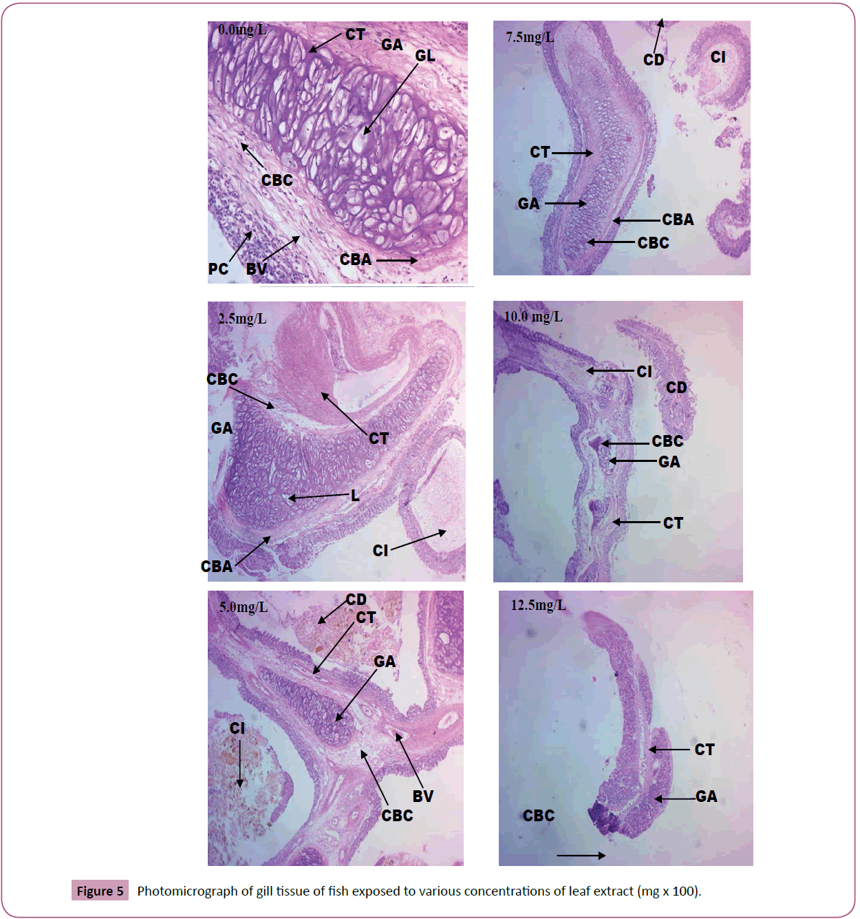
Figure 5: Photomicrograph of gill tissue of fish exposed to various concentrations of leaf extract (mg х 100).
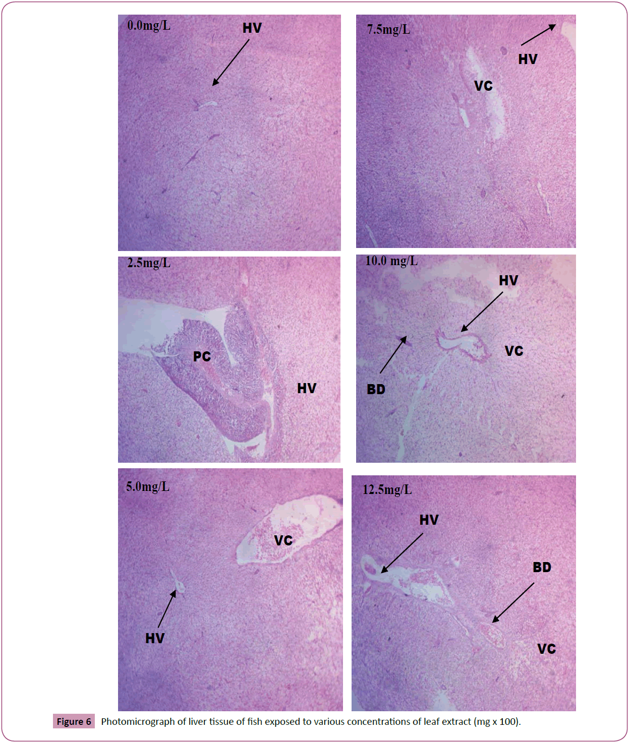
Figure 6: Photomicrograph of liver tissue of fish exposed to various concentrations of leaf extract (mg х 100).
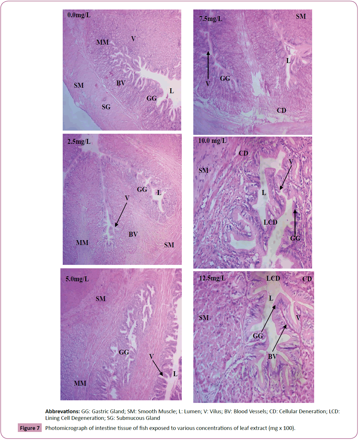
Abbrevations: GG: Gastric Gland; SM: Smooth Muscle; L: Lumen; V: Vilus; BV: Blood Vessels; CD: Cellular Deneration; LCD: Lining Cell Degeneration; SG: Submucous Gland
Figure 7: Photomicrograph of intestine tissue of fish exposed to various concentrations of leaf extract (mg х 100).
Discussion
The use of haematological techniques in fish culture is of growing importance to toxicological research, environmental monitoring and fish health conditions [30]. Fish are so intimately associated with the aqueous environment, often physical and chemical changes in the environment are rapidly reflected as measurable physiological changes in the fish [17,30]. Haematological parameters and physiological profile can be useful indicators of the physiological disturbances in animals and so can be crucial in providing vital information on the general well-being of fish [31]. Blood cell indices (RBC, WBC, etc.) are good indicators of system response to external stimulus and any changes are therefore reflected in their morphology and distribution in the blood. Thus detailed information can be obtained on general metabolism and physiological status of fish in different groups of age and habitat [32]. Therefore, blood serves as a good indicator to determine the health of an organism and acts as a pathological reflector of the whole body. Hence haematological parameters are important in diagnosing the functional status of animals exposed to toxicants [30].
In this study, behavioural responses observed in exposed fish were related to concentration of the extract as more of the responses were observed at higher concentrations of the extract [22,27,33]. The observed behavioural abnormalities are attributed to respiratory impairment, resulting from the effects of the toxicant on the gills of the exposed fish [12]. The reductions in blood cell indices and tissue deformation observed from the chronic bioassay are in line with findings of the study on behavioural, haematological and histopathological changes in C. gariepinus exposed to 2,4-dichlorophenoxyacetic acid [29]. Similar observations have also been reported in a study on exploitation of ethanol extract of Adenium obesum stem bark as a potent organic piscicide [22]. In a study conducted by Olusegun and Adadayo on Haematological Responses, Serum Biochemistry and Histology of C. gariepinus, exposed to sublethal concentrations of cold water fresh root bark extract of Plumbago zeylanica (Leadwort), it was reported that, exposure of fish to the toxicant for 21 days resulted in remarkable anaemic condition in C. gariepinus resulting from reductions in values of haematological parameters (RBC, Hb, PCV) in the juveniles of C. gariepinus exposed to P. zeylanica extract [30]. The study also reported histopathological alterations observed in the brain, gills, liver, intestine and muscle/flesh of the C. gariepinus juveniles [30].
Stressors evoke non-specific responses in fish which enables the fish to cope with the disturbance and maintain its homeostatic state [34]. If severe or long lasting, the response then becomes maladaptive and threatens the fish health and well-being. Therefore, in the presence of stressors (contaminants/pollutants), blood parameters and blood chemistry can be employed as standard laboratory test to determine diseased conditions and metabolic disturbances in fish [34]. Blood is a tissue fluid and serves as transport medium whose primary function is to supply oxygen and nutrients as well as constitutional elements to tissues and to remove waste products. Blood also enables hormones and other substances to be transported between tissues and organs. Blood is basically composed of the plasma, red blood cells, white blood cells and platelets, each with sub constituents which collectively contribute to the overall functioning of the blood [34,35].
The plasma is made up of 90% water, 7-8% soluble protein, 1% CO2 and 1% plasma salt all to maintain blood osmotic integrity. RBCs contain Hb which transports oxygen from the respiratory organ to the body. Hb consists of four subunits each made up of a haem, a porphyrin ring containing iron and a polypeptide [35,36]. As a haem takes up an O2 molecule, there is a rearrangement of the subunits that facilitates the uptake of additional O2. The amount of O2, in blood is determined by the amount dissolved, the Hb concentration and the affinity of Hb for O2 [35]. WBCs (Basophils, Eosinophils, Neutrophils, Monocytes, B-and T-cell Lymphocytes) synthesize, store and release histamine which is important for allergic reactions. The WBCs help the immune system and as well remove toxins, waste, foreign bodies and abnormal or damaged cells by phagocytosis. Platelets secrete factors that increase local platelet aggregation e.g., Thromboxane A, enhance vasoconstriction e.g., serotonin and promote blood coagulation e.g., Thromboplastin [37].
These observations in blood parameters confirm concentration related toxicity of the toxicant to the test organism [28,29]. The observed reduction in the respective blood parameters results in diseased conditions such as haemolytic anaemia, leukemia and lymphopaenia in exposed fish resulting from interference with blood cells formation process (erythropoiesis) and destruction of blood cells by the toxicants [29,35]. The reduction in MCV and MCH is attributed to the reduction in cellular blood iron denoting iron deficiency in blood of exposed fish [27]. The observed reduction in blood parameters is also attributed to haemolysis resulting from haemodilution, a mechanism for diluting the concentration of pollutants in the circulatory system [28].
From Pearson correlation matrix, changes in physicochemical parameters of culture media showed good correlations with changes in blood parameters in fish treated with the extract. However, temperature showed no correlation with any of the blood parameters measured. DO and pH had positive correlation while TDS and conductivity on the other hand negative correlation with the measured blood parameters. Conductivity and pH showed poor correlation (-0.43 and 0.22 respectively) with RBC, for treatments with flower extract). These observations show that changes in the blood parameters of the exposed fish was not only due to the toxicity of the extract as a single factor but acted in synergy with the resulting changes in physicochemical parameters of test media. This is in line with the findings of the study on effect of bioactive compounds extracted from euphorbious plants on haematological and biochemical parameters of Channa punctatus, which reported correlation of changes in blood cell distribution with changes in environmental conditions [27].
Physicochemical factors of the extracellular environment influence the chemistry of the intracellular fluid and accounts for the osmotic integrity of the intracellular environment [10]. Reduction in factors like DO leading to reduced availability of O2 for blood activities could result in hypoxaemic respiratory failure in exposed fish. O2 saturation of haemoglobin has been reported to have a direct proportional relationship with the pH [35]. Thus, the observed decreased in pH of the test media inhibited the uptake of O2 in blood of exposed fish, hence contributed to the hypoxaemic respiratory failure in exposed fish. On the other hand histological changes caused by the toxicant in exposed fish ulters the molecular configuration of Hb constituents affecting their affirnity for O2 and the overall O2 carrying capacity of the blood [35,37]. Cellular abnormalities revealed from tissue analysis were also observed to increase in severity with concentration of the respective treatments, denoting a concentration related toxicity of the extract [29,33,37]. The above observations established disease condition in exposed fish including oedema (reported as inflammation), kanjopyknosis, cellular necrosis, cellular degeneration, vascular congestion, nuclear fragmentation, Cellular disintegration etc. The observed changes in the histology of the tissues of exposed fish are in line with the general responses of fish organs to environmental pollutants [38].
Fish gills are the prime target organ of all pollutants due to their extensive surface in contact with the external medium and the reduced distance between the external and internal medium. Gill morphology and morphometric are important biomarkers providing a rapid method for detection of the effects of pollutants [39]. In histopathological studies, gills have also been reported to act as storehouse for bio accumulating toxicants [39]. The general morphological changes in the gills recorded in this study have been reported in Astyanax sp. after 96 h brief exposure to water soluble fraction of crude oil and C. gariepinus under brief or prolonged exposure to plant extract [39]. In some of the studies brief exposure to toxicants for about 96 h have produced irreversible changes in the gills [2,40]. The changes in the gills were adaptations by the fish to cope with challenge of the toxicant [28].
Vascular changes in gills of exposed fish could be attempts by the fish to supply more blood to the gills to increase oxygen uptake and supply to the internal organs. The gill of fish is a multipurpose organ that, in addition to providing for aquatic gaseous exchange, plays a dominant role in osmotic and ionic regulation, acid-base regulation and excretion of nitrogenous waste. Thus, despite the fact that all fish groups have functional kidneys, the gill epithelium is the site many processes that are mediated by the renal epithelia in terrestrial vertebrates. Hence, impairment of the gill functions by the overall effect of the pathological changes in the gill of exposed fish will have grave consequences for the fish with respect to the normal functions of the gills [28]. Photomicrograph of histologic sections of the intestine from the respective treatments revealed comprehensive cellular profile of the intestine with cellular abnormalities observed. The observed abnormalities include vascular congestion, cellular inflammation, linning cells degeneration and cellular disintegration.
Results obtain for Liver tissue could be due to high detoxification activity of the organ against the toxic effects of the treatments, which could have been the reason for the slight portal constriction, vascular congestion, hepatocytosis, etc. resulting from excessive work done by the organ in detoxifying the toxicity of the toxicant. The observed deformations are attributed to the accumulation of lipids and glycogen due to liver dysfunction as a result of exposure to toxicants. Increased level of vacuolation of hepatocytes has been reported as a signal to degenerating process that suggest metabolic damage possibly related to exposure to contaminated water [17]. The liver of freshwater fish is known to accommodate a high level of toxicants [17,32].
The toxicity of the studied plant extracts to the test organism is due to its bioactive constituents [16,41]. These bioactive constituents have various chemical properties and interfere with the physiology of the test organism. Saponins which are directly absorbed through the gills, haemolyses erythrocytes, due to their ability to lower the surface tension between the aqueous and the lipid phases of the membranes of erythrocytes resulting in the emulsification of their lipid components with Na+, water influx and K+ efflux until they rupture and release their haemoglobin content [1,2,5]. The toxicity might have also been through impairment of oxygen consumption in the exposed fish as saponins are also reported to lower the surface tension of reconstituted extracts with the formation of colloidal substances within them. Saponins are also known to cause structural damages in the respiratory and intestinal epithelia of exposed fish [22].
Tannins have been reported to have protein coagulating property on gill epithelia, causing respiratory failure or asphyxiation in the exposed fish. Observed toxicity might also be due to binding and subsequent inhibition of endogenous proteins by tannins in addition to causing intestinal damage and interference with iron absorption [2]. The toxicity could also result from impaired oxygen consumption by tannins, via its inhibition of oxidative phosphorylation involving the blockage of mitochrondrial enzyme [14]. This is in agreement with the reported inhibition of pyridine-linked substrates oxidation suggestive of interference with oxidative phosphorylation in ticks exposed to aqueous extract of Adenium obesum stem bark [22]. The observed toxicity is also attributed to the inhibition of Na+/K+-Atpase pump caused by cardiac glycosides resulting in a variety of severe arrhythmias, subsequent blockage of cardiac activities, decreased cardiac output and death in some of the exposed fish [35,36]. Alkaloids constituent of the extracts is known to impair oxygen consumption via inhibition of the activities of NADH ubiquinone reductase and subsequent inhibition of oxidative phosphorylation in exposed fish [7].
Conclusion and Recommendations
From the present study, the levels of Ni and Fe are all lower in the fruit vegetables than those in the leafy vegetables. It is found that the Ni levels in the vegetables are highly correlated with the three geochemical and NR fractions of the habitat top soils. The Fe levels in the vegetables are highly correlated with the ‘acid-reducible’ fraction of the habitat top soils. The positive relationships indicated the potential of edible vegetables as good biomonitors of Ni pollution in the habitat top soils. For the health risk assessment, al the THQ values for Ni and Fe in the 18 vegetables investigated in both adult and children are all below 1.00. This indicated that there was no non-carcinogenic risk of Ni and Fe to the consumers for both adults and children. Nevertheless, regular monitoring and management of the vegetable farms is still needed.
Conclusion
From the present study, it is seen that S. siamea is toxic to C. gariepinus fingerlings. Ethanol extract of leaves of S. siamea influence the physicochemistry of the culture media, affect the chemistry and overall osmotic integrity of the blood and cause tissue (gill, intestine and liver) damages in C. gariepinus fingerlings, resulting in observable behavioural abnormalities and eventual death of exposed fish. These confirm substantial piscicidal potential of S. siamea to C. gariepinus fingerlings for use in aquaculture.
Recommendations
Based on findings from the present study, it is recommended as follows:
• S. siamea has high piscicidal potential to C. gariepinus and as such is suitable for use by fish farmers as piscicidal plant.
• 96 h LC50 of 31.62 mg/L for leaf extract of S. siamea is recommended for use as piscicide by fish farmers.
• MATC of 1.581 mg/L for leaf extract of S. siamea to C. gariepinus.
• Further investigation should be conducted by researchers on the ichthyotoxicity of other parts of the plants and to other aquatic species.
References
- Hornby AS (2010) Oxford advanced learners dictionary. 8th edn., Oxford University Press, USA. 1-97.
- David P (1983) Toxicology studies in biology no. 149. Institute of Science and Technology, University of Wales 3-15.
- Ojutiku RO, Asuwaju, FP, Kolo RJ, Obande RA, Agbelege OO (2013) Haematological Effect of acute concentration of cypermethrin on juveniles of Claria gariepinus. Int J Eng Sci Inven 2: 33-41.
- Fafioye OO, Adebisi AA, Fagade SO (2012) Toxicity of Parkia biglobosa and Raphia vinifera extracts on Clarias gariepinus Juveniles. Afr J Biotechnol 3: 627-630.
- Thongsaard W, Chainakul S, Bennett GW (2001) Determination of barakol extracted from cassia siamea by HPLC with electrochemical detection. J Pharm Biomed Anal 25: 853-859.
- Veerachari U, Bopaiah AK (2011) Preliminary phytochemical evaluation of the leaf extract of five cassia species. J Chem Pharm Res 3: 574-583.
- Mamadou KC, N’goran MK, Aminata A, N’guessan ARY, Henri MD (2014) Ethnobotany, phytochemistry, pharmacology and toxicology profile of Cassia siamea Lam. J Pharmacol 3: 57-76.
- Abalaka SE (2013) Evaluation of the haematology and biochemistry of Clarias gariepinus as biomakers of environmental pollution in Tiga dam, Nigeria. Braz Arch Biol Technol 56: 1-7.
- Olaifa FE, Olaifa AK, Lewis OO (2003) Toxic stress of lead on Clarias gariepinus (African catfish) fingerlings. Afr J Biomed Res 6: 101-104.
- Akinsanya B, Otubanjo OA (2006) Helminth parasites of Clarias gariepinus (clariidae) in lekki lagoon, Nigeria. Rev Biol Tropics 54: 93-99.
- Okechi JK (2004) Profitability assessment: A case study of African catfish (Clarias gariepinus) in the Lake Victoria basin, Kenya. Fisheries Training Programme, The United Nations University, Iceland. Pp: 17-22.
- Ogundiran MA, Fawole OO, Adewoye SO, Ogundiran TA (2009) Pathological lesions in the gills of Clarias gariepinus exposed to sub lethal concentrations of soap and detergent effluents. J Cell Anim Biol 3: 78-82.
- Doughari JH (2012) Phytochemicals: Extraction methods, basic structures and mode of action as potential chemotherapeutic agents. InTech Open, Europe. Pp: 14-32.
- Harborne J (1973) Phytochemical methods. Champman and Hall Ltd., London. Pp: 49-88.
- Gracelin DH, Britto AJ, Kumar PB (2013) Qualitative and quantitative analysis of phytochemicals in five pteris species. Int J Pharm Pharm Sci 5: 105-107.
- Obdoni B, Ochuko P (2001) Phytochemical studies and comparative efficacy of the crude extracts of some homeostatic plants Edo and Delta state of Nigeria. Global J Pure Appl Sci 8: 203-208.
- Musa SO, Omeregie E (1999) Haematological changes in the mud fish, Clarias gariepinus (Burchell) exposed to malachite green. J Aquat Sci 14: 37-42.
- Kumaran A, Karunakara R (2006) Anti-oxidant and free radical scavenging activity of an aqueous extract of coleus aromatic. Food chemistry 97: 109-114.
- Ayuba VO, Ofojekwu PC, Musa SO (2012) Acute toxicity of Clarias gariepinus exposed to Datura innoxia leaf extract. J Med Plants Res 6: 2453-2457.
- American Public Health Association (APHA), American Water Works Association (AWWA) and Water Environment Federation WEF (2005) Standard methods for the examination of water and wastewater water. 21st edition, American Public Health Association, Washington DC. Pp: 81-89.
- Tyler SA, Gurian J (1950) Determination of the LD50 by use of probit, angular and logit transformations. University of Chicago, Chicago. Pp: 4-27.
- Abalaka SE, Fatihu MY, Ibrahim NDG, Ambali SF (2013) Exploitation of ethanol extract of Adenium obesum stem bark as a potent organic piscicide. Res J Biol Sci 8: 143-149.
- Boyd CE (2005) LC50 calculations help predict toxicity. Sustain Aquac Practices 1: 84-87.
- Ewald G (1995) Chronic measures of toxicant-induced effects on fish. Ann Zool Fennici 32: 311-316.
- Da’Rocha AB, Lopes RM, Schwartsmann G (2001) Natural products in anticancer therapy. Curr Opin Pharmacol 1: 364-369.
- Interafrican Bureau for Animal Resources (IBAR) (2015) African catfish Clarias gariepinus. FAO, Rome. Pp: 6.
- Shahi J, Singh A (2011) Effect of bioactive compounds extracted from euphorbious plants on haematological and biochemical parameters of Channa punctatus. Rev Inst Med Trop Sao Paulo 53: 259-263.
- Gabriel UU, Amakiriand EU, Ezeri GNO (2007) Haematology and gill pathology of Clarias gariepinus exposed to refined oil, kerosine under laboratory conditions. J Anim Vet Adv 6: 461-465.
- Okogwu OI, Anionwo Q, Anoke DC, Ugwuezi PO (2015) Behavioural, haematological and histopathological changes in the African catfish, Clarias gariepinus exposed to 2,4-dichlorophenoxyacetic acid (2,4-D). Niger J Biotechnol 30: 26-35.
- Olusegun AA, Adedayo OO (2014) Haematological responses, serum biochemistry and histology of Clarias gariepinus (Burchell, 1822) exposed to sublethal concentrations of cold water fresh root back extracts of Plumbago zeylanica (Leadwort). J Aquac Res Dev 5: 282-288.
- Tavares-Dias, M, Moraes FR (2007) Leucocyte and thrombocyte reference values for channel catfish (Ictaluruspunctatus Raf.) with an assessment of morphological, cytochemical and ultrastructural features. Vet Clin Pathol 36: 49-54.
- Ojutiku RO, Asuwaju FP, Kolo RJ, Obande RA, Agbelege OO (2013) Haematological effect of acute concentration of cypermethrin on juveniles of Claria gariepinus. Int J Eng Sci Inven 2: 33-41.
- Bobmanuel NO, Gabriel UU, Ekweozor IK (2006) Direct toxic assessment of treated fertilizer effluents to Oreochromis niloticus and catfish hybrid (Hetero clarias). Afr J Technol 5: 642-653.
- Celik ES (2004) Blood chemistry (electrolytes, lipoproteins and enzymes) values of black scorpion fish (Scorpaena porcus) in the Dardanelles, Turkey. J Biol Sci 4: 716-719.
- Crook MA (2012) Clinical biochemistry and medicine. 8th edition, Hodder & Stoughton Ltd. Pp: 74-81.
- Akpan IA, Francis AJ (2017) Kinetic potentials of the effect of ethanol on iron content of ashed cow blood. J Basic Appl Res 3: 10-15.
- Aqra A (2012) Human medical physiology: Blood physiology. John Wiley & Sons Inc., USA. Pp: 365-371.
- Fernandes MN, Mazon AF (2003) Environmental pollution and fish gill morphology. In: Val AL, Kapoor BG (Eds), Fish Adaptations. USA, Science Publishers. Pp: 203-231.
- Fafioye OO, Adebisi AA, Fagade SO (2012) Toxicity of Parkia biglobosa and Raphia vinifera extracts on Clarias gariepinus Juveniles. Afr J Biotechnol 3: 627-630.
- Ceiqueira CC, Fernandes MN (2001) Gill tissue recovery after copper exposure and blood parameter responses of the tropical fish, Prochilodus scrofa. Ecotoxicol Environ Saf 52: 83-91.
- Nadembega P, Boussim JI, Nikiema JB, Poli F, Antognoni F (2011) Medicinal Plants in Baskoure, Kourittenga province, Burkina Faso: An Ethnobotanical Study. J Ethnopharmacol 133: 378-395.