Keywords
Gastric ulcer; Ethanol; Punicalagin; Hydrochloric acid
Introduction
Gastric ulcer occurs mainly due to the imbalance between the destructive and protective factors to the mucosal barrier. The destructive factors include stomach Hydrochloric Acid (HCl), free oxygen radicals, ethanol, Helicobacter pylori and Non-Steroidal Anti-Inflammatory Drugs (NSAIDs) that encourage the gastric mucosal injury leading to gastric ulceration [1]. Alcohol consumption has been commonly related to mucosal inflammation, gastric ulcer and even gastric carcinoma [2].
Ethanol induces its gastrointestinal toxicity through several mechanisms such as stimulation of acid secretions [3], proinflammatory cytokines, oxidative stress [4], invasion of activated neutrophils and apoptosis as well as exhaustion of mucosal cytoprotective moieties, including Nitric Oxide (NO) and Prostaglandin E2 (PGE2) [5].
Punicalagin (PCG) is ellagitannin found in pomegranate juice which extracted from Punica granatum L. (Lythraceae). It has strong antioxidant, chemopreventive effect and angiogenic activities in addition to its beneficial effects in attention of prostate cancer cell growth, management of diabetes, myocardial ischemia reperfusion injury, and liver toxicity, tissue repairing and wound healing [6-12].
Therefore, herbal drugs may be promising alternative medications for the development of new drugs to organize gastric ulcer. Although previous studies supporting the beneficial effect of punicalagin, its gastroprotective effect against gastric ulcer induced by ethanol has not been yet thoroughly investigated. Accordingly, this study was designed to examine gastroprotective action of PCG against ethanol induced gastric ulcer.
Materials and Methods
Animals
Adult male Wistar albino rats weighing 170-200 g were placed in polyethylene cages in groups of 6 and the experiment was performed under controlled laboratory conditioning where food and water were provided ad libitum. Handling and procedures were carried out according to the Guide for the Care and Use of Laboratory Animals published by the US National Institute of Health (NIH publication No. 85-23, revised 1996). The experimental protocols were approved by the institutional research ethics committee at Faculty of pharmacy, Damanhur University.
Drugs and Chemicals
Punicalagin and ranitidine hydrochloride (Sigma Chemical Co., MO, USA) was dissolved in distil waster. All other chemicals used were of good quality and analytical grade.
Experimental design
Animals were randomly divided into three experimental series, each consisting of 4 groups (n=6); first group act as normal control received vehicle orally by intra-gastric gavage, second group administered of 5 ml/kg of absolute ethanol orally [13], third group (Ran+ Ethanol): Rats received ranitidine (Ran) (used as a standard reference drug) orally (50 mg/kg) [14] 2 h before the administration of ethanol and group 4 (PCG +Ethanol): Treated with Punicalagin (PCG) orally (4 mg/kg) [15] for 2 h before the administration of ethanol. All animals were fasted 24 h prior to ethanol administration except for water to exclude exogenous dietary effect.
Animals in the first set were anesthetized 3 h after ethanol administration and the abdomen were opened with pylorus ligation to collect gastric juice were and their stomach were removed, opened along the greater curvature, and washed with cold saline. The extent of gastric lesions (ulcer index, UI) was calculated by the formula: UI=10X (total ulcerated area/ total mucosal area) [16] and the Preventive Index (PI) was calculated by the equation PI=[(UI of ethanol-UI of pretreated drug)/UI of ethanol] × 100 [17] then part of the stomach was processed for histopathological examination and other was rapidly scraped with two glass slides to extract gastric mucosa.
Determination of free, total acidity and pH of gastric juice
Gastric juice was collected and centrifuged for 5 min at 2000 × g. Immediately the volume and pH of supernatant gastric juice were determined then titrated with 0.01 N NaOH by means of methyl orange as an indicator until yellowish orange colour appeared and the result indicated free acidity. Subsequently phenolphthalein was added as an indicator and continues titrating until red colour reappeared however the total volume of alkali added indicated total acidity as we described before [14].
Determination of gastric mucosal oxidative stress and enzymatic antioxidant activity
Gastric mucosa levels of Thiobarbituric Acid Reactive Species (TBARs) and reduced Glutathione (GSH) were estimated as well as Glutathione Peroxidase Activity (GPx) using commercially available ELISA kits according to manufacturer's instructions (Bio-diagnostic, Egypt) as markers of enzymatic antioxidant activity.
Determination of gastric mucosal levels of inflammatory markers
Gastric mucosa were homogenized with cold Phosphate Buffered Saline (PBS) then centrifuged. The supernatants were utilized for assessment of levels of inflammatory cytokines; TNF-α and IL-1β as well as anti-inflammatory cytokines IL-10 using commercial available ELISA (abcam, Cambridge, MA, USA). Also, IFNγ was estimated by IFNγ ELISA Kit (Boster biological technology Co. Ltd, USA).
Evaluation of gastric tissue levels of myeloperoxidase (MPO) activity
Gastric mucosal myeloperoxidase activity was determined according to the method described by Abdel-Raheem [18] as an index of neutrophil infiltration. Briefly, Extracted mucosa was homogenized in 20 mM phosphate buffer (pH 7.4) then centrifuged at 10,000 × g for 10 min and the consequential pellet was resuspended in 50 mM phosphate buffer (pH 6.0) with 0.5% Hexadecyl Trimethyl Ammonium Bromide (HTAB). Four cycles of freezing and thawing then 1 min sonication were carried out. Consequently, the samples were centrifuged and the supernatants were incubated with the reaction mixture in 37ºC for 110 s. The reaction mixture consisted of the 1.6 mM tetramethylbenzidine, 80 mM sodium phosphate buffer (pH 5.4) and 0.3 mM hydrogen peroxide then, the reaction was terminated by with 0.18 M H2SO4. The myeloperoxidase value was evaluated by determining the absorbance at 450 nm (OD value). The myeloperoxidase activity was expressed as relative values calculated by the following formula: (myeloperoxidase value recovered from treated animal)/myeloperoxidase value recovered from control rat) × 100 (%).
Determination of gastric NFκB protein expression using western blotting
Western blot analysis was performed as we described before [19]. Briefly, gastric mucosa were homogenized in cold RIPA buffer together with inhibitors for proteases and phosphatases (Sigma Chemical Co., MO, USA) then protein was assessed by Bradford method (Bio-Rad, Hercules, CA, USA). 50 μg protein samples were separated by SDS-PAGE then transferred onto a nitrocellulose membrane, blocked with nondairy milk and incubated with primary antibodies NFκB-p65 (abcam, Cambridge, MA, USA) and β-actin (Sigma Chemical Co., MO, USA) were distinguished with a horseradish peroxidase-conjugated antibody and ECL chemiluminescence (Amersham BioSciences, Buckinghamshire, UK). Intensity of immunoreactivity was determined by densitometry.
Determination of genes expression using real time polymerase chain reaction (PCR)
RNA isolation kit (Qiagen Inc., USA) was used for isolation RNA from Samples of gastric mucosa. PCR was carried out in Applied Biosystems Real-Time PCR System (Life Technologies, USA) with TaqMan Fluorescein one step PCR master mix and ready-made primer and probe sets of rat TNF-α (Catalogue. Rn99999017_m1), B-cell lymphoma 2 (Bcl-2) (Rn99999125_m1), caspase 9 (Rn00581212_m1) and housekeeping reference primer glyceraldehyde 3-phosphate dehydrogenase (GAPDH) (Rn01775763_g1) (applied biosystem Inc., USA). The thermal cycling conditions were retention time step 48°C for 15 min then Enzyme activation step 95°C for 15 min, then 40 cycles of 95°C for 15 s and 60°C for 1 min. Analysis of relative gene expression data were conducted using cycle threshold (CT) method and normalized to housekeeping reference gene GAPDH.
Evaluation of gastric mucosal apoptosis
Gastric mucosal caspase-3 activity was measured by caspase 3 ELISA kit (ab39401, abcam, Cambridge, MA, USA). Caspase 3 activities were expressed as relative values calculated by the following formula: (optical density 405 from treated animal)/ (optical density 405 value from control animal) × 100 (%).
Determination of mucosal level of nitric oxide
Gastric mucosa was filtered by Amicon ultra centrifugal filter units 30 kDa (Sigma Chemical Co., MO, USA) then 40 μl of the filtrate was used for determination of NO content by measuring its stable metabolites nitrite (NO2) and nitrate (NO3) using a commercially available ELISA kit (Cayman, Ann Arbor, MI, USA).
Determination of gastric mucosal prostaglandin E2
Gastric mucosa of the second set were scratched and soaked in 100% ethanol and 0.1 M indomethacin then homogenized and centrifuged at 12,000 × g for 10 min and the level of prostaglandin E2 (PGE2) were assessed using a commercially available ELISA kit according to manufacturer's instructions (Cayman, Ann Arbor, MI, USA).
Determination of gastric mucin barrier content
Animals in the third set as explained before [20]. In brief, 3 h after ethanol administration, animals were anesthetized and the lesser curvature of the stomach were cut, weighed and immersed for 2 h in 0.1% alcian blue 8GX dissolved in 0.16 M sucrose buffered with 0.05 sodium acetate then after 2 h washed twice with 0.25 M sucrose solution. The dye was extracted with 10 ml solution of 0.5 M magnesium chloride by intermittent shaking for 2 h then diethyl ether was added and the blue density of the aqueous phase was measured at 580 nm.
Histopathological evaluation
Gastric tissue samples were fixed in buffered 10% formalin and processed for histopathological examination as we described before [14]. Briefly, five micrometre-thick paraffin sections were prepared and stained with haematoxylin and eosin for light microscope examination (magnification × 20) by double blind pathologist.
Statistical analysis
All data are presented as mean ± standard error and analyzed with one-way ANOVA followed by Tukey's post hoc test for multiple group comparisons. Analyses were performed using GraphPad Prism Version 4.0 software (GraphPad Software Inc., La Jolla, CA). For all comparisons, P<0.05 was considered as statistically significant.
Results
Evaluation of antiulcer features of PCG
Ingestion of ethanol caused gastric mucosal lesions indicated by an increase in ulcer index vs. control group however pre-treatment with PCG or Ran reduced severity of gastric lesions in the term of ulcer index vs. ethanol treated animals.
Pre-treatment with PCG or Ran produced preventive index 74.9% and 79% of ethanol induced gastric ulcer, respectively with no significant difference between pre-treatment with PCG and reference drug Ran (Table 1) as well histopathological results confirmed the ability of PCG to recover ethanol-induce gastric ulcer in the gastric mucosa (Figure 1A). Sections from ethanol treated group showed extensive gastric lesion ulceration with inflammatory cells infiltration in submucosal layer (Figure 1B) however, sections from rats pre-treated with PCG or Ran showed noticeably reduced gastric lesions and inflammatory cells infiltration compared to ethanol alone-treated group (Figure 1C and 1D).
| |
Control |
Ethanol |
Ran+Ethanol |
PCG+ethanol |
| Ulcer index |
0 |
0.81 ± 0.05* |
0.15 ± 0.05$ |
0.20 ± 0.07$ |
| Preventive index (%) |
100 |
0 |
79.39 ± 7.95$ |
74.97 ± 9.84$ |
| Gastric volume (ml) |
1.10 ± 0.14 |
2.48 ± 0.13* |
1.46 ± 0.14$ |
2.01 ± 0.12* |
| Gastric juice pH |
3.20 ± 0.09 |
2.10 ± 0.07* |
2.27 ± 0.07*$ |
2.23 ± 0.09*# |
| Free acidity (Meq/l) |
27.43 ± 3.63 |
80.03 ± 4.05* |
34.83 ± 4.54$ |
69.13 ± 3.61*# |
| Total acidity (Meq/l) |
35.50 ± 4.70 |
87.95 ± 3.57* |
45.48 ± 4.82$ |
78.78 ± 4.31*# |
Table 1: Effect of Punicalagin (PCG) or Ranitidine (Ran) on ulcer index, preventive index, gastric juice volume, pH, free acidity and total acidity in ethanol-induced gastric ulcer. Data are expressed as mean ± standard error (n=6; *P<0.05 versus control group, $P<0.05 versus ethanol group and #<0.05 versus Ran+ethanol group).
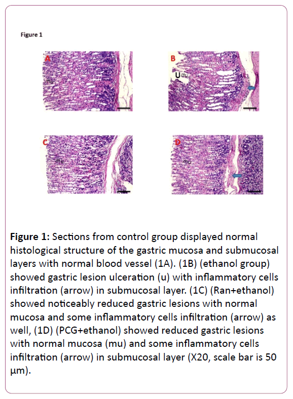
Figure 1: Sections from control group displayed normal histological structure of the gastric mucosa and submucosal layers with normal blood vessel (1A). (1B) (ethanol group) showed gastric lesion ulceration (u) with inflammatory cells infiltration (arrow) in submucosal layer. (1C) (Ran+ethanol) showed noticeably reduced gastric lesions with normal mucosa and some inflammatory cells infiltration (arrow) as well, (1D) (PCG+ethanol) showed reduced gastric lesions with normal mucosa (mu) and some inflammatory cells infiltration (arrow) in submucosal layer (X20, scale bar is 50 μm).
Effect of PCG on volume of gastric secretions and acidity
Oral treatment with ethanol evoked increase in gastric juice volume, free acidity and total acidity while it decreased its pH. Although, pre-treatment with Ran decreased gastric juice volume, free acidity and total acidity, pre-treatment with PCG failed to change gastric juice volume, free and total acidity induced by ethanol.
Effect of PCG on mucosal redox status and mucosal myeloperoxidase activity
In ethanol group, there was an increase in mucosal level of TBARs however pre-treatment with PCG or Ran reduced it indicating a reduction in oxidative stress (Figure 2A). On contrary, there was a significant decrease in mucosal levels of GSH and GPx in ethanol group while pre-treatment with PCG or Ran elevated mucosal levels of GSH and GPx demonstrating antioxidant action. Moreover, PCG produced significant decreased in mucosal level of TBARs vs. Ran+ethanol group as well as there was an increased in mucosal levels of GSH and GPx in PCG+ethanol group vs. Ran+Ethanol group indicating superior in antioxidant action of PCG than Ran (Figures 2B and 2C).
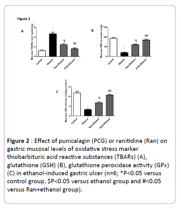
Figure 2: Effect of punicalagin (PCG) or ranitidine (Ran) on gastric mucosal levels of oxidative stress marker thiobarbituric acid reactive substances (TBARs) (A), glutathione (GSH) (B), glutathione peroxidase activity (GPx) (C) in ethanol-induced gastric ulcer (n=6; *P<0.05 versus control group, $P<0.05 versus ethanol group and #<0.05 versus Ran+ethanol group).
Effect of PCG on gene expression and mucosal levels of inflammatory cytokines
Figures 3 and 4 showed the involvement of inflammatory signals and neutrophil infiltration in gastroprotective action of PCG against ethanol.
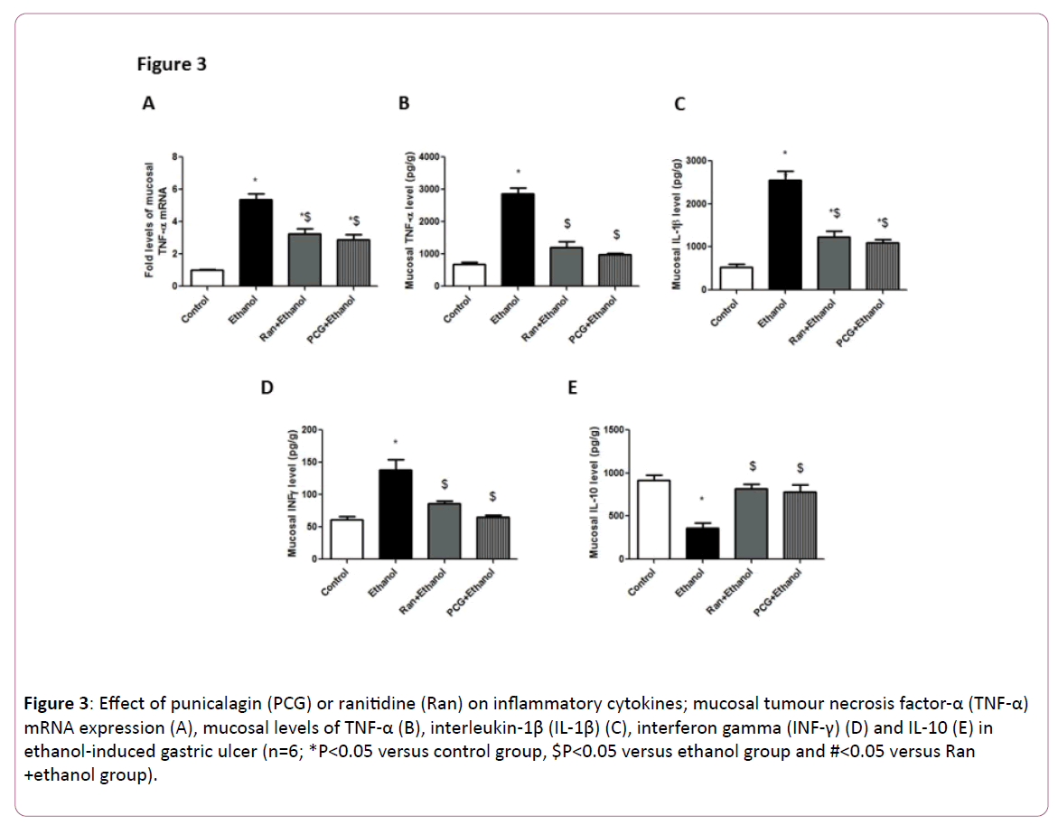
Figure 3: Effect of punicalagin (PCG) or ranitidine (Ran) on inflammatory cytokines; mucosal tumour necrosis factor-α (TNF-α) mRNA expression (A), mucosal levels of TNF-α (B), interleukin-1β (IL-1β) (C), interferon gamma (INF-γ) (D) and IL-10 (E) in ethanol-induced gastric ulcer (n=6; *P<0.05 versus control group, $P<0.05 versus ethanol group and #<0.05 versus Ran +ethanol group).
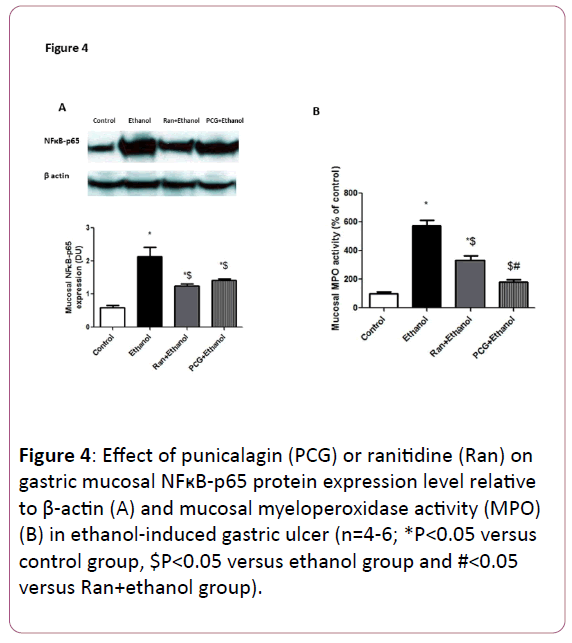
Figure 4: Effect of punicalagin (PCG) or ranitidine (Ran) on gastric mucosal NFÃ’ÂÂÂÂB-p65 protein expression level relative to β-actin (A) and mucosal myeloperoxidase activity (MPO) (B) in ethanol-induced gastric ulcer (n=4-6; *P<0.05 versus control group, $P<0.05 versus ethanol group and #<0.05 versus Ran+ethanol group).
In ethanol group; mucosal TNF-α gene expression was up regulated as well as TNF-α, IL-1β, IFNγ levels (Figure 3) and MPO activity (Figure 4B) were significantly elevated than in normal control. Similar to Ran, pre-treatment with PCG diminished TNF-α gene expression, levels of mucosal TNF-α, IL-1β, IFNγ (Figure 3) and MPO activity (Figure 4B) induced by ethanol. In the different way, ingestion of ethanol produced a significant decrease in anti-inflammatory cytokines IL-10 while pre-treatment with PCG or Ran normalized this alteration (Figure 3). Although, ethanol has an ability to up regulate mucosal NFkB-p65 protein expression. Pre-treatment with Ran or PCG significantly decrease its level indicating involvement of NFkB pathway in the mechanism of PCG’s gastroprotective action against ethanol (Figure 4A).
Effect of PCG on gastric mucosal apoptosis
Oral administration of ethanol caused up regulation of mucosal gene expression of caspase 9 and caspase 3 activities while it down regulated antiapoptotic Bcl-2 gene expression vs. normal control group. Analogous to Ran, pre-treatment with PCG down regulated mucosal gene expression of caspase 9 and caspase 3 activity however it increased Bcl-2 gene expression produced by ethanol (Figure 5).
Figure 5: Effect of punicalagin (PCG) or ranitidine (Ran) on gastric mucosal apoptosis; mucosal mRNA expression of caspase 9 (A), B-cell lymphoma 2 (Bcl-2) (B) and caspase 3 activity (C) in ethanol-induced gastric ulcer (n=6; *P<0.05 versus control group, $P<0.05 versus ethanol group and #<0.05 versus Ran+ethanol group).
Effect of PCG on mucosal levels of NO, PGE2 and mucin barrier content
Ethanol evoked depletion of NO, PGE2 and mucin barrier content while pre-treatment with PCG improved levels of NO and mucin barrier content but with no ability to change mucosal PGE2 level. However, pre-treatment with Ran increased levels of NO, PGE2 and mucin barrier content (Figure 6).
Figure 6: Effect of punicalagin (PCG) or ranitidine (Ran) on cytoprotective factors such as mucosal levels of nitric oxide (NO) (A), prostaglandin E2 (PGE2) (B) and mucus barrier content (C) in ethanol-induced gastric ulcer (n=6; *P<0.05 versus control group, $P<0.05 versus ethanol group and #<0.05 versus Ran+ethanol group).
Discussion
Ethanol induced gastric ulcer is a key experimental model commonly utilized for preclinical evaluation of agents with potential anti-ulcer action as ethanol has been considered an important cause of gastric ulcer in humans [21]. Drugs currently offered in the market against gastric diseases are often correlated with severe side effects [22]. The present study focused for the first time, on the protective actions of punicalagin against ethanol induced gastric ulcer.
Pre-treatment with PCG reduced ulcer index with preventing 74.9% of ethanol induced gastric ulcer with no significant vs. the standard reference drug (ranitidine). Also, histopathological findings confirmed these results by showing normal mucosal layer with reduction in gastric lesions in PCG pre-treated group vs. ethanol group. Ethanol produced gastric ulcer through different mechanisms including generation of oxidative stress, initiation of lipid peroxidation and inflammation, infiltration of neutrophils, induction of apoptosis, and inhibition of prostaglandin E2 synthesis.
Ethanol generated gastric ulcer through unbalance between aggressive factors and decreased cytoprotective action. In the current study, ethanol increased aggressive factor by increasing acid secretions leading to decreasing gastric juice pH. Although, pre-treatment with Ran decreased gastric acid secretions vs. ethanol group due to histamine (H2) receptor blocking action, pre-treatment with PCG failed to change acid secretions indicating passive action of PCG on gastric acid secretions induced by ethanol administration.
Moreover, oxidative stress and inflammation are the key mediators in ethanol’s gastric ulcer pathways. The current study revealed that ethanol provoked free radicals and depletion in antioxidant activities while pre-treatment with PCG normalized these alterations. Unlikely, Ran which reduced these alterations without normalization indicating the superior antioxidant property of PCG to the standard drug; Ran. Previous studies also displayed the antioxidant property of PCG [11,12,23-25].
Ingestion of ethanol induced the inflammatory response as verified by up regulation of gene expression and mucosal level of pro inflammatory cytokine TNF-α as well as elevation in mucosal levels of IL-1β and IFNγ. These were associated with a decline of the anti-inflammatory cytokine IL-10. These findings are in consistent with previous reports [4,25,26]. Meanwhile, inflammatory cytokines TNF-α, IL-1β and IFNγ stimulate neutrophils and macrophages infiltration [27,28] as well TNF-α restrains gastric microcirculation around ulcerated mucosa and delays its healing [29].
In the same context, NFκB is a transcription factor that intervenes crucial inflammatory actions in ethanol produced gastric ulcer including the expression of several downstream proinflammatory targets such as TNF-α, chemokines and adhesion molecules such as intercellular adhesion molecule 1 (ICAM-1) [20]. NFκB consists of p65 and p50 subunits while NFκB-p65 subunit has been commonly regarded as a marker for NFκB activation [25]. NFκB is activated when its inhibitor, IκB, is phosphorylated by oxidative stress or/and inflammatory cytokines. Consequently NFκB is released which then translocates toward the nucleus to initiate transcription of target dependant inflammatory genes [29]. Ingestion of ethanol up regulated protein expression of NFκB-p65 while pre-treatment with PCG reduced protein expression of NFκBp65 and TNF-α gene expression as well as levels of inflammatory cytokines TNF-α, IL-1β and IFNγ in addition to myeloperoxidase activity induced by ethanol indicating antiinflammatory action of PCG through NFκB pathway. PCG decreased protein expression of NFκB-p65 by reducing phosphorylation of IκBα [30]. Also, Kim and his colleagues reported inhibitory effect of PCG on lipopolysaccharide induced inflammation via inhibition of NFκB [31].
Apoptosis is an important mechanism for maintaining cellular homeostasis. The mitochondrial pathway (intrinsic pathway) of apoptosis is mediated with cytochrome c and caspase 9 which then activate caspase 3 which considered the central gate of apoptosis [32]. The current study revealed induction of mucosal apoptosis by ethanol signified by up regulation of mucosal gene expression of caspase 9 and caspase 3 activity with down regulated anti-apoptotic Bcl-2 gene expression. Meanwhile, pre-treatment with PCG suppressed gene expression of caspase 9 and caspase 3 activity plus augmented the anti-apoptotic Bcl-2, indicating attenuation of gastric mucosal apoptosis via intrinsic pathway since it was noted that excessive exposure of gastric mucosa to oxidative stress and TNF-α has been reported to enhance gastric epithelial apoptosis.
The present data also indicated that administration of ethanol depleted cytoprotective mediators; PGE2, Nitric Oxide (NO) and mucin barrier content. These violent actions lead to stasis of blood flow and disruption of gastric microvessel in addition to mucosal friability and cellular exfoliation [21]. On the other hand, although pre-treatment with PCG significantly elevated mucosal level of NO, it failed to change level of PGE2, indicating involvement of NO and passive role of PGE2 in the mechanism of PCG’s gastroprotective effects. PCG improved NO-cGMP signaling [33] as well it quenched of the superoxide anion which consumes NO for the generation of the cytotoxic peroxynitrite [34]. Furthermore, NO enhanced maintenance of mucosal blood flow [35] resulting in improving in gastric mucosal defense as shown in the current study.
In conclusion, oral pre-treatment with punicalagin produced significant gastroprotective effects in ethanol induced gastric ulcer via suppression of mucosal oxidative stress and inflammation through NFκB pathway as well as replenishing of nitric oxide and mucin content lacking the effects on acid secretions and prostaglandins. Punicalagin could have the prospective for additional progress as a promising drug for ulcer treatment.
Acknowledgement
We are appreciative to Dr. Adel Bakeer, professor of Pathology, Faculty of Veterinary Medicine, Cairo University, for his cooperation in histopathological studies.
References
- Al-Wajeeh NS, Hajerezaie M, Suzita MN, Mohammed FH, Nawal AH, et al. (2017)The gastro protective effects of Cibotium barometz hair on ethanol-induced gastric ulcer in Sprague-Dawley rats. BMC Vet Res 13: 27.
- Franke A, Teyssen S, Singer MV(2005) Alcohol-related diseases of the esophagus and stomach. Dig Dis 23: 204-213.
- Laloo D1, Prasad SK, Krishnamurthy S, Hemalatha S (2013)Gastroprotective activity of ethanolic root extract of Potentilla fulgens Wall. J Ethnopharmacol 146: 505-514.
- Mei X, Xu D, Xu S, Zheng Y, Xu S (2012) Novel role of Zn(II)-curcumin in enhancing cell proliferation and adjusting proinflammatory cytokine-mediated oxidative damage of ethanol-induced acute gastric ulcers. Chem Biol Interact 197: 31-39.
- Antonisamy P, Subash-Babu P, Alshatwi AA, Aravinthan A, Ignacimuthu S, et al. (2014)Gastroprotective effect of nymphayol isolated from Nymphaea stellata (Willd.) flowers: contribution of antioxidant, anti-inflammatory and anti-apoptotic activities. Chem Biol Interact 224: 157-163.
- Rao F, Zhai Y, Sun F (2016)Punicalagin Mollifies Lead Acetate-Induced Oxidative Imbalance in Male Reproductive System. Int J Mol Sci 17.
- Carneiro CC, da Costa Santos S, de Souza Lino R Jr, Bara MT, Chaibub BA, et al. (2016) Chemopreventive effect and angiogenic activity of punicalagin isolated from leaves of Lafoensia pacari A. St.-Hil. Toxicol Appl Pharmacol 310: 1-8.
- Heber D (2008)Multitargeted therapy of cancer by ellagitannins. Cancer Lett 269: 262-268.
- Banihani S, Swedan S, Alguraan Z (2013)Pomegranate and type 2 diabetes. Nutr Res 33: 341-348.
- Ding M, Wang Y, Sun D, Liu Z, Wang J, et al. (2017)Punicalagin Pretreatment Attenuates Myocardial Ischemia-Reperfusion Injury via Activation of AMPK. Am J Chin Med 45: 53-66.
- Fouad AA, Qutub HO, Al-Melhim WN (2016) Nephroprotection of punicalagin in rat model of endotoxemic acute kidney injury. Toxicol Mech Methods 26: 538-543.
- Fouad AA, Qutub HO, Al-Melhim WN (2016) Punicalagin alleviates hepatotoxicity in rats challenged with cyclophosphamide. Environ Toxicol Pharmacol 45: 158-162.
- Park SW, Oh TY, Kim YS, Sim H, Park SJ, et al. (2008) Artemisia asiatica extracts protect against ethanol-induced injury in gastric mucosa of rats. J Gastroenterol Hepatol 23: 976-984.
- Soliman NA, Zineldeen DH, Katary MA, Ali DA, et al. (2016) N-acetylcysteine a possible protector against indomethacin-induced peptic ulcer: crosstalk between antioxidant, anti-inflammatory, and antiapoptotic mechanisms. Can J Physiol Pharmacol 14: 1-8.
- Shah TA, Parikh M, Patel KV, Patel KG, Joshi CG, et al. (2016) Evaluation of the effect of Punica granatum juice and punicalagin on NFkappaB modulation in inflammatory bowel disease. Mol Cell Biochem 419: 65-74.
- Khare S, Asad M, Dhamanigi SS, Prasad VS (2008) Antiulcer activity of cod liver oil in rats. Indian J Pharmacol 40: 209-214.
- Hano J, Bugajski J, Danek L, Wantuch C (1976) The effect of neuroleptics on the development of gastric ulcers in rats exposed to restraint-cold stress. Pol J Pharmacol Pharm 28: 37-47.
- Abdel-Raheem IT (2010) Gastroprotective effect of rutin against indomethacin-induced ulcers in rats. Basic Clin Pharmacol Toxicol 107: 742-750.
- Katary MM, Pye C, Elmarakby AA (2016) Meloxicam fails to augment the reno-protective effects of soluble epoxide hydrolase inhibition in streptozotocin-induced diabetic rats via increased 20-HETE levels. Prostaglandins Other Lipid Mediat 16: 30105-30108
- El-Abhar HS (2010) Coenzyme Q10: a novel gastroprotective effect via modulation of vascular permeability, prostaglandin E(2), nitric oxide and redox status in indomethacin-induced gastric ulcer model. Eur J Pharmacol 649: 314-319.
- Arab HH, Salama SA, Omar HA, Arafa el-SA, Maghrabi IA(2015) Diosmin protects against ethanol-induced gastric injury in rats: novel anti-ulcer actions. PLoS One 10: e0122417
- Boligon AA, de Freitas RB, de Brum TF, Waczuk EP, Klimaczewski CV, et al. (2014) Antiulcerogenic activity of Scutia buxifolia on gastric ulcers induced by ethanol in rats. Acta Pharm Sin B 4: 358-367.
- Pathakoti K, Goodla L, Manubolu M, Tencomnao T (2017) Metabolic Alterations and the Protective Effect of Punicalagin against Glutamate-Induced Oxidative Toxicity in HT22 Cells. Neurotox Res.
- Xu Y, Shi C, Wu Q, Zheng Z, Liu P, et al. (2017) Antimicrobial Activity of Punicalagin Against Staphylococcus aureus and Its Effect on Biofilm Formation. Foodborne Pathog Dis.
- Verma S and Kumar VL (2016) Attenuation of gastric mucosal damage by artesunate in rat: Modulation of oxidative stress and NFkappaB mediated signaling. Chem Biol Interact 257: 46-53.
- Wang S, Ni Y, Liu J, Yu H, Guo B, et al. (2016) Protective effects of Weilikang decoction on gastric ulcers and possible mechanisms. J Nat Med 70: 391-403.
- Vinolo MA, Rodrigues HG, Hatanaka E, Hebeda CB, Farsky SH, et al. (2009) Short-chain fatty acids stimulate the migration of neutrophils to inflammatory sites. Clin Sci (Lond) 117: 331-338.
- Hasgul R, Uysal S, Haltas H, Akyol S, Yuksel Y, et al. (2014) Protective effects of Ankaferd blood stopper on aspirin-induced oxidative mucosal damage in a rat model of gastric injury. Toxicol Ind Health 30: 888-895.
- Lawrence T (2009) The nuclear factor NF-kappaB pathway in inflammation. Cold Spring Harb Perspect Biol 1: a001651.
- Xu X, Yin P, Wan C, Chong X, Liu M, et al. (2014) Punicalagin inhibits inflammation in LPS-induced RAW264.7 macrophages via the suppression of TLR4-mediated MAPKs and NF-kappaB activation. Inflammation 37: 956-965.
- Kim YE, Hwang CJ, Lee HP, Kim CS, Son DJ, et al. (2017) Inhibitory effect of punicalagin on lipopolysaccharide-induced neuroinflammation, oxidative stress and memory impairment via inhibition of nuclear factor-kappaB. Neuropharmacology 117: 21-32.
- Potthoff A, Ledig S, Martin J, Jandl O, Cornberg M, et al. (2002) Significance of the caspase family in Helicobacter pylori induced gastric epithelial apoptosis. Helicobacter 7: 367-377.
- Shao J, Wang P, Liu A, Du X, Bai J, et al. (2016) Punicalagin Prevents Hypoxic Pulmonary Hypertension via Anti-Oxidant Effects in Rats. Am J Chin Med 44: 785-801
- Crow JPand Beckman JS (1995) Reactions between nitric oxide, superoxide, and peroxynitrite: footprints of peroxynitrite in vivo. Adv Pharmacol 34: 17-43.
- Wallace JL, Miller MJ (2000) Nitric oxide in mucosal defense: a little goes a long way. Gastroenterology 119: 512-520.

