Bahram Amouoghli Tabrizi1* and Pouria Lorzadeh2
1Department of Clinical Science, Islamic Azad University, Tabriz, Iran
2Department of Veterinary Medicine, Islamic Azad University, Tabriz, Iran
*Corresponding Author:
Bahram Amouoghli TabrizDepartment of Clinical Science,
Islamic Azad University,
Tabriz,
Iran,
Tel: +989161127255
E-mail:b_tabrizi@iaut.ac.ir;
Received Date: September 06, 2021; Accepted Date: September 20, 2021; Published Date: September 27, 2021
Citation: Tabrizi BA, Lorzadeh P (2021) Evaluation of Changes in Catalase and Malondialdehyde Antioxidants in the Blood of Cats with Feline calcivirus Disease in Tabriz, Iran. J Vet Med Surg Vol.5 No.S2:004.
Keywords
Feline calcivirus; Catalase; Malondialdehyde; Antioxidants; Herpesvirus
Introduction
Feline calcivirus is a virus from the calciviridae family that causes disease in cats. This disease, along with alpha herpesvirus, is one of the two major viral causes of respiratory infections in cats. The virus can be isolated from about 50% of cats with upper respiratory infections [1]. Feline calcivirus has a high elasticity in its genome, which makes it more adaptable to environmental pressures and it not only makes vaccine more difficult to produce, but also allows more viral strains to be created and it not only makes vaccine more difficult to produce, but also allows more viral strains to be created. The prevalence of Feline calcivirus varies depending on the environment. In domestic cats, the prevalence of Feline calcivirus is about 10%,
which is present in the active or carrier state, while the prevalence in stray cats is between 25 and 40% [2]. Early clinical signs include discharge from the eyes and nose, mouth ulcers, anorexia, and lethargy, and occur within the first one to five days. Subsequent symptoms include fever, swelling of the hands and feet and face, jaundice, and dysfunction syndrome reported that calcivirus multiplies in oral and respiratory tissues and is secreted in saliva, feces, urine, and respiratory secretions [3,4]. The virus can be transmitted through the air, orally, and on contaminated food. Oxidative stress is involved in the pathogenesis of many diseases and inflammatory conditions. This results in an oxidant / antioxidant imbalance or an excessive accumulation of oxidants or a decrease in antioxidants [5]. Oxidative stress is a secondary aggravator in most diseases. In addition, a significant degree of negative correlation has been observed between total antioxidant capacity and total oxidant capacity in animals suffering from diseases that cause oxidative stress [6]. Oxidative defense mechanisms against reactive oxygen species, although active, may not be sufficiently effective and clinical signs of disease may be observed [7]. Catalase is an antioxidant that is widely distributed in various tissues and is most active in the liver and red blood cells. This enzyme breaks down hydrogen peroxide and prevents the active effect of the active hydroxyl radical [8]. Malondialdehyde is a reliable and commonly used marker of the general level of lipid peroxidation and the presence of oxidative stress. Reactive oxygen species, on the other hand, are potentially toxic and are produced through natural oxidative metabolism and are required in low concentrations for some physiological processes [9]. Oxidative stress has been reported in several infectious or non-infectious diseases of domestic animals. However, no report has been published on the possibility of oxidative stress in Flynn Calcium virus in cats in Iran or the sources are very limited reported that catalase is an oxidoreductase enzyme because it plays an important role in the suppression of reactive oxygen species, hydrogen peroxide, which is often produced as a by-product of aerobic respiration [3,10]. Hence, it acts as an antioxidant and protects the cell against oxidative stress. In a study, the disease of middle ear infection and its relationship with the distribution of catalase in the middle ear in an animal model was investigated. The results showed that the amount of catalase in control ears was lower than infected ears, although this difference was not statistically significant [11]. Reported that the enzyme catalase is found in a wide range of aerobic and anaerobic organisms [12]. Catalase is a tetramer of four polypeptide chains, each with a length of more than 500 amino acids. It also contains four groups of porphyrins (iron) that allow the enzyme to react with hydrogen peroxide. Reported that catalase interacts with the Beta-lactamase (BLA) genes [13]. Mice leukemia virus infection reduces catalase activity in the lungs, heart and kidneys of mice. Reported that catalase has the highest environmental circulation among all enzymes because it has the ability to break down more than one million molecules of hydrogen peroxide per molecule of enzyme [14]. Therefore, it rapidly decomposes the injected hydrogen peroxide into gaseous oxygen and water. Reported that bovine mastitis is one of the major diseases affecting the livestock industry today [15]. The catalase test has been used as an indicator of milk quality to separate contaminated milk from healthy milk. This test involves placing 15 ml of milk for testing in the tub and adding 5 ml of water to the milk. The catalase in milk destroys the underlying layer and breaks down hydrogen peroxide into oxygen and water. The amount of oxygen collected acts as an indicator of milk quality because a high value of 1.5 to 2.5 is abnormal. Malondialdehyde is found in the tissue sections of the joints of patients with osteoarthritis. The level of malondialdehyde can also be used (as a marker of lipid peroxidation) to assess membrane damage in sperm. This is very important because oxidative stress affects sperm function by altering membrane fluidity, permeability, and impaired sperm activity [16]. Lipid peroxidation is a chain phenomenon that leads to the formation of various active compounds that lead to cell damage. Biological monitoring of malondialdehyde is used in living and laboratory studies as a key marker for various disease patterns including hypertension, diabetes, atherosclerosis, heart failure and cancer. Higher levels of malondialdehyde have been reported in patients in different groups including lung cancer patients, patients with complex regional pain syndrome and glaucoma patients. Findings show the validity of malondialdehyde assay as a reliable tool in finding oxidative stress in pathologies of various diseases [17]. Higher levels of malondialdehyde are seen in patients with lung cancer [18]. The high levels of malondialdehyde are involved in atherogenesis in rabbit aorta were reported [19]. Oxidative stress in dogs with cancer was assessed by assessing serum malondialdehyde levels. The results showed that serum malondialdehyde levels in sick dogs were much higher than control dogs but the level of red blood cells and hemoglobin in sick dogs was lower than control dogs. However, white blood cells were higher in sick dogs. There was no significant difference between creatinine and aminotransferase levels in patients and controls [20]. Serum malondialdehyde levels in dogs with babesia disease there was no significant difference between the malondialdehyde levels of sick dogs and control dogs [21].
The aim of this study was to investigate the effect of changes in the antioxidants of catalase and malondialdehyde on Feline calcivirus disease and also to investigate the changes of these antioxidants on this disease in Tabriz city in Iran.
Materials and Methods
In the study, the frequency of Feline calcivirus disease in cats referred to veterinary clinics in Tabriz (including domestic and stray cats) and changes in blood antioxidants in cats involved in calcivirus disease were investigated. For this purpose, 50 cats referred to the clinic, including 25 healthy cats as controls and 25 cats infected with calcivirus as a treatment whose disease was diagnosed by the Randox commercial kit according to the manufacturer's instructions were selected. Changes in the amount of antioxidants, which included catalase and malondialdehyde, were investigated. The activity of antioxidant enzymes was evaluated using commercial kits( RANDOX Laboratories Ltd., U.K.) according to the manufacturer's instructions. The antioxidant activity of catalase enzyme was measured as (k index gram per hemoglobin) and the activity of malondialdehyde enzyme was measured as (nano mol per ml).
Method of measuring catalase enzyme
The enzyme catalase is found in most cells and is mainly found in peroxisomes. Catalytic activity is the main activity of this enzyme, during which hydrogen peroxide is decomposed into water and oxygen. Catalase activity was evaluated by changes in the concentration of hydrogen peroxide at a wavelength of 240 nm. In this method, the catalase enzyme in the serum sample by hydrogen peroxide analysis reduced the absorption of this substance at a wavelength of 240 nm and on the difference in absorption per unit time at a wavelength of 240 nm, the activity of the catalase enzyme activity was measured) [8].
Method of measuring malondialdehyde enzyme
Thiobarbituric acid testing is a direct quantitative method for measuring malondialdehyde in biological samples. Samples containing malondialdehyde and malondialdehyde standards are first reacted with thiobarbituric acid at 95°C. After a few minutes of incubation, samples and standards can be measured with a spectrophotometer or fluorimeter. The amount of malondialdehyde in unknown samples can be determined by comparing it with the standard curve of malondialdehyde [22]. Oral lesions associated with calcivirus infection in disease cats. As shown in Figure 1 oral lesions in sick cats and the oral lesions associated with calcivirus infection in disease cats.

Figure 1: Oral lesions in sick cats.
Results
Findings and results regarding the effect of catalase enzyme
As shown in Figure 2 the activity of catalase enzyme rate in the control group of cats was 52.91 (k/gr Hb) and in the group of treated cats was 40.64 (k/gr Hb).
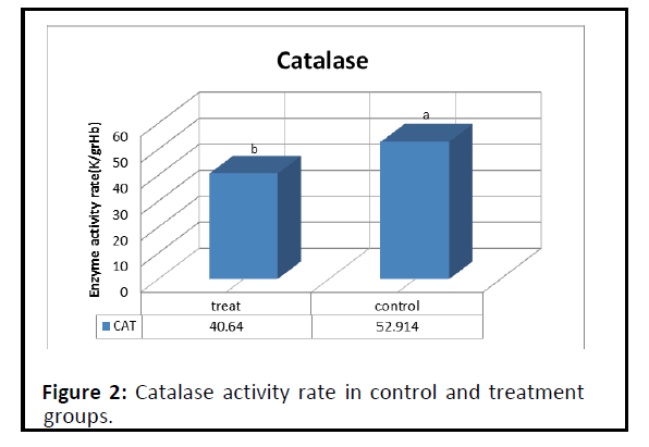
Figure 2: Catalase activity rate in control and treatment groups.
As shown in Table 1 it is described as serum catalase activity rate of tested cats.
Findings and results regarding the effect of malondialdehyde
| Treatment group |
Control group |
Serum sample tested |
| 6.75b ± 40.64 |
14.31a ± 52.91 |
Activity enzyme rate (k/gr Hb) |
ab: Non-identical letters in the table indicate a statistically significant difference between the recorded values(p?0.05)
Table 1: Serum catalase activity rate of tested cats (mean ± SD) during blood sampling.
As shown in Figure 3 the activity of malondealdehyde enzyme rate in the control group of cats was 1.68 (nmol/ml) and in the group of treated cats was 2.3 (nmol/ml).
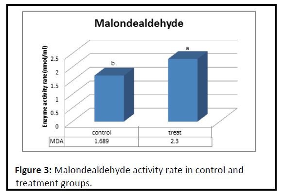
Figure 3 : Malondealdehyde activity rate in control and treatment groups.
As shown in Table 2 we can know about the non-identical letters in the table indicate a statistically significant difference between the recorded values.
| Treatment group |
Control group |
Serum sample tested |
| 0.505b ± 2.3 |
1.68 ± 0.37a |
Activity enzyme rate (nmol/ ml) |
ab: Non-identical letters in the table indicate a statistically significant difference between the recorded values (p?0.05)
Table 2: Serum malondealdehyde activity rate of tested cats (mean ± SD) during blood sampling.
Discussion
Findings from the activity of catalase and malondialdehyde show that the activity of these two enzymes after blood sampling in the treatment group showed a significant decrease compared to the control group (control) and this difference in level 5% was significant (Figures 1 and 2). Following the onset of the disease, the animal's appetite decreases and food consumption stops, and the body consumes fats for energy and fat metabolism increases, which increases the amount of oxidants. It also weakens the animal's immune system and ultimately reduces the amount of antioxidants in the patient's body, such as catalase, glutathione peroxidase and superoxide dismutase. The antioxidant malondialdehyde, which is high in cell membrane structure, is released following membrane damage and increases in serum In the discussion of this result, it can be said that various chemical and biochemical reactions and interactions (metabolism) in the body cause the production of many free radicals that are deficient in electrons in their last electron layer. These free radicals are very destructive and can cause toxicity and disrupt the normal functioning of the body. These free radicals are more than oxygen species such as superoxide, peroxide and hydroxyl, etc. To reach a stable surface, these free radicals must attack the free electroncontaining materials that are available and reach the stable surface by capturing the electron, resulting in severe damage to the organs or components of the cell from which the electron was taken. The most important source of electrons in the body is unsaturated fatty acids (double-bonded fatty acids) that are located in cell membranes. Free radicals act on these fatty acids and capture electrons, converting them to saturated fatty acids. Unsaturated fatty acids are fluid and liquid, which in the process become saturated, which is solid and brittle. Antioxidants are one of the most important elements of the body's defense system against these oxidants, which can prevent these effects by trapping these substances and inactivating them [23]. The results of this study with the results obtained and was Correspond [11,13,20].
Conclusion
Feline calcivirus is one of the most important and common respiratory diseases in cats. The role of antioxidant compounds such as catalase and malondialdehyde in this disease has been investigated in this study. Oxidative stress is involved in the pathogenesis of many diseases and inflammatory conditions.
This results in an oxidant/antioxidant imbalance or an excessive accumulation of oxidants or a decrease in antioxidants. Oxidative stress is a secondary aggravator in most diseases. In addition, a significant degree of negative correlation has been observed between total antioxidant capacity and total oxidant capacity in animals suffering from diseases that cause oxidative stress. The enzyme catalase breaks down hydrogen peroxide and blocks the action of active hydroxyl radicals. These free radicals are highly reactive and cause severe damage to macromolecules such as fats, proteins, carbohydrates and nucleic acids. Antioxidants such as catalase and malondialdehyde are one of the most important elements in the body's immune system against these oxidants, which can prevent these effects by trapping free radicals and inactivating them.
References
- Fenner J, Gibbs E, Paul J, Murphy A (1993) Veterinary Virology (2nd Edn.).
- Radford AD, Coyne KP, Dawson S, Porter CJ, Gaskell RM (2007) Feline calicivirus. Vet Res 38:319-335.
- Rodriguez CC, Menge FW, Ceron JC (2011) Oxidative Stress in Veterinary Medicine. Vet Medi Inter.
- Foley J, Hurley K, Pesavento PA, Poland A (2006) Virulent systemic feline calicivirus infection: local cytokine modulation and contribution of viral mutants. J Fel Med Surg 8:55-61.
- MacNee W (2000) Oxidants/antioxidants and COPD. Chest 117:303-317.
- White SD, Rosychuk RA, Janik TA, Denerolle P, Schultheiss P(1992) Plasma cell stomatitis-pharyngitis in cats: 40 cases (1973-1991). J Ameri Vet Medi Ass 200:1377-1380.
- Gutteridge JMC (1993) Free radicals in disease processes: a compilation of cause and consequence. Free Radi Res Commun 19:141-58.
- Aebi H (1984) Catalase in vitro. Methods Enzymol 105:121-126.
- Kohen R, Nyska A (2002) Oxidation of biological systems: oxidative stress phenomena, antioxidants, redox reac¬tions, and methods for their quantification. Toxicol Pathol 30:620-650.
- Abbott DA, Suir E, Duong GH, Hulster P Pronk JT (2009) Catalase overexpression reduces lactic acid induced oxidative stress in Saccharomyces cerevisiae. Appl Environ Microbiol 75:2320-2325.
- Parks RR, Huang CC, Haddad J (1996) Middle ear catalase distribution in an animal model of otitis media. Eur Arch Otorhinolaryngol 253:445-449.
- Diaz A, Loewen PC, Fita I, Carpena X (2012) Thirty years of heme catalases structural biology. Arch Biochem Biophys 525:102-210.
- Rossi T, Mazzilli F, Delfino M, Dondero F (2001) Improved human sperm recovery using superoxide dismutase and catalase supplementation in semen cryopreservation procedure. Cell Tissue Bank 2: 9-13.
- Sharma A, Kumar P, Rehman MB (2014) Biodegradation of Diesel Hydrocarbon inSoil by Bioaugmentation of Pseudomonas aeruginosa: A Laboratory Scale Study .Inter J Enviro Bioremedi & Biodegra 2:202-212.
- Geciova J, Bury D, Jelen P(2002) Methods for disruption of microbial cells for potential use in the dairy industry-a review. Int Dai J 12:541-553.
- Collodel G, Moretti E, Micheli L, Menchiari A (2015) Semen characteristics and malondialdehyde levels in men with different reproductive problems. Andrology 3:280-286.
- Singh Z, Karthigesu IP, Singh P, Kaur R (2014) Use of Malondialdehyde as a Biomarker for Assessing Oxidative Stress in Different Disease Pathologies: a Review. Ira J Pub Healt 43:7-16.
- Sahin HS (2009) Determination of malondialdehydein human blood by headspace-solid phase micro-extraction gas chromatography mass spectrometry after derivatization with 2,2,2-trifluoroethylhydrazine. J Chromatogr B Analyt Technol Biomed Life Sci 877:3707-3711.
- Jeny C, Goy J, Fechner J, Loeper J (1986) Study of lipid peroxidation by the assay of malondialdehyde in human and experimental atheroma. Presse Med 15:1131-1133.
- Macotpet A, Suksawat F, Sukon P, Pimpakdee K (2013) Oxidative stress in cancer-bearing dogs assessed by measuring serum malondialdehyde. BMC Vet Res 9: 101-106.
- Crnogaj M, Petlevski R, Mrljak V. (2010) Malondialdehyde levels in serum of dogs infected with Babesia canis. Veterinarni Medicina 55:163-171.
- DelRio D, Stewart AJ, Pellegrini N (2005) A review of recent studies on malondialdehyde as toxic molecule and biological marker of oxidative stress. Nutr Metab Cardiovasc Dis 15:316-328.
- Jalili Tabrizi N, Amouoghli Tabrizi B (2020) Evaluating the effect of a single dose of Crack on the activity of serum antioxidant enzymes in the rat. Vet Clini Pathol 13:411-421.