Case Report - (2023) Volume 8, Issue 2
Dysarthria without hepatic manifestation in children revealing Wilson's disease
Rabiy El Qadiry,
Imane Fetoui*,
Houda Nassih,
Aicha Bourrahouat and
Iman Ait Sab
Department of Pediatric, University Hospital Mohammed VI, Marrakech, Morocco
*Correspondence:
Imane Fetoui, Department of Pediatric, University Hospital Mohammed VI,
Marrakech,
Morocco,
Received: 02-Oct-2022, Manuscript No. IPJHCC-22-14473;
Editor assigned: 04-Oct-2022, Pre QC No. IPJHCC-22-14473 (PQ);
Reviewed: 18-Oct-2022, QC No. IPJHCC-22-14473;
Revised: 22-Mar-2023, Manuscript No. IPJHCC-22-14473 (R);
Published:
29-Mar-2023, DOI: 10.36846/2472-1654-8.2.8011
Abstract
Wilson's disease is the consequence of a congenital error in copper’s metabolism. In children, its basic symptom is liver damage. It can be combined with various extra-intestinal manifestations, including neurological ones, which must suggest the diagnosis. We report two cases of dysarthria, with asymptomatic liver injury, revealing Wilson's disease and we detail clinical signs that should make this diagnosis in the child.
Case 1: 12-years-old boy, the first child of a non-consanguineous couple, had normal psychomotor development and schooling. He had consulted for dysarthria, hyper-salivation and tremor of the upper limbs associated with a micrographic started two months before hospitalization. Clinical examination revealed an extrapyramidal syndrome, abnormal movements were noted, associating a kinetic-rigid Parkinson syndrome and a dystonia of the limbs without other signs in particular neither hepatomegaly nor jaundice. Wilson's disease was suspected and confirmed by biological tests, ophthalmologic examination and encephalic MRI.
Case 2: An 11-years-old female patient from first degree marriage with good psychomotor development and normal schooling, who had experienced gait abnormalities, dysarthria and dystonia for the last 6 months. The neurological examination had found an extrapyramidal syndrome, tremor of the extremities and pseudobulbar signs such as dysarthria. Cerebral MRI showed signal abnormalities of the basal ganglia. In front of the starting age, clinical data and radiological appearance, Wilson's disease was suspected. Ophthalmological examination revealed the presence of Kayser fleischer ring and copper assessment confirmed the diagnosis of Wilson's disease.
Conclusion: The diagnosis of Wilson's disease with neurological symptoms is difficult. It must be thought of in front of any dysarthria in young patients.
Keywords
Wilson's disease; Asymptomatic liver injury; Dysarthria; Children; Micrographia
Introduction
Wilson's disease is an autosomal recessive disorder, the consequence of a congenital error in copper’s metabolism. In children, it mainly causes liver damage from the age of 5 to 6 years and can be combined with various extrahepatic manifestations, including neurological manifestations that should make the diagnosis suspected [1]. Neurological damage can be translated as neurological and psychiatric disorders progressively appearing with existent hepatic involvement that may be asymptomatic. We report two cases of dysarthria in children, with asymptomatic liver injury, revealing Wilson's disease and we detail clinical signs that should make this diagnosis in the child.
Case Presentation
Patients and Observations
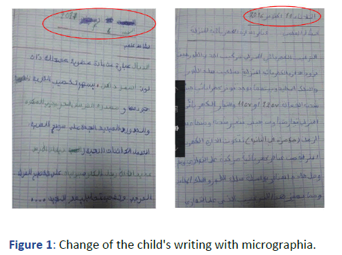
Case 1: S.S a 12-years-old boy, the first child of a non-consanguineous couple, had normal psychomotor development and schooling. He had consulted for dysarthria, hyper-salivation and swallowing disorder, upper limb tremor with micrographia (Figure 1). Progressing two months prior to his hospitalization.

Figure 1: Change of the child's writing with micrographia.
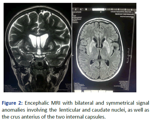
Clinical examination found an extrapyramidal syndrome, abnormal movements were noted, associating an akinetic-rigid Parkinson syndrome and a dystonia of the limbs without other signs. Abdominal, mucocutaneous and osteoarticular examinations were normal. An encephalic MRI revealed bilateral and symmetrical signal abnormalities affecting the lenticular and caudate nuclei, as well as the crus anterius of the two internal capsules, in hypo T1 signal, in hyper T2 signal and Flair (Figure 2).

Figure 2: Encephalic MRI with bilateral and symmetrical signal anomalies involving the lenticular and caudate nuclei, as well as the crus anterius of the two internal capsules.
Slit lamp examination had shown the existence of the bilateral Kayser-fleischer peri-corneal ring. Hepatic assessment showed moderate hepatic cytolysis (ASAT at 85 IU, ALT at 147 IU/l) contrasting with collapsed PT (59%) and abdominal ultrasound had noted the presence of an atrophic and heterogeneous liver without any pictured nodules. The cupric assessment confirmed the diagnosis of Wilson's disease. The child was put under D-penicillamine at an initial dose of 300 mg/day with gradual increase of the dose up to 1800 mg/day for one month then put under Zinc Acetate. After a 3-year follow-up, the progress was fortunately a stabilization of his neurological status, but the abnormal movements and oral-facial disorders remained. At the same time, family investigation found that his 9-year-old brother, in good general status, had moderate hepatic cytolysis without hepatocellular insufficiency and a slightly elevated 24-hour urine copper test.
Case 2: H.S is an 11-years-old daughter of first-degree marriage with good psychomotor development and normal schooling. She presented with gait disorders, dysarthria and dystonia that appeared six months before her hospitalization. Neurological examination had revealed a walk of little steps with the head and neck forward, tremor at extremities, plastic hypertonia at four-limbs level with cogwheel phenomenon, pseudobulbar signs with dysarthria and generalized dystonia, the rest of clinical examination was without particularity precisely, no hepatomegaly.
MRI was performed, revealing signal abnormalities of lenticular nuclei, thalami, caudate nuclei, cerebral peduncles and quadriluminal tubercles, in hyper-signal T2, flair and discreetly in diffusion and hyposignal T1 without detectable contrast enhancement. Moreover, there are no morphological abnormalities, abnormal signal or contrast enhancement in the cerebral parenchyma. In front of the age of beginning, clinical data and radiological appearance, Wilson's disease was suspected. The ophthalmological examination revealed the presence of a kayser-Fleischer ring and cupric assessment showed a level of 4.4 Umol/L, serum ceruloplasmin level of 0.06 g/L and 24 hour urine copper test at 220 micrograms. Hepatic assessment showed hepatic cytolysis with ASAT at 593 IU/l (15 × N), ALAT at 178 IU/l (5 × N) and a PT at 56.1%. The diagnosis was that of Wilson's disease. The child was put under D-penicillamine and zinc acetate with symptomatic treatment of dystonia and hepatocellular insufficiency. The progress noted neurological worsening with that start of swallowing disorders and status dystonicus disorder, finally, the child died in an array of cachexia and hepatocellular insufficiency. MRI was performed, revealing signal abnormalities of lenticular nuclei, thalami, caudate nuclei, cerebral peduncles and quadri luminal tubercles, in hyper signal T2, flair and discreetly in diffusion and hyposignal T1 without detectable contrast enhancement. Moreover, there are no morphological abnormalities, abnormal signal or contrast enhancement in the cerebral parenchyma. In front of the age of beginning, clinical data and radiological appearance, Wilson's disease was suspected. The ophthalmological examination revealed the presence of a kayser-Fleischer ring and cupric assessment showed a level of 4.4 Umol/L, serum ceruloplasmin level of 0.06 g/L and 24 hour urine copper test at 220 micrograms. Hepatic assessment showed hepatic cytolysis with ASAT at 593 IU/l (15 × N), ALAT at 178 IU/l (5 × N) and a PT at 56.1%. The diagnosis was that of Wilson's disease. The child was put under D-penicillamine and zinc acetate with symptomatic treatment of dystonia and hepatocellular insufficiency. The progress noted neurological worsening with that start of swallowing disorders and status dystonicus disorder, finally, the child died in an array of cachexia and hepatocellular insufficiency.
Discussion
Neurologically revealing forms of Wilson's disease are rare and account for about 35% of cases. The clinical picture is very heterogeneous; typically, the neurological start is characterized by a change in the character and abnormalities of mimicry (facial expression). This heterogeneity of clinical signs often causes a delay in diagnosis and management, which has a negative impact on the prognosis of this disease [2]. The first symptoms are often misleading as a drop in school performance [3], teachers must then be aware that the decline in school and the change of writing with micrographia might be the way to diagnose Wilson's disease. Neurologic examination revealed ataxia with dysarthria in 90% of cases, extrapyramidal syndrome and abnormal mixed movements (generalized orofacial dystonia, attitude and movement tremor and akineto-rigid parkinsonian syndrome). We must therefore think of Wilson's disease in front of Parkinson's syndrome in a child or adolescent. Diagnostic confirmation remains easy and inexpensive, based on a disrupted liver test and an ophthalmological examination with a slit lamp consistently objectifying the Fleischer kayser ring. Cerebral MRI is an important diagnostic tool for the disease, usually showing symmetrical involvement of basal ganglia. This involvement must retrospectively suggest the diagnosis of Wilson's disease, which is not clinically suspected [4]. Once Wilson's disease is confirmed, systematic screening of siblings should be performed even if they are asymptomatic. The prognosis of Wilson's disease is at its best as long as neurological and hepatic involvement is not pronounced, the ideal is to confirm the diagnosis at a pre-symptomatic stage [5].
Conclusion
The diagnosis of Wilson's disease with neurological expression is difficult. It must be suggested before any parkinsonian syndrome and dysarthria in young patient. Delayed diagnosis is the main factor of bad prognosis. The treatment is difficult because of the inconsistent improvement of the symptoms or even their irreversible worsening especially under D-penicillamine.
Competing Interests
The authors declare no competing interests.
Author Contributions
All the authors have read and agreed to the final manuscript.
References
- Mak CM, Lam CW (2008) Diagnosis of Wilson's disease: A comprehensive review. Crit Rev Clin Lab Sci. 45(3):263-290.
[Crossref] [Google Scholar] [PubMed]
- Lorincz MT (2010) Neurologic Wilson’s disease. Ann N Y Acad Sci. 1184:173-187.
[Crossref] [Google Scholar] [PubMed]
- Wagner S, Brunet AS, Bost M, Lachaux A, Broussolle E, et al. Diagnosis and care of Wilson disease with neurological revelation. Arch Pediatr. 2012. 19:271-276.
[Crossref] [Google Scholar] [PubMed]
- Roh JK, Lee TG, Wie BA, Lee SB, Park SH, et al. (1994) Initial and follow-up brain MRI findings and correlation with the clinical course in Wilson’s disease. Neurology. 44(1):1064-1068.
[Crossref] [Google Scholar] [PubMed]
- Basir A, Bougteba A, Kissani N (2009) Torticollis revealing Wilson’s disease. Arch Pediatr. 16(4):402-404.
[Crossref] [Google Scholar] [PubMed]
Citation: El Qadiry R, Fetoui I, Nassih H, Bourrahouat A, Sab IA (2023) Dysarthria without Hepatic Manifestation In Children Revealing Wilson's Disease. J Healthc Commun. 8:8006
Copyright: © 2023 El QadiryI R, et al. This is an open-access article distributed under the terms of the Creative Commons Attribution License, which permits unrestricted use, distribution and reproduction in any medium, provided the original author and source are credited.


