Research Article - (2023) Volume 8, Issue 1
Cyto Histopathological Correlation of Soft Tissue Tumors
Fathima Nifra M Mohamed Nizar*
Department of Health Care Communications, University of Delhi, Delhi, India
*Correspondence:
Fathima Nifra M Mohamed Nizar,
Department of Health Care Communications, University of Delhi, Delhi,
India,
Email:
Received: 02-May-2022, Manuscript No. IPJHCC-22-12456;
Editor assigned: 05-May-2022, Pre QC No. IPJHCC-22-12456 (PQ);
Reviewed: 19-May-2022, QC No. IPJHCC-22-12456;
Revised: 23-Jan-2023, Manuscript No. IPJHCC-22-12456 (R);
Published:
30-Jan-2023, DOI: 10.36846/ 2472-1654-8.1.8002
Abstract
Soft tissue can be defined as non-epithelial extra skeletal tissue of the body, exclusive of the reticuloendothelial system, glia and supporting tissue of various parenchymal organs. This was a prospective study undertaken at the department of pathology, Balaji medical college and hospital, chrome pet. A total number of 105 cases clinically suspected as soft tissue tumour were subjected to FNAC and compared with histopathology. A total number of 105 cases clinically suspected as soft tissue tumours were subjected to FNAC and compared with histopathology. In this study the age of the patients ranged from 4 to 80 years.
Keywords
Histopathological; Aspiration cytology; Anatomical; Skeletal tissue; Parenchymal organs
INTRODUCTION
Soft tissue can be defined as non-epithelial extra skeletal
tissue of the body, exclusive of the reticuloendothelial system,
glia and supporting tissue of various parenchymal organs. The
diagnostic role of Fine Needle Aspiration Cytology (FNAC) in
evaluating soft tissue tumour remains controversial as many
of these lesions, especially the sarcomas have overlapping
histopathological and cytomorphologic features associated
with morphologic heterogeneity present in some of these
mass lesions FNAC is a useful tool in distinguishing accurately
between neoplastic and non-neoplastic lesions [1,2]. The two
fundamental requirements on which the success of FNAC
depends are representativeness and adequacy of sample and
high quality of preparation [3]. FNAC is a painless procedure,
easy to perform, safe, and cost effective, which does not
require anesthesia, and acts as a useful diagnostic technique
in the initial diagnosis of tumour [4]. It produces a speedy
result. It can be easily repeated if necessary in the same
sitting. Unfortunately FNAC has a few disadvantages,
especially in identifying borderline lesions. Aspirates from
densely collagenase or sclerotic masses or highly vascular
lesions, provide sparse cellularity [5,6]. Hence there is
absolute necessity to integrate surgeon, radiologist and
pathologists opinion. This study aims to study the utility of
Fine Needle Aspiration Cytology (FNAC) in the diagnosis of
soft tissue tumour and to correlate FNAC with
histopathological examination [7-10].
Materials and Methods
This was a prospective study undertaken at the department of
pathology, Balaji medical college and hospital, chromepet. A
total number of 105 cases clinically suspected as soft tissue
tumour were subjected to FNAC and compared with
histopathology. After taking the history and evaluating the
patient clinically, they were subjected to FNAC and later
biopsy, taking the consent of the patient. Descriptive statistics
were reported as mean (SD) for continuous variables,
frequencies (percentage) for categorical variables. Data were
statistically evaluated with IBM SPSS statistics for windows,
version 20.0., IBM corp., and Chicago, IL.
Results
A total number of 105 cases clinically suspected as soft tissue
tumours were subjected to FNAC and compared with
histopathology. In this study the age of the patients ranged
from 4 to 80 years, the youngest patient being 4 years old and
the oldest 80 year old. The maximum number of cases was
found in the fourth decade followed by third decade. Male:
Female ratio was 1:1.1. Soft tissue masses were found most
commonly in the lower extremities followed by upper
extremities, trunk, head and neck and retro
peritoneum (Table 1 and 2).
| Slno |
Variable |
Frequency |
Percentage |
| 1 |
Age ( in years ) |
| 0-10 |
2 |
1.9 |
| 11-20 |
12 |
11.4 |
| 21-30 |
21 |
20 |
| 31-40 |
22 |
20.9 |
| 41-50 |
26 |
24.8 |
| 51-60 |
15 |
14.3 |
| 61-70 |
4 |
3.8 |
| 71-80 |
3 |
2.9 |
| 2 |
Anatomical distribution |
| Head and neck |
17 |
16.2 |
| Upper extremity |
28 |
26.7 |
| Lower extremity |
33 |
31.4 |
| Trunk |
22 |
20.9 |
| Peritoneum |
5 |
4.8 |
| 3 |
Diagnosis of soft tissue tumours on FNAC |
| Benign |
88 |
83.8 |
| Malignant |
17 |
16.2 |
Table 1: Distribution of variables among the study participants (N=105).
| Slno |
Variable |
Frequency |
Frequency |
(%) |
| 1 |
Myxoid |
2 |
Myxoid malignant fibrous histiocytoma |
2 |
1.9 |
| 2 |
Spindle cell |
56 |
Benign spindle cell |
9 |
53.3 |
| Neoplasm |
- |
- |
| Benign fibrous histiocytoma |
1 |
- |
| Benign nerve sheath |
17 |
- |
| Tumor |
- |
- |
| Haemangioma |
11 |
- |
| Giant cell tumor of tendon sheath |
- |
- |
| Benign mesenchymal |
- |
- |
| tumor |
- |
- |
| Spindle cell sarcoma |
- |
- |
| MPNST |
- |
- |
| Malignant fibrous histiocytoma |
- |
- |
| Dermatofibrosarcoma |
- |
- |
| protuberance |
- |
- |
| 3 |
Pleomorphic |
2 |
Pleomorphic lipoma |
1 |
1.9 |
| Pleomorphic malignant fibrous histiocytoma |
1 |
- |
| 4 |
Round cell |
1 |
PNET |
1 |
1.9 |
| 5 |
Miscellaneous |
44 |
Lipoma |
44 |
41.9 |
Table 2: Cytologic diagnosis of so t tissue tumors, grouped according to their appearance ine needle aspiration in (N=105).
The spindle cell tumours were the most common tumours
constituting 53.3% of cases followed by miscellaneous tumours chiefly of lipomas constituting of 41.9 % of the cases
(Table 3).
| Slno |
Diagnosis |
Number of cases |
Percentage (%) |
| 1 |
Lipoma |
38 |
36.1 |
| 2 |
Fibrolipoma |
2 |
1.9 |
| 3 |
Pleomorphic lipoma |
1 |
1 |
| 4 |
Angiofibrolipoma |
1 |
1 |
| 5 |
Angiomyolipoma |
1 |
1 |
| 6 |
Angiolipoma |
1 |
1 |
| |
Lipoma variants in total |
44 |
|
| 7 |
Giant cell tumour of tendon sheath |
4 |
3.8 |
| 8 |
Neurofibroma |
4 |
3.8 |
| 9 |
Schwannoma |
13 |
12.3 |
| 10 |
Hemangioma |
12 |
11.4 |
| 11 |
Fibromatosis |
4 |
3.8 |
| 12 |
Desmoid fibromatosis |
2 |
1.9 |
| 13 |
Benign fibrous histiocytoma |
3 |
2.8 |
| 14 |
Primitive neuroectodermal tumour |
1 |
1 |
| 15 |
Pleomorphic malignantfibrous histiocytoma |
1 |
1 |
| 16 |
Synovial sarcoma |
1 |
1 |
| 17 |
Dermatofibrosarcoma protuberance |
1 |
1 |
| 18 |
Bednar tumour |
2 |
1.9 |
| 19 |
Hemangioendothelioma |
1 |
1 |
| 20 |
Malignant fibrous histiocytoma |
3 |
2.8 |
| 21 |
Myxoid MFH |
2 |
1.9 |
| 22 |
Malignant peripheral nerve sheath tumour |
7 |
6.6 |
Table 3: Analysis of lesions on histopathology (N=105).
Discussion
The age range in the present study was 4 to 80 years which
was comparable to where the range was between 4 years to
94 years. Where the range was 12 years to 88 years. The male
to female ratio was 1:1. The soft tissue neoplasm was most
commonly seen in the fourth decade followed by the third
decade. Present study showed most common site of tumors
were lower limb and upper limb which was consistent with
also found lower extremity as the most common site followed
by upper extremity [11-17]. While in study trunk was the
most common site. In the present study we have used the
classification recommended by dividing the aspirates into six
categories and (Table 4).
| Study |
Categories |
| 1 |
2 |
3 |
4 |
5 |
6 |
| Miralles, et al. |
Low grade sarcoma |
Myxoid sarcomas |
Monomo rphic sarcomas |
Round cell sarcomas |
Pleomorp hic sarcomas |
-- |
| Gonzalez Campor R, et al. |
Myxoid |
Round cell |
Spindle cell |
Pleomorphic cell |
Polygonal cell |
Well differentiated |
| Umarani, et al. |
Myxoid rich |
Round cell |
Spindle cell |
Pleomorphic |
Epithelial -like |
Mature like cell |
| Geisinger and Abdul-Karim |
Myxoid |
Spindle cell |
Pleomorphic |
Polygonal |
Round cell |
Miscellaneous |
Table 4: Various cytomorphological classifications proposed by different authors.
We had two cases of myxoid tumour in our study. Smears of
amyloid tumour studied showed high cellularity showing two
populations of cells consisting of spindle cells and round to
polygonal cells arranged in loose clusters and in singles. The
nuclei showed hyperchromasia with pleomorphic. The
presence of foamy histolytic and scattered bizarre
multinucleated giant cells was typical of the tumour. Grossly
showed a well circumscribed tumour with multilobulation and
myxoid degeneration. Histopathology sections studied
showed a neoplasm composed of elongated cells and giant
cells with dark staining nuclei arranged in interlacing bundles
and fascicles in an abundant myxoid background. Tumour
giant cell, thin walled blood vessels, focal areas of hyalinization and occasional mitotic figures were seen
(Figures 1 and 2).
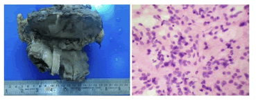
Figure 1: a) Gross image showing cut section of the tumour with myxoid degeneration; b) Cytology image of myxoid MFH (H and E 10 x 10).
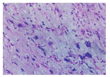
Figure 2: Myxoid MFH H and E (10 x 40) showing a neoplasm composed of elongated cells and giant cells with dark staining nuclei arranged in interlacing bundles and fascicles in an abundant myxoid background.
Fifty three cases of spindle cell tumours were studied, out of
which 43 were diagnosed as benign tumours and 10 as
malignant spindle cell tumours on cytology. Seventeen cases
of benign neural tumour were diagnosed on FNAC. Fifteen
cases of benign neural tumour were diagnosed correctly on
cytology consisting of 3 cases of neurofibroma and 13 cases of
schwannoma. One case was discordant which was diagnosed
as MPNST on HPE. Smears studied showed low to moderate
cellularity with cells lying in tight cohesive clusters and
bundles. The predominant cell type was spindle cells. The
nuclei were spindle to ovoid with few being wavy or bent,
having a bland chromatin pattern. Background showed a
distinct eosinophilia fibrillary stroma. Sections studied
showed a benign neoplasm composed of spindle cells with
wavy nuclei in fascicles surrounded by fibro collagenous tissue
with areas of myxoid changes (Figure 3).
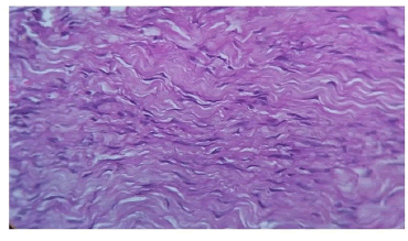
Figure 3: Neurofibroma: spindle cells with characteristic wavy nuclei with sort fusiform nuclei (H and E 10 x 40).
Thirteen cases of schwannoma were correctly diagnosed on
FNAC. Smears studied showed moderate to high cellularity
with spindle cells being arranged in cohesive clusters,
interlacing bundles. The nuclei were wavy with few being
ovoid to fusiform. Multiple sections studied showed a
capsulated tumour with spindle cells arranged in interlacing
bundles and fascicles. The spindle cells had vesicular nuclei
with nuclear palisading. Antoni A and antoni B areas with areas of hyalinization and cystic degeneration were seen
(Figure 4). Two cases of ancient schwannoma showed
degenerative features.
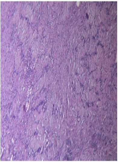
Figure 4: Ancient schwannoma. Spindle shaped cells with elongated nuclei in a fibrillary background, nuclear palisading, verrucay bodies are also seen H and E (10 x 10).
Out of 9 cases diagnosed as benign spindle cell tumour on
cytology, only 7 cases were correlated remaining two were
discordant. Two cases were of malignant or borderline
category with one as dermatofibrosarcoma protuberant and
one as haemangioendothelioma, on histopathology.
Malignant spindle cell tumours of neural origin (MPNST),
around 7 cases were diagnosed on HPE. One case showed
incorrect diagnosis which was diagnosed as benign nerve
sheath tumour on FNAC. Aspirates were moderately to highly
cellular consisting of tumour cells arranged in interlacing
fascicles, clusters and singles. Individual tumour cells had
eosinophilia, fibrillary cytoplasm with nuclear shape being
varied i.e. ovoid, wavy, fusiform and spindle. Chromatin
pattern was vesicular to hyperchromaticwith nuclear
pleomorphic. One case showed mitotic figures and
multinucleated giant cells. Sections studied show a cellular
neoplasm composed of spindle shaped cells with elongated,
wavy and fusiform nuclei with prominent nucleoli, arranged in
whorls, fascicles, and interlacing bundles. Focal cartilaginous
metaplasia, epithelioid cells and occasional mitotic figures
were also seen (Figure 5).
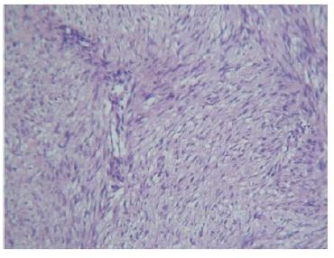
Figure 5: MPNST fascicles and bundles of spindle shaped cells in a fibrillary background H and E (10 x 10).
Twelve cases of vascular tumours were diagnosed accurately
on cytology and confirmed by histopathology. Eight cases
were of haemangioma and four cases of cavernous
haemangioma one case of haemangioendothelioma were
diagnosed as benign spindle cell tumour on cytology. The
smears of haemangioma studied showed low to moderate
cellularity. The spindle cells were arranged in fragments,
clusters and singles. The spindle cells had ovoid to spindle
shaped nuclei showing bland chromatin pattern with bipolar
eosinophilia cytoplasm. Background was bloody. In one case
of intramuscular cavernous haemangioma only blood was
aspirated on repeated FNAC. Histopathology sections studied
showed large, open vascular spaces separated by fibrous
tissue septate in a case of cavernous haemangioma.
Haemangioendothelioma histopathology sections studied
showed a vascular lesion composed of proliferating
endothelial lined spaces and capillaries. The tumour cells
were seen infiltrating skeletal muscles and widely separating
it. Cells were round to oval with scanty cytoplasm and hyper
chromatic nuclei, showing mild pleomorphic. Few mitotic
figures and occasional tumour giant cells were seen (Figure 6).
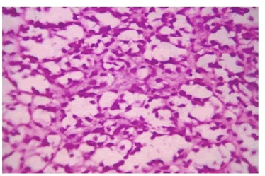
Figure 6: Haemangioendothelioma: Tumor cells lining tiny vascular like spaces. Tissue section (H and E, 10 x 40).
The desmoids tumour was over diagnosed as malignant
fibrous histiocytoma. One case diagnosed as MFH had spindle cells admixed with polygonal cells having abundant
eosinophilia cytoplasm. Nuclei showed mild pleomorphic with
finely dispersed chromatin and prominent nucleoli. Few
mitotic figures were seen. Sections studied showed a spindle
cell neoplasm composed of elongated cells with vesicular
nucleus arranged in interlacing bundles and fascicles in a fibro
collagenous background thin walled blood vessels, focal
myxoid changes, cartilaginous metaplasia and occasional
mononuclear cells were seen in the stroma. The interlacing
bundles of spindle cells were seen with entrapped muscle
fibers. One case of benign fibrous histiocytoma was
diagnosed accurately on cytology whereas two cases were
diagnosed as benign spindle cell tumor. Out of 4 cases of
malignant fibrous histiocytoma, 3 were accurately diagnosed
on cytology. One case was inaccurately diagnosed, which was
confirmed as fibromatosis on histopathology. One case in our
study, which was inaccurately diagnosed, showed a low
cellularity with cell clusters consisting of spindle cells with
ovoid, bland nuclei. Histiocyte like cells which aids in the
diagnosis, were not seen. So a diagnosis of only a benign
spindle cell tumour was possible. Sections studied showed a
tumour arranged in short fascicles. Malignant fibrous
histiocytoma Cytology Smears studied were highly cellular
showing two populations of cells consisting of spindle cells
and round to polygonal cells. The cells were arranged in
scattered loose clusters and in singles. The spindle cells had
spindle nucleus with bipolar eosinophilia cytoplasm. The
nuclei showed hyper chromatic or coarse chromatin with
moderate to marked pleomorphic. Multinucleated giant cells
were seen in all the cases of MFH. The presence of foamy
histiocyte like cells and population of cells with
multinucleated giant cells were typical of the tumour. Cut
section showed yellowish white circumscribed tumour with
focal areas of hemorrhage. Sections studied showed a cellular
neoplasm composed of spindle tumour cells arranged in
interlacing bundles, sheets and focal storiform pattern. The
spindle cells showed nuclear pleomorphic. Good number of
tumour giant cells and mitotic figures were noted, along with
areas of myxoid change and necrosis. One case of
Dermatofibrosarcoma protuberant was incorrectly diagnosed
as benign spindle cell tumour on cytology. On cytology
showed a moderately cellular aspirate with cells arranged in
clusters and singles. The cells were spindle to oval in shape,
with oval nuclei showing mild pleomorphic. Chromatin
pattern was bland and nucleoli were seen in many. Cells had
indistinct borders with eosinophilia cytoplasm. Background
showed few myxoid stromal fragments. Gross showed two
nodular grey-pink masses protruding above the skin. Cutsection
showed a fleshy grey-white, soft tumour. Sections
studied showed a tumour in the sub epithelium arranged in
storiform pattern. The tumour cells were spindle shaped
having scanty cytoplasm and a spindle shaped nuclei with
inconspicuous nucleoli. Few mitotic figures seen. Seven cases
of spindle cell sarcoma were diagnosed on FNAC and the
same was diagnosed on HPE. Two cases of Bednar tumour, 2
cases of MPNST, 2 cases MFH, and one case of synovial
sarcoma. Gross- cut section showed hemorrhagic areas.
Sections studied showed skin overlyingfibrocollagenous
dermis enclosing a spindle cell lesion composed of elongated cells with pleomorphic nuclei. Focal storiform pattern was
also seen. Synovial sarcoma (biphasic type) histopathology
sections studied showed an eoplasm composed of spindle
shaped cells with elongated dark staining nuclei arranged in
interlacing bundles and fascicles admixed with nests of round
to oval cells with vesicular nuclei. Focal areas of hemorrhage,
necrosis, and myxoid degeneration were also seen.
Pleomorphic tumours-pleomorphic lipoma on cytology
showed floret like giant cells and spindle cells. Sections
studied showed mixture of fat cells, floret like giant cells and
ropy collagen. Pleomorphic tumour cells in loose aggregates
with bizarre tumour giant cells were seen. Gross: Cut section
showed whitish, hemorrhagic, yellowish brown and areas of
necrosis. Sections studied showed a cellular neoplasm
composed of pleomorphic spindle cells with hyper chromatic
nuclei arranged in interlacing bundles, fascicles and focal
storiform pattern. Pleomorphic tumour giant cells,
multinucleated cells, bizarre giant nuclei and mitotic figures
were seen (Figure 7).
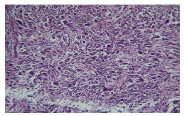
Figure 7: Pleomorphic MFH: Cellular spindle cell neoplasm composed of pleomorphic spindle cells arranged in fascicles with pleomorphic tumour giant cells. H and E (10 x 10).
Forty four cases of Lipoma were diagnosed by cytology and
forty four were confirmed by histopathology as lipoma. Most
cases showed a moderately cellular aspirate with mature fat
cells being arranged in small clusters. Significant amount of
collagen fragments and small clusters of fibrocystic were seen
in one case. A case of intramuscular lipoma showed skeletal
muscle fibers with adipose tissue. On cut section,
characteristically a grey-yellow tumour mass was seen
replacing the skeletal muscle fibers. Sections studied showed
well circumscribed tumour arranged in lobular pattern
separated by thin connective tissue stroma and few
capillaries. The tumour was made up of mature fat cells. One
case showed bundles of hyalinized collagen fibers mixed with
mature fat cells and was diagnosed as fibro lipoma. One case
of angiomyolipoma showed lobules of mature fat with thick
walled blood vessels and bundles of smooth muscle.
Conclusion
Aspiration cytopathology of soft tissue mass lesions using
FNAC can be a cost effective, accurate, minimally invasive and a swift preliminary diagnostic procedure. It can provide initial
pathologic diagnosis of primary benign and malignant soft
tissue tumors. Hence FNAC provides reliable information to
the clinicians for triage of the patients and enable them to
consider management decisions at the earliest.
References
- Soni PB, Verma AK, Chandoke RK, Nigam JS (2014) A prospective study of soft tissue tumorshistocytopathology correlation. Patholog Res Int. 2014:678628.
[Crossref][Googlescholar][Indexed]
- Dey P, Mallik MK, Gupta SK, Vasishta RK (2004) Role of fine needle aspiration cytology in the diagnosis of soft tissue tumours and tumour‐like lesions. Cytopathology. 15(1):32-37.
[Crossref][Googlescholar][Indexed]
- Parajuli S, Lakhey M (2012) Efficacy of fine needle aspiration cytology in diagnosing soft tissue tumors. Pathol Nepal. 2(4):305-308.
[Crossref][Googlescholar]
- Boni LS, Kasturi S, Uma P, Atla B (2019) Role of fine needle aspiration cytology in the diagnosis of soft tissue tumors a prospective study. Trop J Pathol Microbiol. 5(8):535-541.
[Crossref][Googlescholar][Indexed]
- Fathimanifra M, Vijayalakshmi NS, Thanka J (2021) Histopathological Study of Soft Tissue Lesions with Cytology Correlation. J Pharm Res Int. 33(22):101-117.
[Crossref]
- Kim SW, Roh J, Park CS (2016) Immunohistochemistry for pathologists: protocols, pitfalls, and tips. J Pathol Transl Med. 50(6):411-418.
[Crossref][Googlescholar][Indexed]
- Wakely Jr PE, Kneisl JS (2000) Soft tissue aspiration cytopathology: diagnostic accuracy and limitations. Cancer. 25;90(5):292-298.
[Crossref][Googlescholar][Indexed]
- Akerman M, Rydholm A, Persson BM (1985) Aspiration cytology of soft-tissue tumors: the 10 years experience at an orthopedic oncology center. Acta Orthopaedica Scandinavica. 56(5):407-412.
[Crossref][Googlescholar][Indexed]
- Bezabih M, Mariam DW (2003) Determination of etiology of superficial enlarged lymph nodes using fine needle aspiration cytology. East Afr Med J. 80(11):559-563.
[Crossref][Googlescholar][Indexed]
- Nagira K, Yamamoto T, Akisue T, Marui T, Hitora T, et al. (2002) Reliability of fine‐needle aspiration biopsy in the initial diagnosis of soft‐tissue lesions. Diagn Cytopatho. 27(6):354-361.
[Crossref][Googlescholar][Indexed]
- Roy S, Manna AK, Pathak S, Guha D (2007) Evaluation of fine needle aspiration cytology and its correlation with histopathological findings in soft tissue tumours. J Cytol. 24(1):37.
[Googlescholar]
- Abdul‐Karim FW, Weaver MG (1991) Needle aspiration cytology of an oncocytic carcinoma of the parotid gland. Diagn Cytopathol. 7(4):420-422.
[Crossref][Googlescholar][Indexed]
- Miralles P, Berenger J, Ribera JM, Rubio R, Mahillo B, et al. (2007) Prognosis of AIDS related systemic non-Hodgkin lymphoma treated with chemotherapy and highly active antiretroviral therapy depends exclusively on tumor-related factors. J Acquir Immune Defic Syndr. 44(2):167-173.
[Crossref][Googlescholar][Indexed]
- Gonzalez‐campora R, Herrero‐Zapatero A, Lerma E, Sanchez F, Galera H (1986) Hurthle cell and mitochondrion‐rich cell tumors. A clinicopathologic study. Cancer. 15;57(6):1154-1163.
[Crossref][Googlescholar][Indexed]
- Umarani MK, ShuchitaLakra P, Bharathi M (2015) Histopathological Spectrum of Soft Tissue Tumors in a Teaching Hospital. IOSR J Dent Med Sci. 14(4):74-80.
[Crossref][Googlescholar]
- Chakrabarti PR, Chakrabarti S, Pandit A, Agrawal P, Dosi S, et al. (2015) Histopathological study of soft tissue tumors: A three year experience in tertiary care centre. Indian J Pathol Oncol. 2(3):141-149.
[Googlescholar][Indexed]
- Jain P, Shrivastava A, Mallik R (2014) Clinic morphological Assessment of Soft Tissue Tumors. Sch J App Med Sci. 2(2):886-890.
Citation: Nizar FNMM (2022) Cyto Histopathological Correlation of Soft Tissue Tumors. J Healthc Commun. 8:8002
Copyright: © 2023 Nizar FNMM. This is an open-access article distributed under the terms of the Creative Commons Attribution License, which permits unrestricted use, distribution, and reproduction in any medium, provided the original author and source are credited.


