Research Article - (2026) Volume 27, Issue 3
Comparative outcomes of Open and Laparoscopic drainage in Walled off Pancreatic Necrosis
Khamar Jaha Banu1*,
Natasha Mathias2 and
Nameera Tabassum2
1Deptartment of Surgical Gastroenterology, Yenepoya Medical College Hospital, Mangalore, Karnataka, India
2Department of General Surgery, Yenepoya Medical College hospital, Mangalore, Karnataka, India
*Correspondence:
Khamar Jaha Banu, Deptartment of Surgical Gastroenterology, Yenepoya Medical College Hospital, Mangalore, Karnataka,
India,
Email:
Received: 25-May-2026, Manuscript No. IPP-26-23996;
Editor assigned: 27-May-2026, Pre QC No. IPP-26-23996;
Reviewed: 11-Jun-2026, QC No. IPP-26-23996;
Revised: 17-Jun-2026, Manuscript No. IPP-26-23996;
Published:
25-Jun-2026, DOI: 10.36648/1590-8577-27.3.983
Abstract
Introduction: Walled-Off Pancreatic Necrosis (WOPN) develops in approximately 1-9% of patients following acute pancreatitis, typically manifesting 4-6 weeks after the initial episode. Around 15% of patients with severe necrotising pancreatitis go on to develop WOPN. This condition is associated with significant morbidity and occasional mortality. Most necrotic collections ultimately require intervention-either percutaneous, endoscopic, or surgical.
Materials and Methods: A prospective study was conducted involving 51 patients diagnosed with WOPN. Of these, 23 patients underwent open transgastric drainage, while 28 underwent laparoscopic drainage. Demographic data, intraoperative and postoperative outcomes were analysed and compared between the two groups.
Results: Overall, in our study comparing the open versus laparoscopic approach for transgastric drainage of WOPN, the significant finding was a shorter operative time with the open approach. This can be attributed to the use of intracorporeal suturing rather than staplers. Blood loss was lower in the laparoscopic group. Postoperative infections were more common in the open approach. Mortality and morbidity were similar in both groups; hospital stay was significantly shorter in the laparoscopic group.
Conclusion: No single approach is currently considered the gold standard. Therefore, management must be individualised based on the patient’s clinical presentation and available resources. The laparoscopic approach reduces overall complications, particularly infectious complications. The use of staplers may also significantly reduce operative time. A robotic approach may be best, but due to limited avail ability in remote areas, the laparoscopic approach may be considered.
Keywords
Walled Off Pancreatic Necrosis (WOPN); Laparoscopic; Transgastric drainage
Introduction
Acute pancreatitis is a spectrum of disease entities characterised by complex pathophysiology and variable natural history. Pancreatic inflammation triggers the release of cytokines, which can initiate a cascade of events. However, the aetiopathogenesis for the presence or severity of necrosis is largely unknown. Necrosis is a major determinant of outcome in acute pancreatitis [1]. The necrotic collection needs to be addressed in the majority of patients. The management of necrosis depends on the duration of collection and the presence of infection. The Atlanta classification divides necrotic collections into acute necrotic collections (<4 weeks’ duration) and walled-off pancreatic necrosis (>4 weeks’ duration) [2]. Management of these collections involves a multidisciplinary approach, including medical, interventional radiology, endoscopic and surgical approaches. Surgical intervention involves minimally invasive or open necrosectomy. Laparoscopic transgastric necrosectomy is a minimally invasive one-time intervention to manage a lesser sac necrotic collection. In this study, we evaluated the feasibility and outcomes of laparoscopic transgastric necrosectomy for the management of Walled-Off Pancreatic Necrosis (WOPN).
Materials and Methods
It was a prospective comparative study conducted at a tertiary care referral hospital over 4 years. Fifty-one patients who underwent laparoscopic and open transgastric drainage were included in the study. The data were collected on patient demographics, intraoperative events and 30-day postoperative events. Patients were followed up for a minimum of 1 year and recurrence, exocrine and endocrine insufficiency were recorded. Inclusion criteria were symptomatic WOPN or infected WOPN situated in the lesser sac and having an indentation on the stomach. Patients who had prior interventions, such as percutaneous drainage and endoscopic treatment, were excluded from the study. Patients with features of portal hypertension and pseudoaneurysms were also excluded. stomach was noted Contrast-Enhanced Computed Tomography (CECT) was done in all the patients at the onset of symptoms and before surgical intervention. The CECT recorded the size, situation, number and proximity to the stomach. Associated complications, including pulmonary, vascular, biliary and enteric, were noted. Endoscopic Ultrasound (EUS) was performed just prior to surgery to confirm the presence of a collection and any blood vessels between the gastric wall and the collection.
Operative procedure leg split position was used in all the patients, with the surgeon standing between the legs. We used four ports in all patients. One 10mm camera port at the periumbilical region, another 10mm left lower quadrant port and a 5mm right lower quadrant port for dissection, debridement and suturing. Another 5 mm epigastric port for retraction. Diagnostic laparoscopy was done and the impression of the stomach was noted.
Laparoscopic ultrasound may be used if needed. The anticipated gastrostomy site is marked with cautery for about 4-5cm. Stay sutures are taken on the lateral edges of the marked site. Anterior gastrostomy made with a Harmonic device. The collection was reconfirmed with needle aspiration. The fused posterior wall of the stomach and cyst wall is incised for 4-5cm stay sutures of different colours. The cyst wall was excised and sent for histopathological examination. Necrosectomy is done with forceps and the necrosed tissue is placed in a bag. The necrosum was sent for microbiological culture. Thorough saline irrigation of the cyst cavity is done. The cyst wall was thoroughly inspected after the camera was introduced into the cyst. The cyst wall and posterior wall of the stomach are sutured with continuous absorbable sutures to facilitate continuous internal drainage into the stomach. Anterior gastrostomy closed with sutures.
Results
Fifty-one patients were included in the study. Twenty-eight in the laparoscopic group and twenty-three in the open group. 46 were male patients and 5 were female patients. Alcohol-induced acute pancreatitis was the most common cause. The mean duration of illness was 47.3 days are shown in the Table 1.
| |
Laparoscopy (n = 28) |
Open (n = 23) |
|
| Age (Years) |
40.04 ± 7.36 |
40.65 ± 10.17 |
0.8091 |
| Age |
|
|
0.8762 |
| 21-30 Years |
5 (17.9%) |
3 (13.0%) |
|
| 31-40 Years |
11 (39.3%) |
10 (43.5%) |
|
| 41-50 Years |
9 (32.1%) |
6 (26.1%) |
|
| 51-60 Years |
3 (10.7%) |
4 (17.4%) |
|
| Gender |
|
|
0.0562 |
| Male |
23 (82.1%) |
23 (100.0%) |
|
| Female |
5 (17.9%) |
0 (0.0%) |
|
| Aetiology |
|
|
0.0552 |
| Alcohol |
16 (57.1%) |
20 (87.0%) |
|
| Biliary |
9 (32.1%) |
2 (8.7%) |
|
| Tropical |
3 (10.7%) |
1 (4.3%) |
|
| Duration |
41.89 ± 6.50 |
42.74 ± 7.01 |
0.6601 |
| CP (Present) |
5 (17.9%) |
6 (26.1%) |
0.5142 |
| Endocrine insufficiency (Present) |
5 (17.9%) |
8 (34.8%) |
0.1683 |
| Comorbidities (Present) |
5 (17.9%) |
5 (21.7%) |
0.7392 |
| Indications |
|
|
0.3322 |
| Pain |
20 (71.4%) |
12 (52.2%) |
|
| Persistent unwellness |
5 (17.9%) |
5 (21.7%) |
|
| Biliary obstruction |
3 (10.7%) |
4 (17.4%) |
|
| Duodenal obstruction |
0 (0.0%) |
2 (8.7%) |
|
Table 1: Patient demographics: Summary table for association between group and parameters.
The mean age of the study group was 40 years and males were frequently affected. The most common aetiology was alcohol consumption, followed by gallstone disease and tropical pancreatitis. All the patients were operated on after 4 weeks of symptom onset and the mean duration was 42 days. All the collections in proximity to the stomach situated in the lesser sac were included. The WOPN ranged in size from 7 to 21 cm (Figure 1, 2 and 3). The intervening wall was at least 5mm in thickness. Common bile duct obstruction was noted in 7 patients and duodenal obstruction in 2. Pleural effusion was the most common associated complication, observed in 18% of patients. Diabetes mellitus was present at admission in nineteen patients. Other comorbidities like systemic hypertension, cardiac, renal and endocrine ailments were present in 17 patients are shown in Table 2.
| Operative time (Minutes)*** |
117.68 ± 29.52 |
80.83 ± 17.38 |
<0.0014 |
| Blood loss (mL)*** |
95.54 ± 43.83 |
141.30 ± 68.51 |
0.0114 |
| Additional procedures |
|
|
0.1592 |
| No |
15 (53.6%) |
17 (73.9%) |
|
| Cholecystectomy |
9 (32.1%) |
2 (8.7%) |
|
| Additional drainage |
4 (14.3%) |
4 (17.4%) |
|
| Conversion (Yes) |
3 (10.7%) |
0 (NaN%) |
1.0003 |
Table 2. Operative details.
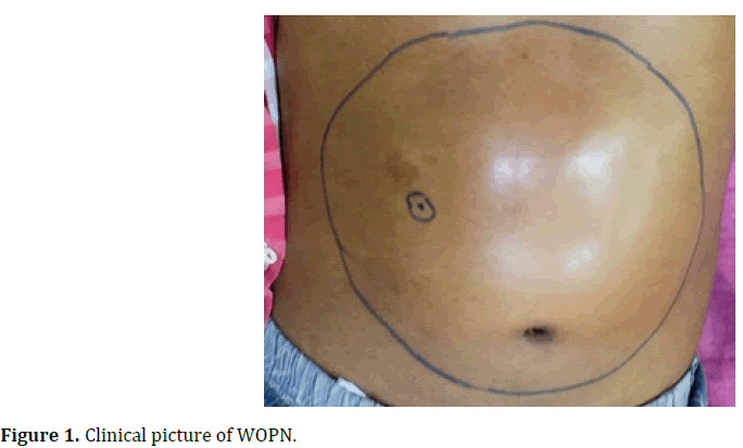
Figure 1: Clinical picture of WOPN.
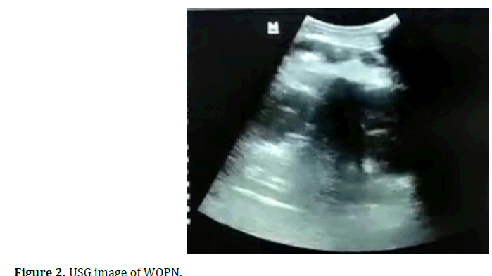
Figure 2: USG image of WOPN.
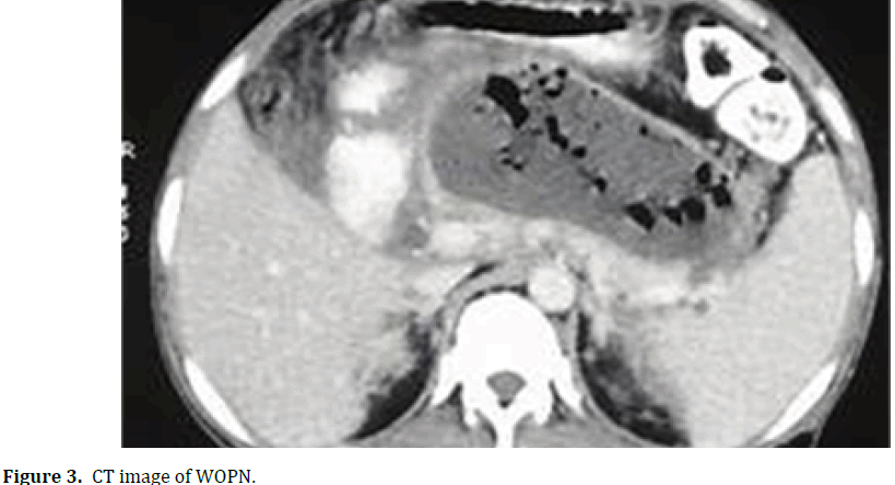
Figure 3: CT image of WOPN.
The most common indication for surgery was pain, persistent unwellness and biliary and duodenal obstruction. Eighteen of the above patients had either fever, tachycardia, or leukocytosis at the time of surgery. The mean duration of surgery was 80 minutes for open surgery and 117 minutes for laparoscopic surgery. Blood loss was greater in the open group (mean-140ml) than in the laparoscopic group (mean-95ml). Eleven patients with biliary pancreatitis underwent cholecystectomy. Four patients in each group underwent simultaneous drainage for retroperitoneal or paracolic collections. Three patients needed conversion to open due to dense adhesions and bleeding are shown in Table 3.
| Overall complications (Present) |
10 (35.7%) |
11 (47.8%) |
0.3823 |
| Infection*** |
|
|
0.0042 |
| Absent |
26 (92.9%) |
15 (65.2%) |
|
| Wound infection |
0 (0.0%) |
6 (26.1%) |
|
| Intra-abdominal abscess |
1 (3.6%) |
2 (8.7%) |
|
| Port site infection |
1 (3.6%) |
0 (0.0%) |
|
| Pancreatic fistula (Present) |
3 (10.7%) |
4 (17.4%) |
0.6872 |
| Bleeding (Present) |
2 (7.1%) |
3 (13.0%) |
0.6472 |
| GI fistula |
|
|
0.7222 |
| Absent |
27 (96.4%) |
21 (91.3%) |
|
| Colonic |
1 (3.6%) |
1 (4.3%) |
|
| Gastric |
0 (0.0%) |
1 (4.3%) |
|
| Pulmonary complications (Present) |
5 (17.9%) |
8 (34.8%) |
0.1683 |
| DVT (Present) |
3 (10.7%) |
3 (13.0%) |
1.0002 |
| Cardiac (Present) |
3 (10.7%) |
6 (26.1%) |
0.2682 |
| Hospital stay*** |
6.14 ± 2.07 |
8.65 ± 3.47 |
0.0024 |
| Readmissions (Yes) |
3 (10.7%) |
7 (30.4%) |
0.1542 |
| New onset DM (Present) |
4 (14.3%) |
4 (17.4%) |
1.0002 |
| Steatorrhoea (Present) |
2 (7.1%) |
2 (8.7%) |
1.0002 |
| Mortality (Present) |
1 (3.6%) |
2 (8.7%) |
0.5832 |
Table 3. Post-operative results.
The overall complication rate was higher in the open group (47%) than in the laparoscopic group (35%). Postoperative infections were significantly higher in the open group. Pulmonary complications were most common, affecting 25% of patients. One patient in the open group needed percutaneous drainage for an infected perisplenic collection. Six patients had external pancreatic fistulae (Grade A and B); these patients were managed conservatively. Two patients developed colonic fistula, which required diversion ileostomy. One patient in the open group developed a gastric fistula, which was managed with Nasojejunal Tube (NJ) tube insertion and octreotide. This patient developed Deep Vein Thrombosis (DVT) in the post-operative period and on Postoperative Day (POD) 11 had a massive bleed from the fistula site and midline wound and succumbed. Four of the five patients with post-operative haemorrhage had a sentinel bleed and emergent referral to Intervention Radiology (IR) was done. All three patients had splenic artery aneurysms and underwent coil embolisation. The fifth patient, before referral, had a massive bleed and succumbed.
Discussion
The necrotic collections of the pancreas need a multidisciplinary approach to management. It includes image-guided percutaneous drainage, endoscopic treatment with stent placement or direct endoscopic necrosectomy. The surgical approach involves open necrosectomy, VARD or minimally invasive, either laparoscopic or now robotic approach [3]. Gagner described three types of laparoscopic approaches to WOPN. First, the retrogastric, retrocolic route, which can also be used in early intervention if warranted. Second is the retroperitoneal approach and third is the transgastric route [4]. Apart from reduced pain and an earlier return to work, the advantage of laparoscopic intervention is that a single procedure usually suffices. Assessing all abdominal compartments facilitates drainage of remote collections inaccessible by percutaneous or endoscopic approaches. Simultaneous cholecystectomy can be done. Wide drainage can be achieved by cystogastrostomy [5].
Open procedures have high rates of pancreatic fistula, endocrine and exocrine insufficiency, enteric fistula (up to 20%) and incisional hernia (up to 25%) [6,7]. However, in our study, we did not notice such a pattern. We did not study the incidence of incisional hernia. Percutaneous drainage is the least invasive technique, but its success rate is low in the presence of a thick necrotic mass. The presence of an external catheter may predispose to recurrent infections and the need for multiple catheters makes this approach less appealing [8]. The endoscopic intervention has the disadvantages of requiring multiple interventions and multiple imaging modalities. Endoscopic intervention cannot access all the collections and patients with biliary pancreatitis need additional laparoscopic cholecystectomy [9]. Patients who had combined endoscopic and percutaneous intervention needed the external catheter for 83-189 days. Hospital stay was 26-55 days and the need for reimaging was 14.3 times [10].
Indications for laparoscopic transgastric drainage include symptomatic WOPN, persistent unwellness, new onset organ failure and biliary or enteric obstruction. The most common cause for intervention in this study was persistent pain and persistent unwellness. The prerequisites for the procedure include proximity to the stomach and the absence of blood vessels in the intervening wall [1]. The vascular involvement in acute necrotic pancreatitis is 22% of the portosplenic-mesenteric venous axis [11].
In our study, WOPN was common in males in the fourth and fifth decades of life. The most common aetiology is alcohol consumption and gallstones [13]. Eleven patients had associated Chronic pancreatitis and nineteen patients had Diabetes mellitus. However, the association of these conditions with morbidity or mortality is unknown. The mean surgical duration was longer in open surgery than in laparoscopic surgery. The mean duration of laparoscopic transgastric drainage in the study by Worhunsky DJ, et al. was 170 minutes and in the study by Kevin, et al. was 219 minutes [14,15]. The operative complications were more common in the open group (47%) than in the laparoscopic group. In a study by Munene, et al. the overall complication rate was 33% [16]. The major complication rate with the laparoscopic approach was 24%, similar to our study and the study done by Worhunsky DJ, et al. [14].
In a meta-analysis by Luo D, et al. overall clinical resolution, postoperative bleeding, endocrine and exocrine dysfunction and mortality are similar between endoscopic and laparoscopic techniques [16]. In a study conducted in three high-volume centres with 178 patients undergoing both open and laparoscopic transgastric necrosectomy, the intra-abdominal infection rate was 11%, the pancreatic fistula rate was 4% and the readmission rate was 20%, similar to our study [13].
In a study of large or extended WOPN, open surgery (47.4%) had significantly more complications than laparoscopic procedures (9.1%). In the same study, mortality was also significantly higher in the open group [17]. In critically ill patients, the most common complication was pulmonary infections [18]. The conversion rate in our study was 6%, similar to those reported in other studies [19,20]. However, robotic transgastric necrosectomy showed a lower conversion rate (3.5%) in 57 patients [21]. The operative time and conversion rates were low when using staplers to create a cystogastrostomy [22,23]. Overall, in our study comparing the open versus laparoscopic approach for transgastric drainage of WOPN, the significant finding was a shorter operative time with the open procedure. This can be attributed to the use of intracorporeal suturing rather than staplers. Blood loss was lower in the laparoscopic group. Postoperative infections were more common in the open approach. Mortality and morbidity were similar in both groups; hospital stay was significantly shorter in the laparoscopic group.
Conclusion
No single approach is currently considered the gold standard. Therefore, management must be individualised based on the patient’s clinical presentation and available resources. The laparoscopic approach reduces overall complications, particularly infectious complications. The use of staplers may also significantly reduce operative time. A robotic approach may be best, but due to limited availability in remote areas, the laparoscopic approach may be considered.
Acknowledgements
None
Conflict of Interest
The authors have no conflicts of interest to declare.
References
- Petrov MS, Shanbhag S, Chakraborty M, Phillips AR, Windsor JA. Organ failure and infection of pancreatic necrosis as determinants of mortality in patients with acute pancreatitis. Gastroenterology. 2010;139:813e820.
[Crossref] [Google Scholar] [PubMed]
- Banks PA, Bollen TL, Dervenis C, Gooszen HG, Johnson CD, Sarr MG, et al. Classification of acute pancreatitis 2012: Revision of the Atlanta classification and definitions by international consensus. Gut 2013;62(1):102-111.
[Crossref] [Google Scholar] [PubMed]
- Sion MK, Davis KA. Step-up approach for the management of pancreatic necrosis: A review of the literature. Trauma Surg Acute Care Open. 2019;4(1):e000308.
[Crossref] [Google Scholar] [PubMed]
- Gagner M. Laparoscopic Treatment of Acute Necrotizing Pancreatitis. Semin Laparosc Surg. 1996;3(1):21-28.
[Crossref] [Google Scholar] [PubMed]
- Freeman ML, Werner J, van Santvoort HC, Baron TH, Besselink MG, Windsor JA, et al. Interventions for necrotizing pancreatitis: Summary of a multidisciplinary consensus conference. Pancreas. 2012;41(8):1176-1194.
[Crossref] [Google Scholar] [PubMed]
- Zyromski, N.J. Laparoscopic Transgastric Pancreatic Debridement. Curr Surg Rep 2013;1(2):114-120.
[Crossref] [Google Scholar]
- Baron TH, Thaggard WG, Morgan DE, Stanley RJ. Endoscopic therapy for organized pancreatic necrosis. Gastroenterology. 1996;111:755-764.
[Crossref] [Google Scholar] [PubMed]
- Ammori BJ. Laparoscopic transgastric pancreatic necrosectomy for infected pancreatic necrosis. Surg Endoscopy 2002;16:1362.
[Crossref] [Google Scholar] [PubMed]
- Ramia JM, de la Plaza R, Quiñones-Sampedro JE, Ramiro C, Veguillas P, García-Parreño J. Walled-off pancreatic necrosis. Neth J Med. 2012;70(4):168-71.
[Google Scholar]
- Bakker OJ, van Santvoort HC, van Brunschot S, Geskus RB, Besselink MG, Bollen TL, et al. Endoscopic transgastric vs surgical necrosectomy for infected necrotizing pancreatitis: A randomized trial. JAMA. 2012;307(10):1053-1061.
[Crossref] [Google Scholar] [PubMed]
- Gluck M, Ross A, Irani S, Lin O, Hauptmann E, Siegal J, et al. Endoscopic and percutaneous drainage of symptomatic walled-off pancreatic necrosis reduces hospital stay and radiographic resources. Clin Gastroenterol Hepatol. 2010;8(12):1083-1088.
[Crossref] [Google Scholar] [PubMed]
- Easler J, Muddana V, Furlan A, Dasyam A, Vipperla K, Slivka A, et al. Portosplenomesenteric venous thrombosis in patients with acute pancreatitis is associated with pancreatic necrosis and usually has a benign course. Clin Gastroenterol Hepatol. 2014;12(5):854-862.
[Crossref] [Google Scholar] [PubMed]
- Driedger M, Zyromski NJ, Visser BC, Jester A, Sutherland FR, Nakeeb A, et al. Surgical Transgastric Necrosectomy for Necrotizing Pancreatitis: A Single-stage Procedure for Walled-off Pancreatic Necrosis. Ann Surg. 2020;271(1):163-168.
[Crossref] [Google Scholar] [PubMed]
- Worhunsky DJ, Qadan M, Dua MM, Park WG, Poultsides GA, Norton JA, et al. Laparoscopic transgastric necrosectomy for the management of pancreatic necrosis. J Am Coll Surg. 2014;219(4):735-743.
[Crossref] [Google Scholar] [PubMed]
- Simo KA, Niemeyer DJ, Swan RZ, Sindram D, Martinie JB, Iannitti DA. Laparoscopic transgastric endolumenal cystogastrostomy and pancreatic debridement. Surg Endosc. 2014;28(5):1465-1472.
[Crossref] [Google Scholar] [PubMed]
- Munene G, Dixon E, Sutherland F. Open transgastric debridement and internal drainage of symptomatic non-infected walled-off pancreatic necrosis. HPB (Oxford). 2011;13(4):234-239.
[Crossref] [Google Scholar] [PubMed]
- Luo D, Liu X, Du J, Liu J, Chen X, Zhou P, et al. Endoscopic Transgastric Versus Surgical Approach for Infected Necrotizing Pancreatitis: A Systematic Review and Meta-Analysis. Surg Laparosc Endosc Percutan Tech. 2019;29:141-149.
[Crossref] [Google Scholar] [PubMed]
- Szentkereszty Zs, Krasnyánszky N, Kammili A, Balog K, Berhés M, Sápy P Gy. Comparative Study of Conventional and Transgastric Necrosectomy for Wide Extended Walled-of Pancreatic Necrosis. Pancreat Disord Ther. 2020;9:199.
- Parekh D. Laparoscopic-assisted pancreatic necrosectomy: A new surgical option for treatment of severe necrotizing pancreatitis. Arch Surg. 2006;141(9):895-902.
[Crossref] [Google Scholar] [PubMed]
- Mathew MJ, Parmar AK, Sahu D, Reddy PK. Laparoscopic necrosectomy in acute necrotizing pancreatitis: Our experience. J Minim Access Surg. 2014;10(3):126-131.
[Crossref] [Google Scholar] [PubMed]
- Saleh, Abdel F, Al Sageer, Emad, Mahran, Khaled, et al. Laparoscopic anterior cystogastrostomy. Egypt J Surg 2018;37(4):549-555.
[Google Scholar]
- Wang Y, Yoshino O, Driedger MR, Beckman MJ, Vrochides D, Martinie JB. Robotic pancreatic necrosectomy and internal drainage for walled-off pancreatic necrosis. HPB (Oxford). 2023;25(7):813-819.
[Crossref] [Google Scholar] [PubMed]
- Yetisir F, Güzel K. Laparoscopic anterior transgastric cystogastrostomy for the treatment of pancreatic pseudocysts. Ann Clin Gastroenterol Hepatol. 2020;4:6-10.
[Google Scholar]
Citation: Banu KJ, Mathias N, Tabassum N (2026) Comparative outcomes of Open and Laparoscopic drainage in Walled off Pancreatic Necrosis. J Pancreas. 27:983.
Copyright: This is an open access article distributed under the terms of the Creative Commons Attribution License, which permits unrestricted use, distribution, and reproduction in any medium, provided the original work is properly cited.