Keywords
Colitis; Cadmium chloride; Malondialdehyde
Introduction
Inflammatory bowel diseases are chronic diseases of the bowel which includes Crohn’s disease and ulcerative colitis. It rises from uncontrolled intestinal inflammation resulting from deranged immune responses [1]. Genetic IBD research has been helpful in identifying a number of genes that are prone to this disease however, none of them alone is sufficient enough to initiate the disease [2,3]. Environmental factors which includes smoking, appendectomy and childhood exposures to antibiotic may be involved in the pathogenesis of IBD [4,5] non-other however, apart from these factors have been identified in the pathogenesis of IBD [6-9].
The continuous increase in the incidences of IBD in industrially developing countries including Nigeria, is a pointer to the potential role of industrialization and its resultant environmental pollutants [10-13] in the pathophysiology of IBD [14,15]. Industrialization has led to an increase in the accumulation of heavy metals in our surrounding ecosystems; one of such metals is cadmium, found in the environment [16].
Cadmium is an element of the group IIb in the periodic table; air, water, food and smoking represent significant sources of cadmium [17], and it has a high rate of soil to plant transference [18]. Several studies have reported the side effects of cadmium exposure on health; erosion of the gastric mucosa, lower gastrointestinal irritation and erosion, vomiting, abdominal pain and diarrhea as well as nervous and cardiovascular disorders after exposure to high levels in experimental and clinical conditions [19-22].
In spite of the known toxic effects of cadmium, its effect on colonic intestinal inflammation is not known; particularly in the pathophysiology of ulcerative colitis. The aim of this study was to investigate the effect(s) of oral cadmium exposure on the healing of experimental colitis in rats.
Materials and Methods
Chemicals and reagents
Cadmium Chloride salts (CdCl2) (Burgoyne Burbidges & Co Mumbai-India), ketamine (Rottexmedica, Germany) and acetic acid (BDH, London).
Animals
Male Wistar rats (100-120 g) obtained from the University animal house and were acclimatized for two weeks prior to commencement of experiment, contained in wire-meshed cages, kept under standard conditions with food (vital feed; crude protein 14%, fat 7%, crude fibre 10%, calcium 1.0%, phosphorous 0.35%) and water made accessible to them freely. The Experimental protocol was approved by the Gastrointestinal Research Group, University of Ibadan. Handling of the animals was in line with international guidelines on the care and use of laboratory animals in research [23].
Experimental design
Animals were randomly assigned into four groups of twenty rats each; Control group was given drinking water, three other groups received cadmium chloride (CdCl2) at 25 ppm, 50 ppm and 100 ppm respectively in drinking water. Colitis was induced in all the groups after four weeks and rats were monitored two weeks post colitis induction; they were sacrificed at predetermined intervals of days 0,3,7 and 14 of colitis and whole colon was resected macroscopic scoring and histology. Weekly weight of rats in all the groups were taken weekly throughout the experiment and blood cadmium levels were assayed after four weeks of exposure.
Induction of colitis
Colitis was induced after cadmium had been appropriately administered for four weeks; a modified method of colitis induction previously described by [24,25] was followed – rats were fasted for 24 hours but were given access to water ad libitum and were thereafter, anaesthetized with 35 mg/kg intraperitoneal ketamine, then an improvised Teflon cannula was inserted intrarectally into the colon 8 cm proximal to the anus of the animals to induce colitis by administering 2 ml of 4% acetic acid, this was retained for 55 seconds, after which it was withdrawn. The rats were thereafter monitored and scored for the presence of diarrhea [26], at predetermined intervals.
Stool consistency scoring
Stool consistency was monitored and scored daily according to the method of [26], with slight modifications as follows: 0- (normal), 1-(soft but still formed), 2-(very soft), 3-(diarrhea).
Collection of blood and tissue samples
Blood samples were obtained from each animal prior to euthanasia, through the retro orbitals plexus using capillary tubes. Animals were thereafter euthanized and colon, kidney, liver and spleen were removed from each animal for organ weights, macroscopic scoring and histopathological analysis as applicable. Colon samples for histopathological analysis were immediately fixed in 10% buffered formalin.
Macroscopic scoring
The macroscopic appearances were scored using a scale of 0-4 as follows:
0=no damage; 1=hyperemia, no ulcers; 2=linear ulcer with no significant inflammation; 3=linear ulcer with inflammation at one site; 4=two or more sites of ulceration/inflammation; 5=two or more major sites or of ulceration and inflammation or one site of ulceration/inflammation extending>1cm along the length of the colon; 6-10=If damage covers>2 cm along the length of the colon, the score is increased by one from each additional centimeter of involvement [27].
Determination of blood cadmium concentration
Blood (1 ml) was digested with HNO3 (2 ml in each) as previously described by [28,29]. Cadmium concentrations were assessed from the filtrates using an atomic absorption spectrophotometer and its concentration was measured in parts per million (ppm).
Other procedures
Malondialdehyde concentration was determined by the method of [30], differential white cell counts by the method described by [31] and histological studies were performed by a slight modification of the method previously described by [32].
Statistical Analysis
Analysis of data was performed using a statistical softwaregraph pad prism (version 5.0). Results were expressed as Mean ± standard error of mean (S.E.M), statistical significance was assessed using one-way analysis of variance (ANOVA). Differences were considered significant at *p<0.05.
Results
Effect of cadmium on body weight after four weeks of exposure
There were significant decreases in weekly percentage weight gain (%) (Figure 1) by weeks 3 and 4 (128.07 ± 3.40 and 123.57 ± 3.34) in Cd100 group when compared with control (141.75 ± 4.75 and 139.65 ± 4.51).
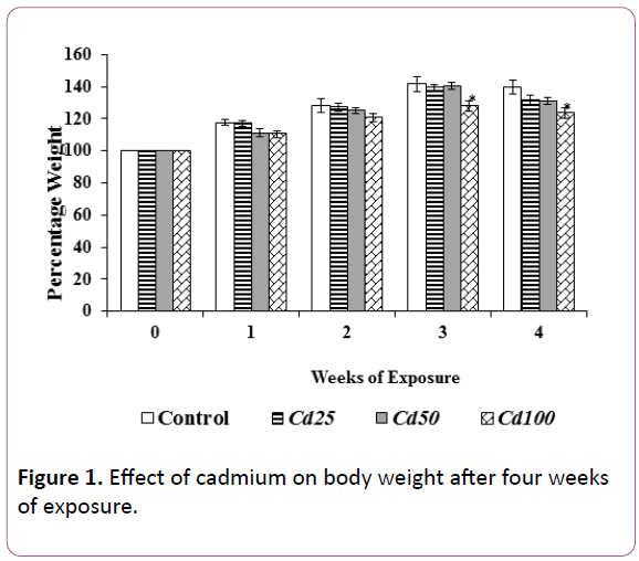
Figure 1. Effect of cadmium on body weight after four weeks of exposure.
Each bar is expressed as mean ± S.E.M of 5 rats. Where Cd25=Cadmium at 25 ppm; Cd50=Cadmium at 50 ppm and Cd100=Cadmium at 100 ppm. *=p<0.05 values differ significantly from control group.
Blood cadmium concentration analyzed by atomic absorption spectrophotometer
Blood cadmium concentration (ppm) (Figure 2) was significantly high in Cd50 (0.04 ± 0.00) and Cd100 group (0.04 ± 0.00) when compared with control (0.03 ± 0.00) by the end of four weeks’ cadmium exposure.
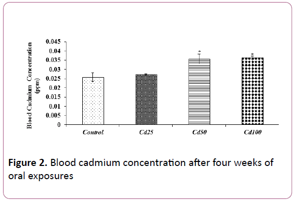
Figure 2: Blood cadmium concentration after four weeks of oral exposures
Each bar is expressed as mean ± S.E.M of 5 rats. Where Cd25=Cadmium at 25 ppm; Cd50=Cadmium at 50 ppm and Cd100=Cadmium at 100 ppm. *=p<0.05 values differ significantly from control group.
Effect of cadmium on kidney weight
There were significant increases in the kidney weight (g) in Cd100 group (0.48 ± 0.02) when compared with control group (0.40 ± 0.01) by day 7 (Figure 3).
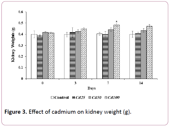
Figure 3: Effect of cadmium on kidney weight (g).
Each value is expressed as mean ± S.E.M of 5 rats. Where Cd25=Cadmium at 25 ppm; Cd50=Cadmium at 50 ppm and Cd100=Cadmium at 100 ppm. *=p<0.05 values differ significantly from control group.
Effect of cadmium on stool consistency post colitis induction
There were significant increases in stool consistency (Figure 4) by day 5 in Cd100 group (2.5 ± 0.22) when compared with control (1.5 ± 0.22).
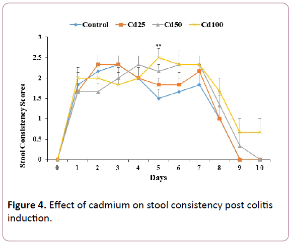
Figure 4: Effect of cadmium on stool consistency post colitis induction.
Each value is expressed as mean ± S.E.M of 5 rats. Where Cd25=Cadmium at 25 ppm; Cd50=Cadmium at 50 ppm and Cd100=Cadmium at 100 ppm. *=p<0.05 values differ significantly from control group.
Effect of cadmium on colon macroscopic scores
Macroscopic scores from the Cd50 and Cd100 by days 0, 3, 7 and 14 were increased when compared with control (Figure 5), this was however not statistically significant.
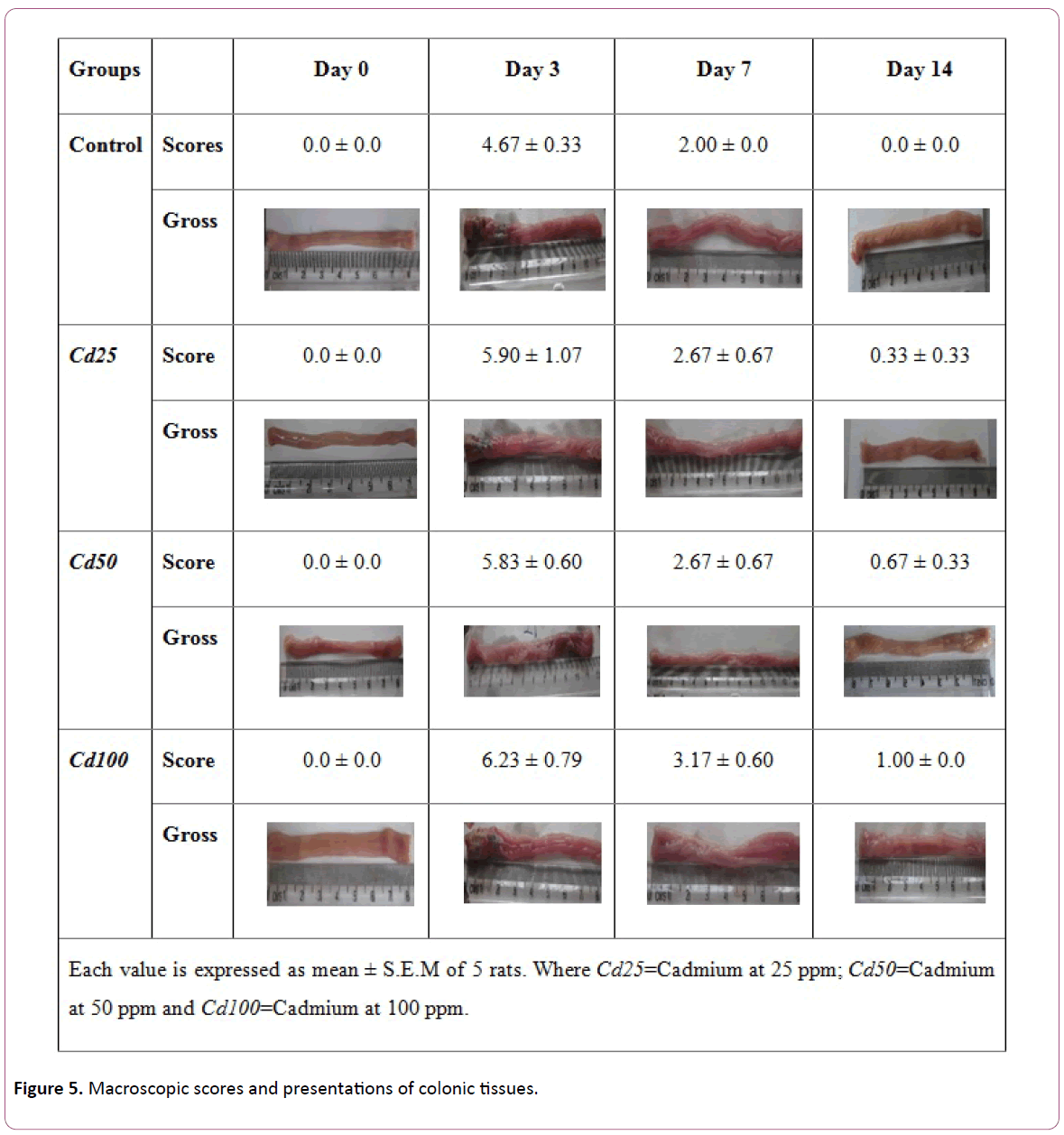
Figure 5: Macroscopic scores and presentations of colonic tissues.
Effect of cadmium on neutrophil/lymphocyte ratio
Neutrophil/Lymphocyte ratio (Figure 6), was significantly increased in Cd50 (0.43 ± 0.04; 0.80 ± 0.06) and Cd100 (0.49 ± 0.03; 0.80 ± 0.03) when compared with control (0.36 ± 0.03; 0.63 ± 0.04) on day 0 and 7, respectively.
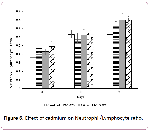
Figure 6: Effect of cadmium on Neutrophil/Lymphocyte ratio.
Effect of cadmium on malondialdehyde concentration
Significant increases in the concentration of malondialdehyde (nmol mg/protein) were seen (Figure 7) at day 7 in Cd25, Cd50 and Cd100 groups (0.56 ± 0.02; 0.61 ± 0.03; 0.65 ± 0.01) and at day 14 (0.41 ± 0.02; 0.52 ± 0.02; 0.60 ± 0.02) when compared with control (0.50 ± 0.02; 0.31 ± 0.00) at days 7 and 14, respectively.
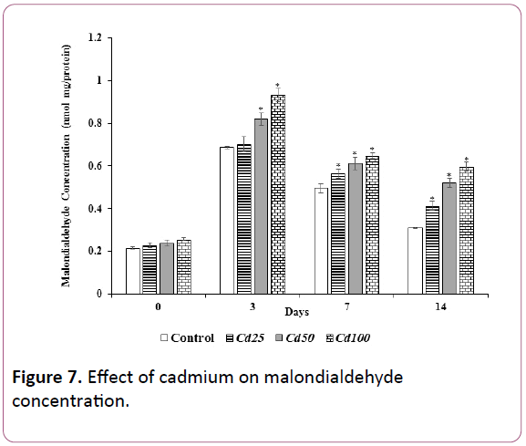
Figure 7: Effect of cadmium on malondialdehyde concentration.
Each bar is expressed as mean ± S.E.M of 5 rats. Where Cd25=Cadmium at 25 ppm; Cd50=Cadmium at 50 ppm and Cd100=Cadmium at 100 ppm. *=p<0.05 values differ significantly from control group.
Each bar is expressed as mean ± S.E.M of 5 rats. Where Cd25=Cadmium at 25 ppm; Cd50=Cadmium at 50 ppm and Cd100=Cadmium at 100 ppm. *=p<0.05 values differ significantly from control group.
Effect of cadmium on colonic histology
Lymphocyte infiltration of different levels in the mucosa, edema, erosion, ulcer and regenerating crypts are seen in Cd25, Cd50 and Cd100 groups when compared with control (Figure 8).
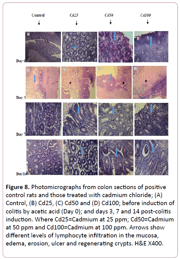
Figure 8: Photomicrographs from colon sections of positive control rats and those treated with cadmium chloride; (A) Control, (B) Cd25, (C) Cd50 and (D) Cd100; before induction of colitis by acetic acid (Day 0); and days 3, 7 and 14 post-colitis induction. Where Cd25=Cadmium at 25 ppm; Cd50=Cadmium at 50 ppm and Cd100=Cadmium at 100 ppm. Arrows show different levels of lymphocyte infiltration in the mucosa, edema, erosion, ulcer and regenerating crypts. H&E X400.
Discussion
In the present study, intrarectal administration of AA caused an increase in stool consistency scores; this was more pronounced in cadmium at 100 ppm group by day 5 post colitis induction. It may be due to inflammation reactions within the colonic mucosa caused by AA in addition to the already accumulating effect from cadmium. However, AA has been documented to induce localized inflammation, desquamation and loss of mucosal integrity leading to epithelial injury [33].
Cadmium decreased weekly percentage weight gain; this finding is consistent with previous reports by [34-37]; they reported that Sprague-Dawley rats that received cadmium by gavage exhibited decreased body weight. These decreases in body weight gain may be due to increased degeneration of lipids and proteins [38], decreased growth rate or decreases in nutrient digestion and absorption [39,40]. Decreased body weight and decreased growth rate have however, been common findings in studies where experimental animals are orally exposed to cadmium [41,42].
The increase in kidney weight observed in this study could be due to cadmium effects. [41,43] had earlier reported kidney swelling after subcutaneous and oral exposures of cadmium in rats.
Increased blood cadmium concentration of the cadmium at 50 ppm and cadmium at 100 ppm groups after four weeks indicates sufficient exposure [44]. Although the concentrations were generally low, reports have shown that exposures to high cadmium concentration orally or intravenously still presents as extremely low in the blood [45].
Neutrophil/Lymphocyte ratio (NLR) is an index that can be used to measure disease progression particularly during inflammation [46]. The observed increase in Neutrophil/ Lymphocyte ratio in cadmium at 50 ppm and cadmium at 100 ppm on days 0 and 7 may be indicative of continuous activation of inflammatory cells by cadmium.
Macroscopic scores are useful in assessing gross lesions or alterations during tissue injury, the increase in these scores may be indicative of ulcers and edematous formations; [47] and [48] had earlier reported such observations.
Earlier researches have documented MDA as one of the products of lipid peroxides and it marks for peroxidation in cells [49]. The notable increase in MDA concentration in cadmium at 50 ppm and cadmium at 100 ppm groups by day 3 may be indicating oxidative stress which is common during cadmium exposures [50-53], while cadmium at 25 ppm group on the same day had MDA values not different from that of control; it may be that cadmium at 25 ppm was not sufficient enough to increase oxidative stress or that endogenous antioxidant enzymes and molecules were stimulated by cadmium and they were able to maintain biochemical homeostasis.
An adaptive response to cadmium by cells may also, have been induced thereby causing resistance to MDA, Hart and colleagues [54-56] found out that repeated exposures to cadmium at low concentrations to alveolar epithelia cells can result in the development of an adaptive survival response. However, increases in MDA seen in cadmium at 25 ppm, cadmium at 50 ppm and cadmium at 100 ppm groups on days 7 and 14 may be as a result of stimulated endogenous enzymes that have been exhausted from cadmium-stimulated reactions.
Histological evaluation showed that cadmium caused erosion of colonic epithelium, increased severity of colonic injury, and delayed healing in cadmium at 50 ppm and cadmium at 100 ppm groups up-till day14 of colitis, this may be indicative of a continuous stimulation of colonic inflammatory cells due to a sustained exposure to cadmium. [57] and [58] reported a similar finding, where oral administration of cadmium compounds caused desquamation of the colonic epithelium.
Conclusion
It can therefore be concluded, that cadmium increased colonic inflammation in rats during colitis through increased neutrophil/lymphocyte ratio and lipid peroxidation thereby delaying healing of acetic acid induced colitis.
References
- Abraham C, Medzhitov R (2011) Interactions Between the Host Innate Immune System and Microbes in Inflammatory Bowel Disease. Gastroenterology 140: 1729-1737.
- Kaser A, Zeissig S, Blumberg RS (2010) Inflammatory bowel disease. Annu Rev Immunol 28: 573-621.
- Kabi A, Nickerson KP, Homer CR, McDonald C (2012) Digesting the genetics of inflammatory bowel disease: Insights from studies of autophagy risk genes. Inflamm Bowel Dis 18: 782-792.
- Pinsk V, Lemberg DA, Grewal K, Barker CC, Schreiber RA, et al. (2007), “Inflammatory bowel disease in the South Asian pediatric population of British Columbia”. American Journal of Gastroenterology, 102:1077–1083.
- Molodecky NA, Soon IS, Rabi DM, Ghali WA, Ferris M, et al. (2012) Increasing Incidence and Prevalence of the Inflammatory Bowel Diseases with Time, Based on Systematic. Gastroenterology 142: 46-54.
- Cosnes J, Carbonnel F, Beaugerie L, Blain A, Reijasse D, et al. (2002) Effects of appendicectomy on the course of ulcerative colitis. Gut 51: 803-807.
- Cosnes J (2004) Tobacco and IBD: relevance in the understanding of disease mechanisms and clinical practice. Best Pract Res Clin Gastroenterol 18: 481-496.
- De Vroey B, De Cassan C, Gower-Rousseau C, Colombel JF (2010) Editorial: Antibiotics Earlier, IBD Later?. Am J Gastroenterol 105: 2693-2696.
- Hviid A, Svanström H, Frisch M (2011) Antibiotic use and inflammatory bowel diseases in childhood. Gut 60: 49-54.
- Egborge ABM (1994) Water pollution in Nigeria. Biodiversity and chemistry of Warri River. Warri: Ben Miller Books.
- Leong RWL, James YL, Sung JJY (2004) The Epidemiology and Phenotype of Crohn’s Disease in the Chinese Population. Inflamm Bowel Dis 10: 646-651.
- Zheng JJ, Zhu XS, Huangfu Z, Gao ZX, Guo ZR, et al. (2005) Crohn's disease in mainland China: a systematic analysis of 50 years of research. Chin J Dig Dis 6: 175-181.
- Ekwunife CN, Nweke IG, Achusi IB, Ekwunife CU (2015) Ulcerative colitis prone to delayed diagnosis in a Nigerian Population: Case series. Annals of Medicine and Health Science Research 5: 311-313.
- Kaplan GG, Hubbard J, Korzenik J, Sands BE, Panaccione R, et al. (2010) The inflammatory bowel diseases and ambient air pollution: a novel association. American Journal of Gastroenterology 105: 2412-2419.
- Ananthakrishnan AN, Khalili H, Higuchi LM, Bao Y, Korzenik JR, et al. (2012) Higher predicted vitamin D status is associated with reduced risk of Crohn’s disease. Gastroenterology 142: 482-489.
- Waalkes MP, Rehm S (1992) Carcinogenicity of oral cadmium in the male Wistar (WF/NCr) rat: effect of chronic dietary zinc deficiency. Fundam Appl Toxicol 19: 512-520.
- Friberg L, Nordberg GF, Vouk VB (1986) Handbook of the toxicology of metals. Amsterdam, Elsevier 2: 130-184.
- Satarug S, Baker JR, Reilly PEB, Moore MR, Williams DJ (2002) Cadmium levels in the lung, liver, kidney cortex and urine samples from Australians without occupational exposure to metals. Arch Environ Health 57: 69-77.
- Järup L, Bellander T, Hogstedt C, Spang G (1998) Mortality and cancer incidence in Swedish battery workers exposed to cadmium and nickel. Occup Environ Med 55: 755-759.
- Ikeda M, Zhang ZW, Moon CS, Shimbo S, Watanabe T, et al. (2000) Possible effects of environmental cadmium exposure on kidney function in the Japanese general population. International Archives of Occupational and Environmental Health 73: 15-25.
- Hooser SB (2007) Cadmium. In: Gupta RC (editors) Veterinary Toxicology: Basic and Clinical Principles. Academic Press; New York.
- Singh P, Mogra P, Bano H, Sankhla V, Deora K, et al. (2012) Protective and preventive effects of curcumin against cadmium chloride induced gastrointestinal toxicity in Swiss albino mice. World Journal of Science and Technology 2: 10-17.
- National Research Council (2011) Guide for the Care and Use of Laboratory Animals. 8th ed. Washington: The National Academies Press.
- MacPherson BR, Pfeiffer CJ (1978) Experimental production of diffuse colitis in rats. Digestion 17: 135-150.
- Millar AD, Rampton DS, Chander CL (1996) Evaluating the antioxidant potential of new treatments for inflammatory bowel disease using a rat model of colitis. Gut 39: 407-415.
- Fukuda M, Kanauchi O, Araki Y, Andoh A, Mitsuyama K, et al. (2002) Prebiotic treatment of experimental colitis with germinated barley foodstuff: a comparison with probiotic or antibiotic treatment. Int J Mol Med 9: 65-70.
- Peran L, Camuesco D, Comalada M, Nieto A, Concha A, et al. (2005) Preventative effects of a probiotic, Lactobacillus salivarius ssp. salivarius, in the TNBS model of rat colitis. World J Gastroenterol 11: 5185-5192.
- Asagba SO, Eriyamremu GE (2007) Oral cadmium exposure alters haematological and liver function parameters of rats fed a Nigerian-like diet. Journal of Nutritional and Environmental Medicine 16: 267-274.
- Goncalves JF, Fiorenzab AM, Spanevellob RM, Mazzanti CM, Bochi GV, et al. (2010) N-acetylcysteine prevents memory deficits, the decrease in acetylcholinesterase activity and oxidative stress in rats exposed to cadmium. Chem Biol Interact 186: 53-60.
- Varshney R, Kale RK (1990) Effect of calmodulin antagonist on radiation induced lipid peroxidation in microsomes. Int J Radiat Biol 58: 733-743.
- Brown BA (1993) Hematology Principles and Procedures, sixth edition, Lea and Febiger, Philadelphia.
- Ogihara Y, Okabe S (1993) Effect and mechanism of sucralfate on healing of acetic acid-induced gastric ulcers in rats. J Physiol Pharmacol 44: 109-118.
- Kandhare AD, Kumar VS, Adil M, Rajmane AR, Ghosh P, et al. (2012) Investigation of gastro protective activity of Xanthum strumarium L. by modulation of cellular and biochemical marker. Oriental Pharmacy and Experimental Medicine 12: 287-299.
- Kotsonis FN, Klaassen CD (1977) Toxicity and distribution of cadmium administered to rats at sublethal doses. Toxicol Appl Pharmacol 41: 667-680.
- Borzelleca JF, Clarke EC, Condcie LW (1989) Short-term toxicity (1 and 10 days) of cadmium chloride in male and female rats: Gavage and drinking water. Journal of American College of Toxicology 8: 377-404.
- Kozlowska D, Brzozowska A, Sulkowska J, Roszkowski W (1993) The effect of cadmium on iron metabolism in rats. Nutrition Research 13: 1163-1172.
- Horiguchi H, Sato M, Konno N, Fukushima M (1996) Long term cadmium exposure induces anaemia in rats through hypo-induction of erythropoietin in the kidney. Arch Toxicol 71: 11-19.
- Erdogan Z, Erdogan S, Celik S, Unlu V (2005) Effects of ascorbic acid on cadmium-induced oxidative stress and performance of broilers. Biol Trace Elem Res 104: 19-31.
- Elsenhans B, Strugala G, Schmann K (1999) Longitudinal pattern of enzymatic and absorptive functions in the small intestine of rats after short term exposure to dietary cadmium chloride. Arch Environ Contam Toxicol 36: 341-346.
- Eriyamremu GE, Asagba SO, Onyeneke EC, Adaikpo MA (2005) Changes in carboxypeptidase A, dipeptidase and Na+/K+ ATPase activities in the intestine of rats orally exposed to different doses of cadmium. BioMetals 18: 1-6.
- Asagba SO, Isamah GK, Ossai EK, Ekakitie AO (2002) Effect of oral exposure to cadmium on the levels of vitamin A and lipid peroxidation in the eye. Bulletin of Environmental Contamination and Toxicology 68: 18-21.
- FEDRIP (Federal Research in Progress) database (2012) Cadmium. Springfield, VA: National Technical Information Service.
- Asagba SO (2010) Alteration in the activity of oxidative enzymes in the tissues of male wistar albino rats exposed to cadmium. Int J Occup Med Environ Health 23: 55-62.
- Jin T, Nordberg M, French W, Dumont X, Bernard A, et al. (2002) Cadmium biomonitoring and renal dysfunction among a population environmentally exposed to cadmium from smelting in China (China Cad). Biometals 15: 397-410.
- Analytical Research Labs (ARL) (2012) Inc., Arizona 85021 USA.
- Celikbilek M, Dogan S, Ozbakir O, Zararsiz G, Kücük H (2013) Neutrophil-lymphocyte ratio as a predictor of disease severity in ulcerative colitis. J Clin Lab Anal 27: 72-76.
- Tahan G, Aytac E, Aytekin H, Gunduz F, Dogusoy G, et al. (2011) Vitamin E has a dual effect of anti-inflammatory and antioxidant activities in acetic acid-induced colitis in rats. Can J Surg 54: 333.
- Somani SJ, Badgujar SB, Sutariya BK, Saraf MN (2014) Protective effect of Delinia indica L. on Acetic Acid induced Colitis in Mice. Indian J Exp Biol 52: 876-881.
- Bagchi D, Bagchi M, Hassoun EA, Stohs SJ (1996) Cadmium induced excretion of urinary lipid metabolites, DNA damage, glutathione depletion and hepatic lipid peroxidation in Sprague–Dawley rats. Biol Trace Elem Res 52: 143-154.
- Shukla GS, Shukla A, Potts RJ, Osier M, Hart BA (2000) Cadmium-mediated oxidative stress in alveolar epithelial cells induces the expression of gammaglutamylcysteine synthetase catalytic subunit and glutathione-S-transferase alpha and pi isoforms: potential role of activator protein-1. Cell Biol Toxicol 16: 347-362.
- Sharma AM, Sainger S, Dwivedi S, Srivastava S, Tripathi RD, et al. (2010) Genotype variation in Brassica juncea L., Czern. Cultivars and growth, nitrate assimilation, antioxidants response and phytoremediation potential during cadmium stress. Journal of Environmental Biology 31: 773-780.
- Kompe YO, Sagiroglu A (2016) The effects of cadmium on the biochemical and physiological parameters of Eruca sativa. Acta Biol Hung 67: 393-402.
- Hart BA (1986) Cellular and biochemical response of the rat lung to repeated inhalation of cadmium. Toxicol Appl Pharmacol 82: 281-291.
- Hart BA, Voss GW, Willean CL (1989) Pulmonary tolerance to cadmium following cadmium aerosol pretreatment. Toxicol Appl Pharmacol 101: 447-460.
- Hart BA, Potts RJ, Watkin RD (2001) Cadmium adaptation in the lung - a double-edged sword? Toxicology 160: 65-70.
- Tarasenko NY, Vorobjeva RS, Spiridinova VS, Sabalina LP (1974) Experimental investigation of toxicity of cadmium and zinc caprylates. J Hyg Epidemiol Microbiol Immunol 18:144-153.
- James HM, Hilburn ME, Blair JA (1985) Effects of meals and meal times on uptake of lead from the gastrointestinal tract in humans. Human Toxicology 4: 401-407.
- Zalups RK, Ahmad S (2003) Molecular handling of cadmium in transporting epithelia. Toxicol Appl Pharmacol 186: 163-188.

