Keywords
Cadmium; Zebrafish; LC50; Bioaccumulation; Histopathology
Introduction
Cadmium (Cd), a common aquatic environmental contaminant, is associated with wide variety of human activities and products like pigments, ceramics, plastics, glasses, vehicle tires and other synthetics [1]. The dispersion of Cd in the environment has increased over the past decades because of its extensive industrial uses such as nickel-cadmium battery production and metal plating [1,2]. For example, it can enter the water environment from industrial operations, chemically agricultural manuring and mining activities [3,4]. It is a highly toxic heavy metal and recognized as one of the most harmful metal contaminants [5]. Cd is a biologically non-necessary, non-biodegradable and persistent heavy metal, thus, it has high toxic possibilities for human and animals’ health [3,6].
Moreover, continuous exposure to low level of Cd may have a gross biological effect comparable to that of frequent exposures of more intensity. Field studies have demonstrated various degrees of pollution to aquatic systems via direct or indirect Cd inputs [4]. Generally, in freshwater fish, Cd absorption is carried out through three ways, namely, through gills, skin and from food via the intestinal wall [7]. Considerable quantity of Cd will be accumulated in various tissues of fishes in aquatic environment, and it is based on style of exposure, diet or waterborne. Yet accumulation of Cd in fish muscle was rarely reported though it can pose severe impact on fish without any visible signs [8,9]. Because of bio magnification of Cd in food chain, when consumed by human, it will become a more important subject for aquatic science [10]. As pointed out by Fraser et al. [11], fish and seafood, a part of healthy diet, all bio accumulates heavy metals. In particular, the bioaccumulation of Cd and other minerals in food chain is causing worries as they can have harmful impacts on human and animal health, and sometimes can cause Alzheimer’s and Parkinson’s diseases [12]. Usually, fish and seafood are one of the key links between heavy metals in the environment and human exposure.
Biomarkers are actions of sub-organismal replies in organisms or exposed bio systems which can prove exposure to, or the impacts of, environmental pollutants. During last two decades, the science of biomarkers has greatly advanced. Histo-cytological responses are relatively easy to identify, and can potentially be relevant to health and fitness of individuals which, in turn, allows further extrapolation to population/ community impacts. A wide variety of histo-cytological modifications in fish have been developed and recommended as biomarkers for monitoring the impacts of contamination [13]. Cd exposure may also cause some patho-physiological harm.
In this context, the present investigation was designed to study the pattern of bioaccumulation of Cd in tissues of zebrafish exposed to sub lethal concentrations of Cd, as well as examining the toxicity of Cd on the fish and histo-pathological responses in brain and skeletal muscle of zebrafish served as primary indicator of exposure to pollutants, especially heavy metals.
Materials and Methods
Animals and acute toxicity testing
Zebrafish (Danio rerio) samples were purchased from the Institute of hydrobiology, Chinese academy of Science, Wuhan, P. R. China, with the body weight from 0.295 to 0.847 g and length of about 3.1-4.6 cm. The fishes were acclimatized to the laboratory conditions in glass aquaria filled with 4 L of de-chlorinated water. Each aquaria contained ten fishes, which were randomly exposed to a range of concentrations of Cd (0, 1, 5, 10, 20, 30 mg L-1) diluted in water. Cd-free water was used as control. Each test concentration was replicated 3 times. All exposures were conducted at 25-28°C with a 12: 12 h light: dark photoperiod. The exposure solution was always aerated and 50% of the contaminated water was changed every 48 h for the duration of the exposure period. The water quality parameters (pH and temperature) were measured daily. During the acute toxicity experiment, fish were not fed and mortality of the fish due to exposure was recorded up to 96 h at every 24 h interval to obtain LC50 values of Cd, following the probity analysis method described by Finney [14].
Bio concentration test
Bioaccumulation of Cd in zebrafish was examined by exposing 400 fish to sub lethal concentrations of Cd in 4 different aquaria. Each aquarium contained 40 L of tap water and 100 zebrafish. Control group were kept in uncontaminated water for Cd bioaccumulation levels and histology analysis. In tank C1, fish were exposed to low contaminated water (at a nominal Cd concentration of 1/15th of the LC50 value, 0.645 mg L-1). In tank C2, fish were exposed to mid contaminated water (at a nominal Cd concentration of 1/10th of the LC50 value, 0.968 mg L-1). In the remaining unit (C3) fish were exposed to highly contaminated water (at a nominal Cd concentration of 1/5th of the LC50 value, 1.936 mg L-1). Then record the Cd values over 0, 15 and 25 days. They were fed twice a day with artificial dry food, after the remainder food was removed; further, one third of the contaminated water was changed every 24 h during the exposure period. The fish were dissected at the end of each exposure period. Then the Cd concentration in the water and entire zebrafish was measured according to the procedure described by Cambier et al. [2]. 1g wet weight of fish tissue was digested with 3 ml of nitric acid (65% HNO3) in a pressurized medium (borosilicate glass tubes) at 100°C for 3 h and then diluted with 20 ml of deionized water before Cd concentration was measured. Water samples were taken from the aquaria at 0, 15 and 25 days to determine the amounts of Cd inside. The 10 ml of water was filtrated and 200 μl of concentrated nitric acid (65% HNO3) was added. Thus, Cd concentrations of water and tissue samples were measured by Inductively Coupled Plasma Mass Spectrometer (ICP-MS) in Faculty of Material and Chemistry, China University of Geosciences, Wuhan, P. R. China.
Histopathological changes analysis
For histological examination, tissues (brain and skeletal muscle) were fixed in paraformaldehyde, dehydrated through a graded series of ethanol, cleared in xylene and embedded in paraffin. Sections of 7 μm thickness were prepared from paraffin blocks using a rotary microtome and then stained with haematoxylin-eosin. Histopathological changes were examined under a Nikon Eclipse Bio microscope. Histological alterations were scored as: (-), no histopathology; (+), histopathology in <20% of the fields; (++), histopathology in 20–60% of the fields; and (+++) histopathology in all fields.
Statistical analysis
Computations were performed using graph pad program and results were given as mean. For all statistical results, a probability of p<0.05 was considered significant. The median Lethal Concentration (LC50) values and 95% confidence limits of Cd to zebrafish during different periods were calculated by probit analysis. The Bio Concentration Factor (BCF) of Cd in zebrafish was estimated using the following equation: BCF=Cf/Cw, where Cf is the concentration of Cd in the fish and Cw is the concentration of Cd in the exposure solution.
Results
Median lethal concentration and behavior variations
None of the fishes died during the course of the experiment. The control fish swam normally without any mark of abnormality. After 15 days of exposure to Cd, the fishes showed erratic swimming and aggressive. In addition, hyperventilation was especially evident in fishes and excess mucus occurred on their opercular surface at 25 days of Cd exposure. The LC50-96 h for Cd on zebrafish was found to be 9.68 mg L-1, with a range (95% confidence limits) of 11.12-8.43 mg L-1.
Table 1 presents the mortality and the LC10, LC50, and LC90 for Cd experimentally found at different periods and varying concentrations of Cd. No mortality was however observed in the control group during the experimental period. As well, The 96 h LC50 concentration is less than those of 24 h, 48 h and 72 h, which shows that the more the duration period is, the less the concentration is required. Thus with the increase of exposure time, LC50 values decreased from 16.73 mg L-1 (24 h) to 12.88 mg L-1 (48 h) to 11.456 mg L-1 (72 h) then to 9.68 mg L-1 (96 h) in Cd (Table 1).
| Concentration (mg L−1) |
24 h |
48 h |
72 h |
96 h |
| Dead fish/ total fish |
Mortality (%) |
Dead fish/ total fish |
Mortality (%) |
Dead fish/ total fish |
Mortality (%) |
Dead fish/ total fish |
Mortality (%) |
| Control |
0/30 |
0 |
0/30 |
0 |
0/30 |
0 |
0/30 |
0 |
| 1 |
0/30 |
0 |
0/30 |
0 |
0/30 |
0 |
0/30 |
0 |
| 5 |
Mar-30 |
10 |
Oct-30 |
36.6 |
Oct-30 |
36.6 |
13/30 |
49.99 |
| 10 |
Jul-30 |
23.33 |
Nov-30 |
26.6 |
Nov-30 |
26.6 |
14/30 |
53 |
| 20 |
15/30 |
50 |
24/30 |
80 |
25/30 |
83.33 |
25/30 |
83.33 |
| 30 |
24/30 |
80 |
30/30 |
86.6 |
30/30 |
96.6 |
100 |
100 |
| LC10 |
7.89 (9.12-6.83) |
6.43 (7.56-5.47) |
5.96 (7.08-5.025) |
5.24 (6.34-4.34) |
| LC50 |
16.73 (20.03-13.98) |
12.88 (14.96-11.08) |
11.46 (13.21-9.94) |
9.68 (11.12-8.43) |
| LC90 |
35.47 (49.63-25.36) |
25.79 (33.48-19.87) |
22.01 (27.62-17.54) |
17.87 (21.60-14.78) |
| Regression line |
y=3.92x + 0.20 |
y=4.24x + 0.29 |
y=4.51x + 0.22 |
y=4.81x + 0.26 |
| R2=0.9745 |
R2=0.9507 |
R2=0.9657 |
R2=0.9654 |
| |
The LC10, LC50 and LC90 values were all with 95% confidence limits |
Table 1: Mortality percentage and LC10, LC50, and LC90 values of Zebrafish at different exposure times and varying concentrations of Cd.
Cd bioaccumulation in fish tissues
Cd concentrations identified in the water and entire zebrafish after different period of exposure showed significant differences among all treatments (P<0.01, Table 2). Therefore, a progressive increase of Cd accumulation in fish were noticed when shifting from the control exposure to the 15 and 25 days exposures, for all treatments, which reached a high level after 25 days. The computed accumulation factors in whole fish body were 14.3, 18.16 and 13.48 in C1, C2 and C3, respectively.
| Time (day) |
C1 |
C2 |
C3 |
| Cf |
Cw |
BCF |
Cf |
Cw |
BCF |
Cf |
Cw |
BCF |
| 0 |
3.2 |
0.15 |
0.48 |
1.74 |
0.28 |
0.48 |
1.38 |
0.52 |
0.72 |
| 15 |
12.57 |
0.14 |
1.76 |
18.92 |
0.22 |
5.24 |
24.25 |
0.52 |
12.66 |
| 25 |
95.33 |
0.15 |
14.3 |
66.04 |
0.27 |
18.16 |
24.42 |
0.55 |
13.48 |
| Cf is the concentration of Cd in the fish, with the unit of μg·g-1 wet weight; Cw is the concentration of Cd in the exposure solution with the unit of mg L-1 |
Table 2: Bio concentration factor (BCF) of Cd in zebrafish exposed to C1, C2 and C3 for 0, 15 and 25 days.
Unlike this projected accumulation pattern, a recession of Cd accumulation in fish body in C3 were observed when shifting from 15 to 25 days exposures. So, the Cd concentration in the treated fish appeared to increase exponentially and culminate at Day 25, which was 95.33, 66.04 and 24.42 μg Cd·g−1 wet weight.
While the concentration of Cd in the water was maintained at a constant level, as the exposure solution was fully renewed every 24 h, and it was detected by (ICP-MS) during different periods (Table 2).
Histopathological variations
The general histological screening indicated a low to high incidence of chemical damage on tissues of zebrafish after 5 and 25 days exposure to Cd. Histopathological alterations were registered in the tissue from brain and skeletal muscle and are summarized in Table 3, shown in Figures 1 and 2. The control group did not show any histological changes in any of the tissues examined. In return, the structures of the brain and skeletal muscle of Cd-treated fish were changed, the longer time of exposure, the more severe. Histopathological changes were not observed in the skeletal muscle tissue of the treated fishes in C1 after 5 days. As shown in Figure 1, the musculoskeletal tissues from zebrafish exposed to other dose groups were detected to be various degrees of swelling, coagulation necrosis accompanied by infiltration of hemocytes and high necrosis. Figure 2 shows the brain of zebrafish also exhibiting different degrees of granule cell loss, degeneration of Purkinje cells aggregation area of gliosis and tissue anarchy in many areas where the cells appeared well spaced due to intercellular spaces of different width.
| Histopathology findings |
5 days |
25 days |
| Skeletal Muscle |
Brain |
Skeletal Muscle |
Brain |
| 0 |
- |
- |
- |
- |
| C1 |
+ |
- |
++ |
++ |
| C3 |
++ |
++ |
+++ |
+++ |
| (−) no histopathology; (+) histopathology in <25% of fields; (++) histopathology in >75% of fields; (+++) histopathology in all fields |
Table 3: Histopathologic analysis of skeletal muscle and brain tissues from zebrafish exposed to Cd for 5 days and for 25 days.
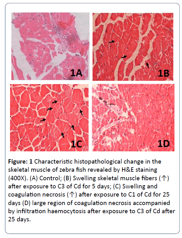
Figure 1: Characteristic histopathological change in the skeletal muscle of zebra fish revealed by H&E staining (400X). (A) Control; (B) Swelling skeletal muscle fibers (↑) after exposure to C3 of Cd for 5 days; (C) Swelling and coagulation necrosis (↑) after exposure to C1 of Cd for 25 days (D) large region of coagulation necrosis accompanied by infiltration haemocytosis after exposure to C3 of Cd after 25 days.
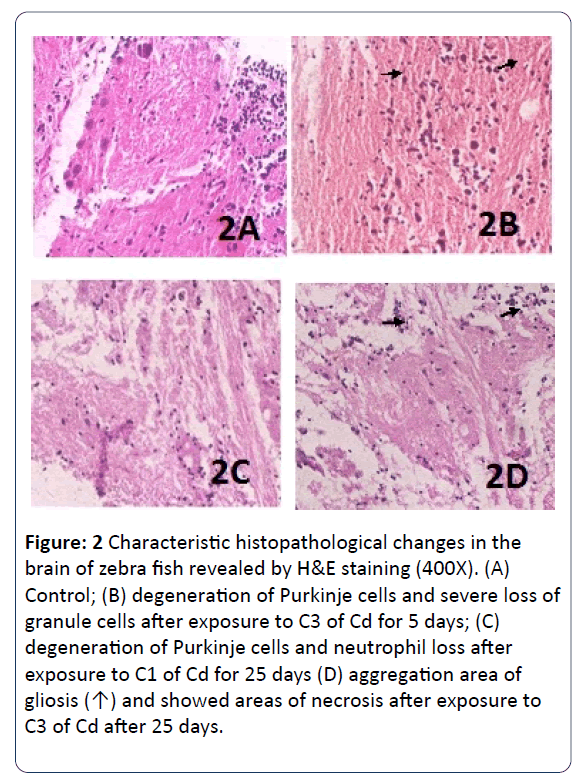
Figure 2: Characteristic histopathological changes in the brain of zebra fish revealed by H&E staining (400X). (A) Control; (B) degeneration of Purkinje cells and severe loss of granule cells after exposure to C3 of Cd for 5 days; (C) degeneration of Purkinje cells and neutrophil loss after exposure to C1 of Cd for 25 days (D) aggregation area of gliosis (↑) and showed areas of necrosis after exposure to C3 of Cd after 25 days.
Discussion
In the present study, LC50 of Cd for zebrafish after 96 h exposure was 9.68 mg L-1. So far, 96 h LC50 of Cd showed considerable differences among fish species. For example, Synechogobius hasta has a 96 h LC50 of 0.79 mg L-1 [5]; Ophiocephalus striatus (Channa) has a 96 h LC50 of 0.63 mg L-1 [15], while edible carp Catla catla has a LC50 value of 4.53 mg L-1 [16]. These values show that Cd is less toxic to zebrafish than other species, similar values are found to Clarias macrocephalus × Clarias gariepinos, 13.6 mg L-1 (LC50-96 h) [17] and to L. rohita 22.92 mg L-1 [18]. However, the differences in the 24, 48, 72 and 96 h LC50 values between zebrafish and other fishes may be attributed to the fact that metal induced changes in physiology might differ from metal to metal, species to species, and also influenced by chemical structure of metal compound and experimental conditions (water temperature, salinity, oxygen content and pH) [19].
Previous studies have evaluated the neurotoxicity of Cd in vitro. Unlike lead, an uncompetitive inhibitor which affected the enzyme-substrate binding affinity, Cd was a noncompetitive inhibitor to acetyl cholinesterase and the activity of this enzyme decreased by 50% when exposed to Cd of 5.7 mM for 77 min [12,20]. Forecasting the toxic impacts of Cd to fishes in natural exposure environments stays tough though many studies have examined the relationship between metal exposure, accumulation and toxicity under laboratory circumstances. Fish accumulate Cd in the tissues mainly through contaminated water and their diet [21]. Furthermore, waterborne Cd can be accumulated in zebrafish more efficiently than dietary Cd. Increasing numbers of research have demonstrated several factors affecting Cd accumulation in fish tissues, including environmental metal concentration and time of exposure [22]. Many authors have shown that animal tissues could accumulate heavy metals in dose and time dependent manner. Fishes have the capacity to accumulate heavy metal in their tissues by gill surface and the wall of gut tract to greater levels than the environmental toxin concentration [23]. In agreement to our results, Malekpouri et al. [24] noted that the accumulation of Cd in muscles of common carp (Cyprious carpio) has been elevated under increased concentration and duration of Cd exposure, as well as the maximum accumulation was in the third treatment (i.e., 100 ppb for dissolved cadmium) after 90 days of exposure. Kim et al. [25] also showed a similar increasing pattern of Cd accumulation in olive flounder muscle. Reynders et al. [26] also indicated that common carp was dependent on the time of exposure and the doses of Cd.
The mineral accumulation that we detected is also clearly the cause of the behavioral response observed in fish after 15 days of exposure. And the change became more pronounced at 25 days, when the zebrafish showed hyperventilation in addition to erratic swimming and excess mucus were secreted on their opercular surface. Similar observation of Paralichthys olivaceus also showed erratic swimming and excessive mucus production on their opercular surface [27], as well as in Channa punctatus [28]. It has been verified that the toxicity of Cd was mainly attributed to the free radicals and oxidative stress produced by this metal. Typically, it can combine with the thiol group of enzymes working in antioxidant processes or occupy the place of magnesium and calcium in certain reactions and disrupt the normal functioning [29]. Finally the intoxicated organism suffered from respiratory, excretory and circulatory disorders as well as neurotoxicity [1]. Behavioral alterations have been established as sensitive indicators of chemically induced stress in aquatic organisms. Moreover, behavioral alterations like erratic swimming, restlessness and surfacing may be a kind of avoiding reaction to the heavy metal narcotic effects or to change in sensitivity of chemoreceptors [19].
Pathological changes in fish are strong indicators of exposure to environmental pressures. Histopathology has been widely used as biomarker in assessing the health of fish exposed to Cd both in laboratory and field, as reported by Annabi et al. [30]. Au [13] confirmed the histopathological evaluation in fish as an extremely valuable tool to determine the toxicopathic impacts of material because they may better reflect the real health state of the animal than other biomarker/diagnosis methods. Our histological results showed that zebrafish exposed to Cd pollution were adversely affected at the tissue level compared to control groups, while the histopathological changes in the tissues were identical to typical histological responses to the contaminants. In our study, Cd caused changes to skeletal muscle structure of the zebrafish, as evidenced by swelling and necrosis of the skeletal muscle. Ramah [21] showed that the pathological changes in the muscle tissue as swelling and necrosis of muscle fibers occurred to grass carp (Ctenopharyngodan idella) when exposed to rice herbicides butachlor 1.5 kg ha-1, oxyfluorfen 0.25 kg ha-1 and thiobencarb 1.5 kg ha-1 for 12 days. Cd exposure caused serious damage to the fish brain, as evidenced by a series of lesions in the brain tissue by degeneration of Purkinje cells and necrosis, which may later affect normal physiological activities. Such changes in brain and skeletal muscle suggest that these tissues are sensitive to Cd toxicity. Similarly, Favorito et al. [31] indicated the histomorphological harm of Cd to zebrafish brain became clearer with increasing Cd accumulation in the brain. In contrast, Xing et al. [32] reported the histological examination with a low to moderate dose, incidence of chemical damage on brain and kidney tissues of common carp after a 40 days exposure to ATR, CPF and ATR/CPF combination. As an indicator of exposure to pollutants, histology represents a powerful tool to evaluate the impacts of contamination, particularly in relation to sub-lethal and chronic effects.
Conclusion
This histological investigation showed that these changes are a typical cellular response to heavy metals and may represent an important parameter for the study of environmental contaminants. Cd-accumulation in fish is affected by some parameters such as Cd-concentration and exposure time. Most of these primary factors must be taken into consideration while estimating ecotoxicological impacts of Cd on fish populations. Furthermore, large accumulations of Cd in fishes are of significant attention. Because the later consumption by human might cause some pathophysiological disturbances in human body. With regard to this, further investigation needs to be done in order to clarify accurate of fish Cd toxicity on human.
Conflict of Interest
The authors declare that there are no conflicts of interest.
References
- Sharma B, Shweta S, Nikhat JS (2014) Biomedical implications of heavy metals induced imbalances in redox systems. BioMed Research International 640754 (Published online).
- Cambier S, Gonzalez P, Durrieu G, Bourdineaud JP (2010) Cadmium-induced genotoxicity in zebra fish at environmentally relevant doses. Ecotox Environ Safe 73: 312-319.
- Zaki MS, Mostafa SO, Fawzi OM, Khafagy M, Bayumi FS (2009) Clinicopathological, biochemical and microbiological change on grey mullet exposed to cadmium chloride. American-Eurasian Journal of Agriculture and Environment Science 5: 20-23.
- Gonzalez P, Baudrimont M, Boudou A, Bourdineaud J-P (2006) Comparative effects of direct cadmium contamination on gene expression in gills, liver, skeletal muscles and brain of the zebrafish (Danio rerio). Biometals 19: 225-235.
- Liu XJ, Luo ZL, Xiong BX, H ZY, Li XD, et al. (2011) Antioxidant responses, hepatic inter mediary metabolism, histology and ultrastructure in Synechogobius hasta exposed to water borne cadmium. Ecotox Environ Safe 74: 1156–1163.
- Al-Sawafi AGA, Yan Y (2013) Alterations of Acetyl cholinesterase Activity and Antioxidant Capacity of Zebrafish Brain and Muscle Exposed to Sub lethal Level of Cadmium. International Journal of Environmental Science and Development 4: 327-330.
- Jayakumar P, Vattapparumbil IP (2006) Patterns of cadmium accumulation in selected tissues of the catfish Clarias batrachus (Linn.) exposed to sublethal concentration of cadmium chloride. Vet arhiv 76: 167-177.
- Gupta A, Rai DK, Pandey RS, Sharma B (2009) Analysis of some heavy metals in the riverine sediments and fish from river Ganges at Allahabad. Environ. monitor. Assess 157: 449-458.
- De Conto Cinier C, Petit-Ramel M, Faure R, Garin D, Bouvet Y (1999) Kinetics of cadmium accumulation and elimination in carp Cyprinus carpio tissues. Comparative Biochemistry and Physiology Part C: Pharmacology, Toxicology and Endocrinology 122: 345-352.
- Mansour SA, Sidky MM (2002) Eco toxicological studies. 3. Heavy metals contaminating water and fish from Fayoum Governorate, Egypt. Food Chem 78: 15-22.
- Fraser M, Surette C, Vaillancourt C (2013) Fish and seafood availability in markets in the Baie des Chaleurs region, New Brunswick, Canada: a heavy metal contamination baseline study. Environ Sci Pollut R 20: 761-770.
- Vivek KG, Shweta S, Anju A, Nikhat JS, Sharma B (2015) Phytochemicals Mediated Remediation of Neurotoxicity Induced by Heavy Metals. Biochemistry research international 534769 (Published online).
- Au DWT (2004) The application of histo-cytopathological biomarkers in marine pollution monitoring: a review. Mar Pollut Bull 48: 817-834.
- Finney DJ (1971) Probit Analysis: 3d Ed: Cambridge University Press.
- Bais UE, Lokhande M (2012) Effect of Cadmium Chloride on histopathological changes in the freshwater fish Ophiocephalus striatus (channa). International Journal of Zoological Research 8: 23-32.
- Sobha K, Poornima A, Harini P, Veeraiah K (2007) A study on biochemical changes in the fresh water fish, (hamilton) exposed to the heavy metal toxicant cadmium chloride. Kathmandu university journal of science, engineering and technology 3: 1-11.
- Pantunga N, Helander KG, Helandera HF, Cheevaporna V (2008) Histopathological alterations of hybrid Walking catfish (Clarias macrocephalus x Clarias gariepinus) in acute and subacute cadmium exposure. Environment Asia 1: 22-27.
- Latif A, Ali M, Kaoser R, Iqbal R, Umer K, et al. (2012) Effect of cadmium chloride and ascorbic acid exposure on the vital organs of freshwater Cyprinid, Labeo rohita. Afr J Biotechnol 11: 8398-8403.
- Kaushal BT, Mishra A (2013) Investigation of acute toxicity cadmium on snakehead fish Channa puntatus-a comparative toxicity analysis on median lethal concentration. International Journal of Advanced Biological Research 3: 289-294.
- Vivek KG, Abhishek K, Nikhat JS, Sharma B (2016) Rat brain acetyl cholinesterase as a biomaker cadmium induced neurotoxicity. Open access J of Tox 1: 555553 (Published online).
- Ramah K (2011) Histopathological study on the effect of rice herbicides on grass carp (Ctenopharyngodan idella). Afr J Biotechnol 10: 1112-1116.
- Annabi A, Said K, Messaoudi I (2013) Cadmium: Bioaccumulation, Histopathology and Detoxifying Mechanisms in Fish. American Journal of Research Communication 1: 60-79.
- Chevreuil M, Carru A-M, Chesterikoff A, Boet P, Tales E, et al. (1995) Contamination of fish from different areas of the river Seine (France) by organic (PCB and pesticides) and metallic (Cd, Cr, Cu, Fe, Mn, Pb and Zn) micropollutants. Sci total environ 162: 31-42.
- Malekpouri P, Moshtaghie AA, Hosseini R, Ebrahimi E (2011) Short and Long-Term Effects of Waterborne Cadmium on Growth and its Muscle Accumulation in Common Carp Fish (Cyprinus carpio), an Experimental Study. Turk J Fish Aquat Sc 11: 587-593.
- Kim S-G, Jee J-H, Kang J-C (2004) Cadmium accumulation and elimination in tissues of juvenile olive flounder, Paralichthys olivaceus after sub-chronic cadmium exposure. Environ Pollut 127: 117-123.
- Reynders H, Van Campenhout K, Bervoets L, De Coen WM, Blust R (2006) Dynamics of cadmium accumulation and effects in common carp (Cyprinus carpio) during simultaneous exposure to water and food (Tubifex tubifex). Environ toxicol chem 25: 1558-1567.
- Yildirim MZ, Benl A, Selv M, Ozkul A, Erkoç F, et al. (2006 ) Acute toxicity, behavioral changes, and histopathological effects of deltamethrin on tissues (gills, liver, brain, spleen, kidney, muscle, skin) of Nile tilapia (Oreochromis niloticus L.) fingerlings. Environ toxicol 21: 614-620.
- Kaushal BT, Mishra A (2011) A comparative toxicity analysis of cadmium compounds on morphological and behavioral aspects in air breathing freshwater fish Channa punctatus. International Journal of Science and Nature 2: 266-269.
- Vivek KG, Rajnish P, Nikhat JS, Sharma B (2015) Acetyl cholinesterase from Human Erythrocytes as a Surrogate Biomarker of Lead induced Neurotoxicity. Enzyme research 370705 (Published online).
- Annabi A, Messaoudi I, Kerkeni A, Said K (2011) Cadmium accumulation and histological lesion in mosquito fish (Gambusia affinis) tissues following acute and chronic exposure. Inter J Env Res 5: 745-756.
- Favorito R, Chiarelli G, Grimaldi MC, De Bonis S, Lancieri M, et al. (2011) Bioaccumulation of cadmium and its cytotoxic effect on zebrafish brain. Chem Ecol 27: 39-46.
- Xing H, Li S, Wang Z, Gao X, Xu S, et al. (2012) Histopathological changes and antioxidant response in brain and kidney of common carp exposed to atrazine and chlorpyrifos. Chemosphere 88: 377-383.